
| 产品编号 | bsm-54266R |
| 英文名称 | Wnt2b Recombinant Rabbit mAb |
| 中文名称 | 信号通路Wnt2B重组兔单抗 |
| 别 名 | WNT13; WNT2B_HUMAN; WNT2B; Protein Wnt-13; WNT2B_MOUSE; |
 |
Specific References (1) | bsm-54266R has been referenced in 1 publications.
[IF=5.736] Lan Zhang. et al. Inhibited HDAC3 promotes microRNA-376c-3p to suppress malignant phenotypes of gastric cancer cells by reducing WNT2b. Genomics. 2021 Jul;: WB ; Human.
|
| 研究领域 | 细胞生物 信号转导 |
| 抗体来源 | Rabbit |
| 克隆类型 | Recombinant |
| 克 隆 号 | 2G5 |
| 交叉反应 | Human,Mouse,Rat |
| 产品应用 | WB=1:500-1000,Flow-Cyt=1:50-100,ICC/IF=1:100-500
not yet tested in other applications. optimal dilutions/concentrations should be determined by the end user. |
| 理论分子量 | 44 kDa |
| 检测分子量 | 44 |
| 细胞定位 | 细胞外基质 分泌型蛋白 |
| 性 状 | Liquid |
| 浓 度 | 1mg/ml |
| 免 疫 原 | Recombinant human Wnt2b |
| 亚 型 | IgG |
| 纯化方法 | affinity purified by Protein A |
| 缓 冲 液 | 0.01M TBS (pH7.4) with 1% BSA, 0.02% Proclin300 and 50% Glycerol. |
| 保存条件 | Shipped at 4℃. Store at -20℃ for one year. Avoid repeated freeze/thaw cycles. |
| 注意事项 | This product as supplied is intended for research use only, not for use in human, therapeutic or diagnostic applications. |
| PubMed | PubMed |
| 产品介绍 |
WNT2B is a member of the wingless-type MMTV integration site (WNT) family of highly conserved, secreted signaling factors. WNT family members function in a variety of developmental processes including regulation of cell growth and differentiation and are characterized by a WNT-core domain. This gene may play a role in human development as well as human carcinogenesis. This gene produces two alternative transcript variants.This gene encodes a member of the wingless-type MMTV integration site (WNT) family of highly conserved, secreted signaling factors. WNT family members function in a variety of developmental processes including regulation of cell growth and differentiation and are characterized by a WNT-core domain. This gene may play a role in human development as well as human carcinogenesis. This gene produces two alternative transcript variants. Function: Ligand for members of the frizzled family of seven transmembrane receptors. Probable developmental protein. May be a signaling molecule which affects the development of discrete regions of tissues. Is likely to signal over only few cell diameters. May be involved in normal development or differentiation as well as in carcinogenesis. Subcellular Location: Secreted, extracellular space, extracellular matrix. Tissue Specificity: Isoform 1 is expressed in adult heart, brain, placenta, lung, prostate, testis, ovary, small intestine and colon. In the adult brain, it is mainly found in the caudate nucleus, subthalamic nucleus and thalamus. Also detected in fetal brain, lung and kidney. Isoform 2 is expressed in fetal brain, fetal lung, fetal kidney, caudate nucleus, testis and cancer cell lines. Post-translational modifications: Palmitoylation at Ser-243 is required for efficient binding to frizzled receptors. It is also required for subsequent palmitoylation at Cys-107. Palmitoylation is necessary for proper trafficking to cell surface (By similarity). Similarity: Belongs to the Wnt family. SWISS: Q93097 Gene ID: 7482 Database links: Entrez Gene: 7482 Human Entrez Gene: 22414 Mouse Omim: 601968 Human SwissProt: Q93097 Human SwissProt: O70283 Mouse Unigene: 27254 Cow |
| 产品图片 |
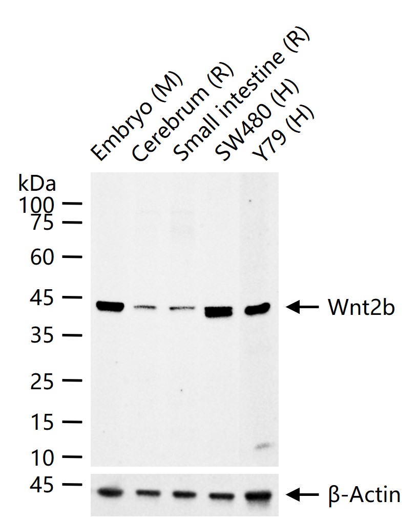
25 ug total protein per lane of various lysates (see on figure) probed with Wnt2b monoclonal antibody, unconjugated (bsm-54266R) at 1:1000 dilution and 4°C overnight incubation. Followed by conjugated secondary antibody incubation at r.t. for 60 min.
|
| 1、抗体溶解方法 | |
| 2、抗体修复方式 | |
| 3、常用试剂的配制 | |
| 4、免疫组化操作步骤 | |
| 5、免疫组化问题解答 | |
| 6、Western Blotting 操作步骤 | |
| 7、Western Blotting 问题解答 | |
| 8、关于肽链的设计 | |
| 9、多肽的溶解与保存 | |
| 10、酶标抗体效价测定程序 | |


