
| 产品编号 | bsm-52869R |
| 英文名称 | CD90/Thy-1 Recombinant Rabbit mAb |
| 中文名称 | CD90重组兔单抗 |
| 别 名 | CD90; CDw90; T25; Thy-1; Thy-1.2; Thy1.1; Thy1.2; CD7; THY1_HUMAN; THY1; Thy-1 antigen; THY1_MOUSE; THY1_RAT; |
| 研究领域 | 细胞生物 免疫学 神经生物学 |
| 抗体来源 | Rabbit |
| 克隆类型 | Recombinant |
| 克 隆 号 | 4C6 |
| 交叉反应 | Human,Mouse,Rat |
| 产品应用 | WB=1:1000-5000,IHC-P=1:50-200,IHC-F=1:50-200,IF=1:50-200
not yet tested in other applications. optimal dilutions/concentrations should be determined by the end user. |
| 理论分子量 | 12 kDa |
| 检测分子量 | 25 |
| 细胞定位 | 细胞膜 |
| 性 状 | Liquid |
| 浓 度 | 1mg/ml |
| 免 疫 原 | KLH conjugated synthetic peptide derived from human CD90/Thy-1 |
| 亚 型 | IgG |
| 纯化方法 | affinity purified by Protein A |
| 缓 冲 液 | 0.01M TBS (pH7.4) with 1% BSA, 0.02% Proclin300 and 50% Glycerol. |
| 保存条件 | Shipped at 4℃. Store at -20℃ for one year. Avoid repeated freeze/thaw cycles. |
| 注意事项 | This product as supplied is intended for research use only, not for use in human, therapeutic or diagnostic applications. |
| PubMed | PubMed |
| 产品介绍 |
Thy-1 or CD90 (Cluster of Differentiation 90) is a 25–37 kDa heavily N-glycosylated, glycophosphatidylinositol (GPI) anchored conserved cell surface protein with a single V-like immunoglobulin domain, originally discovered as a thymocyte antigen. Thy-1can be used as a marker for a variety of stem cells and for the axonal processes of mature neurons. Structural study of Thy-1 lead to the foundation of the Immunoglobulin superfamily, of which it is the smallest member, and led to some of the initial biochemical description and characterization of a vertebrate GPI anchor and also the first demonstration of tissue specific differential glycosylation. Function: May play a role in cell-cell or cell-ligand interactions during synaptogenesis and other events in the brain. Subunit: Cell membrane; Lipid-anchor, GPI-anchor. Tissue Specificity: Abundant in lymphoid tissues. Post-translational modifications: Glycosylation is tissue specific. Sialylation of N-glycans at Asn-93 in brain and at Asn-42, Asn-93 and Asn-117 in thymus. Similarity: Contains 1 Ig-like V-type (immunoglobulin-like) domain. SWISS: P04216 Gene ID: 7070 Database links: Entrez Gene: 7070 Human Entrez Gene: 21838 Mouse Omim: 188230 Human SwissProt: P04216 Human SwissProt: P01831 Mouse Unigene: 644697 Human Unigene: 3951 Mouse Unigene: 108198 Rat |
| 产品图片 |

Sample:
Lane 1: Mouse Cerebrum tissue lysates
Lane 2: Rat Thymus tissue lysates
Primary: Anti-CD90/Thy-1 (bsm-52869R) at 1/2000 dilution
Secondary: IRDye800CW Goat Anti-Rabbit IgG at 1/20000 dilution
Predicted band size: 12 kDa
Observed band size: 25 kDa
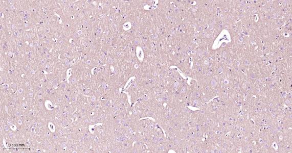
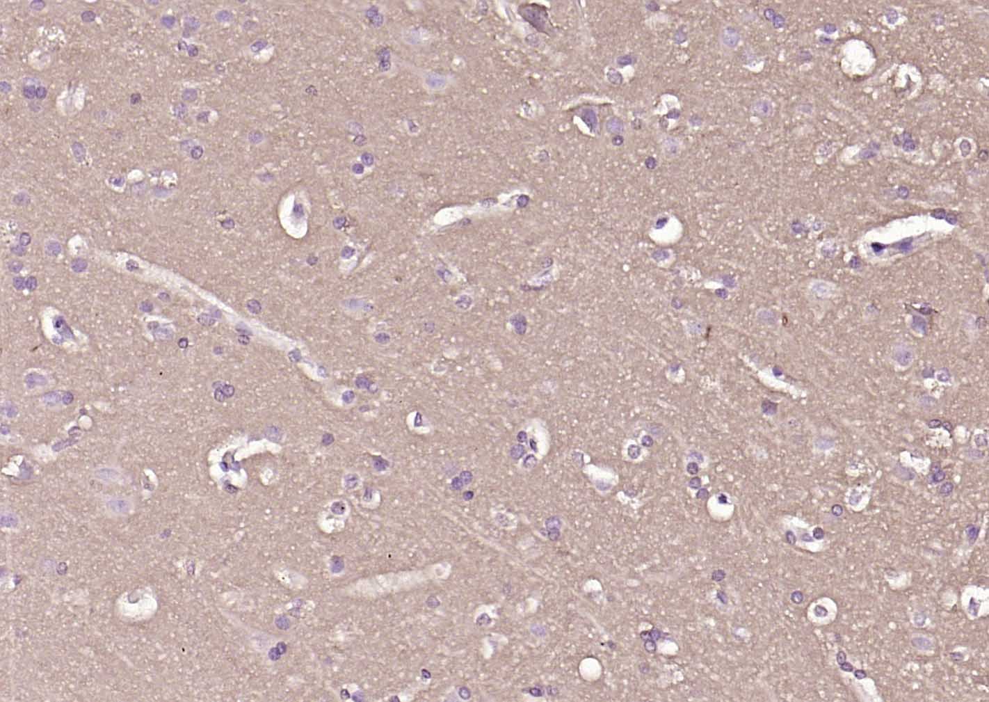
Paraformaldehyde-fixed, paraffin embedded Rat Cerebrum; Antigen retrieval by boiling in sodium citrate buffer (pH6.0) for 15 min; Antibody incubation with CD90/Thy-1 Monoclonal Antibody, Unconjugated(bsm-52869R) at 1:200 overnight at 4°C, followed by conjugation to the bs-0295G-HRP and DAB (C-0010) staining.
Paraformaldehyde-fixed, paraffin embedded Mouse Cerebrum; Antigen retrieval by boiling in sodium citrate buffer (pH6.0) for 15 min; Antibody incubation with CD90/Thy-1 Monoclonal Antibody, Unconjugated(bsm-52869R) at 1:200 overnight at 4°C, followed by conjugation to the bs-0295G-HRP and DAB (C-0010) staining.

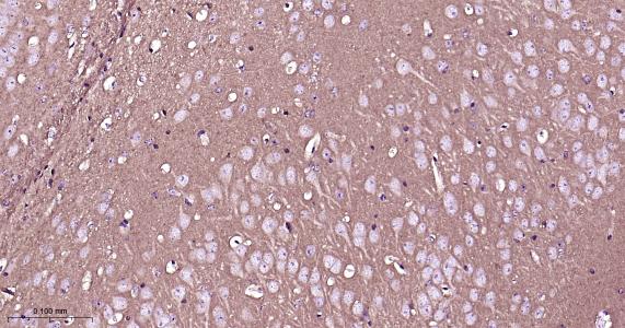
Paraformaldehyde-fixed, paraffin embedded Human Cerebrum; Antigen retrieval by boiling in sodium citrate buffer (pH6.0) for 15 min; Antibody incubation with CD90/Thy1 Monoclonal Antibody, Unconjugated(bsm-52869R) at 1:100 overnight at 4°C, followed by conjugation to the SP Kit (Rabbit, SP-0023) and DAB (C-0010) staining.
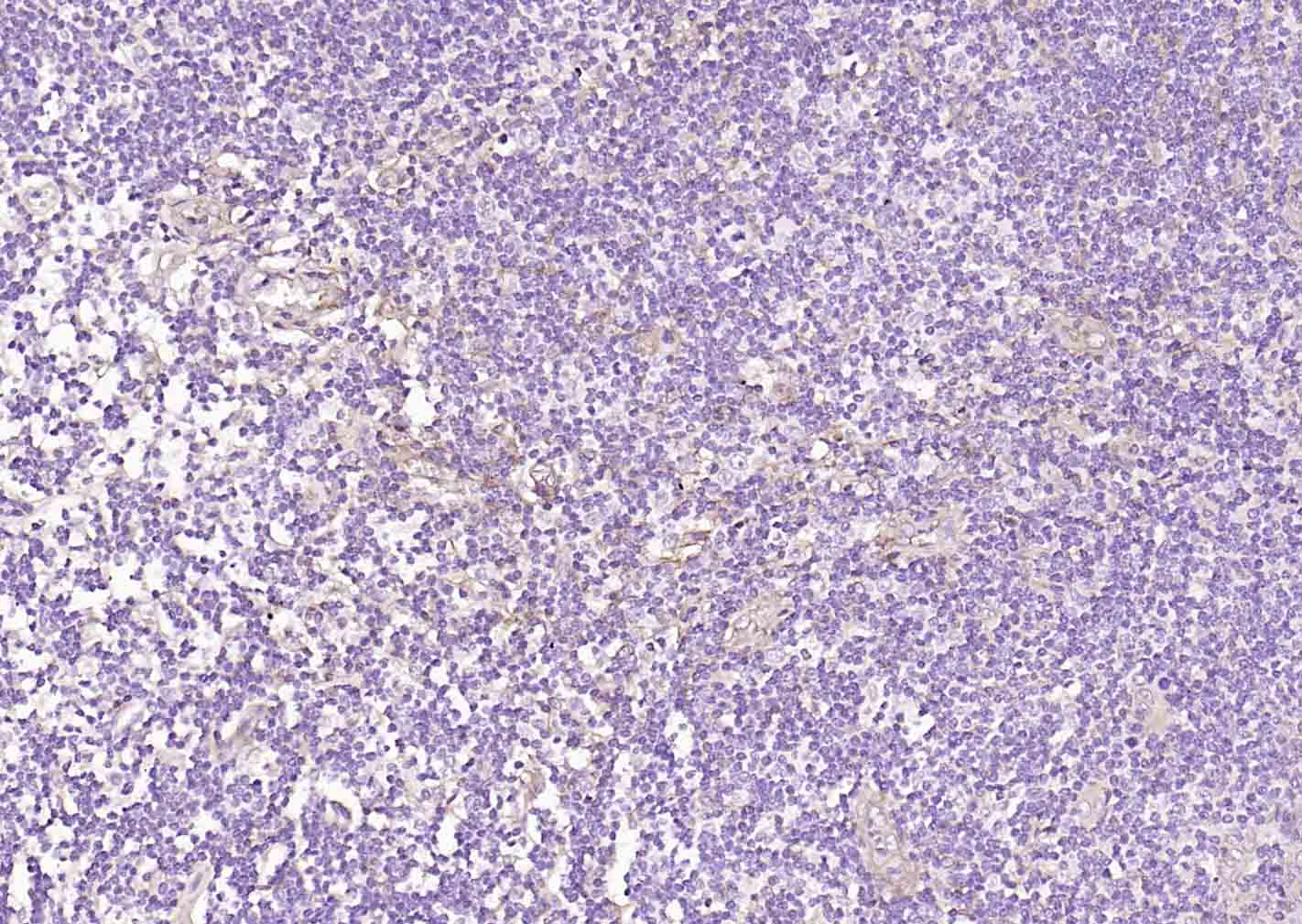
Paraformaldehyde-fixed, paraffin embedded Human Tonsil; Antigen retrieval by boiling in sodium citrate buffer (pH6.0) for 15 min; Antibody incubation with CD90/Thy1 Monoclonal Antibody, Unconjugated(bsm-52869R) at 1:100 overnight at 4°C, followed by conjugation to the SP Kit (Rabbit, SP-0023) and DAB (C-0010) staining.
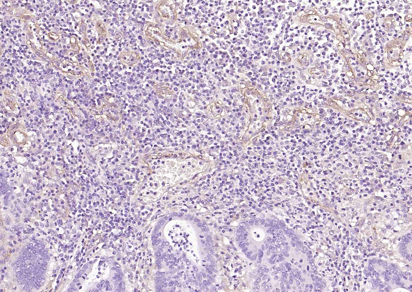
Paraformaldehyde-fixed, paraffin embedded Human Colon Cancer; Antigen retrieval by boiling in sodium citrate buffer (pH6.0) for 15 min; Antibody incubation with CD90/Thy1 Monoclonal Antibody, Unconjugated(bsm-52869R) at 1:100 overnight at 4°C, followed by conjugation to the SP Kit (Rabbit, SP-0023) and DAB (C-0010) staining.
Paraformaldehyde-fixed, paraffin embedded Human Glioma; Antigen retrieval by boiling in sodium citrate buffer (pH6.0) for 15 min; Antibody incubation with CD90/Thy1 Monoclonal Antibody, Unconjugated(bsm-52869R) at 1:100 overnight at 4°C, followed by conjugation to the SP Kit (Rabbit, SP-0023) and DAB (C-0010) staining.
|
| 1、抗体溶解方法 | |
| 2、抗体修复方式 | |
| 3、常用试剂的配制 | |
| 4、免疫组化操作步骤 | |
| 5、免疫组化问题解答 | |
| 6、Western Blotting 操作步骤 | |
| 7、Western Blotting 问题解答 | |
| 8、关于肽链的设计 | |
| 9、多肽的溶解与保存 | |
| 10、酶标抗体效价测定程序 | |


