
| 产品编号 | bsm-52283R |
| 英文名称 | SCARB1/Scavenger Receptor BI Recombinant Rabbit mAb |
| 中文名称 | 高密度脂蛋白受体/清道夫受体重组兔单抗 |
| 别 名 | CD36L1; CLA-1; CLA1; HDLCQ6; HDLQTL6; SR-BI; SRB1; AMRF; CD36L2; EPM4; HLGP85; LGP85; LIMP-2; LIMPII; SR-BII; 9330185J12Rik; LIMP II; MLGP85; CD36; Chohd1; D5Ertd460e; Hdlq1; Hlb398; SR-B1; SRBI; mSR-BI; SCRB2_HUMAN; SCARB2; 85 kDa lysosomal membrane sial |
| 研究领域 | 免疫学 生长因子和激素 细胞膜受体 糖尿病 |
| 抗体来源 | Rabbit |
| 克隆类型 | Recombinant |
| 克 隆 号 | 1C2 |
| 交叉反应 | Human,Mouse,Rat |
| 产品应用 | WB=1:500-2000,IHC-P=1:100-500,IHC-F=1:200-500,IF=1:100-500,ICC/IF=1:50-200
not yet tested in other applications. optimal dilutions/concentrations should be determined by the end user. |
| 理论分子量 | 61 kDa |
| 检测分子量 | 75 |
| 细胞定位 | 细胞浆 细胞膜 |
| 性 状 | Liquid |
| 浓 度 | 1mg/ml |
| 免 疫 原 | KLH conjugated synthetic peptide derived from human SCARB1/Scavenger Receptor BI |
| 亚 型 | IgG |
| 纯化方法 | affinity purified by Protein A |
| 缓 冲 液 | 0.01M TBS (pH7.4) with 1% BSA, 0.02% Proclin300 and 50% Glycerol. |
| 保存条件 | Shipped at 4℃. Store at -20℃ for one year. Avoid repeated freeze/thaw cycles. |
| 注意事项 | This product as supplied is intended for research use only, not for use in human, therapeutic or diagnostic applications. |
| PubMed | PubMed |
| 产品介绍 |
High density lipoproteins (HDLs) play a critical role in cholesterol metabolism and their plasma concentrations are inversely correlated with risk for atherosclerosis. The SR-BI (Scavenger Receptor BI) protein binds HDLs and mediates selective uptake of HDL cholesteryl ester. SR-BI binds HDL with high affinity, is expressed primarily in liver and nonplacental steroidgenic tissues, and mediates selective cholesterol uptake by a distinct mechanism. In mice, it seems that SR-BI plays a key role in determining the levels of plasma lipoprotein cholesterol and the accumulation of cholesterol stores in the adrenal gland. Scavenging Receptor SR-BI plays a critical role in HCV attachment and/or cell entry by interacting with HCV E1/E2 glycoproteins heterodimer. Function: Receptor for different ligands such as phospholipids, cholesterol ester, lipoproteins, phosphatidylserine and apoptotic cells. Probable receptor for HDL, located in particular region of the plasma membrane, called caveolae. Facilitates the flux of free and esterified cholesterol between the cell surface and extracellular donors and acceptors, such as HDL and to a lesser extent, apoB-containing lipoproteins and modified lipoproteins. Probably involved in the phagocytosis of apoptotic cells, via its phosphatidylserine binding activity. Receptor for hepatitis C virus glycoprotein E2. Binding between SCARB1 and E2 was found to be independent of the genotype of the viral isolate. Plays an important role in the uptake of HDL cholesteryl ester. Subunit: Plays a critical role in HCV attachment and/or cell entry by interacting with HCV E1/E2 glycoproteins heterodimer. The C-terminal region binds to PDZK1. Subcellular Location: Cell membrane; Multi-pass membrane protein. Membrane, caveola; Multi-pass membrane protein. Note=Predominantly localized to cholesterol and sphingomyelin-enriched domains within the plasma membrane, called caveolae. Tissue Specificity: Widely expressed. The six cysteines of the extracellular domain are all involved in intramolecular disulfide bonds. Post-translational modifications: N-glycosylated. Similarity: Belongs to the CD36 family. SWISS: Q8WTV0 Gene ID: 950 |
| 产品图片 |
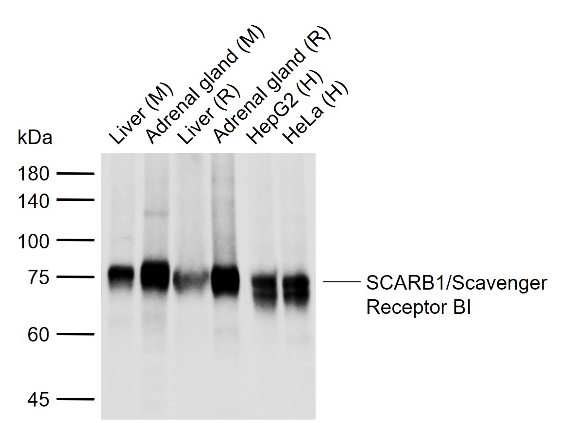
Sample:
Lane 1: Mouse Liver tissue lysates
Lane 2: Mouse Adrenal gland tissue lysates
Lane 3: Rat Liver tissue lysates
Lane 4: Rat Adrenal gland tissue lysates
Lane 5: Human HepG2 cell lysates
Lane 6: Human HeLa cell lysates
Primary: Anti-SCARB1/Scavenger Receptor BI (bsm-52283R) at 1/500 dilution
Secondary: IRDye800CW Goat Anti-Rabbit IgG at 1/20000 dilution
Predicted band size: 61 kDa
Observed band size: 75 kDa
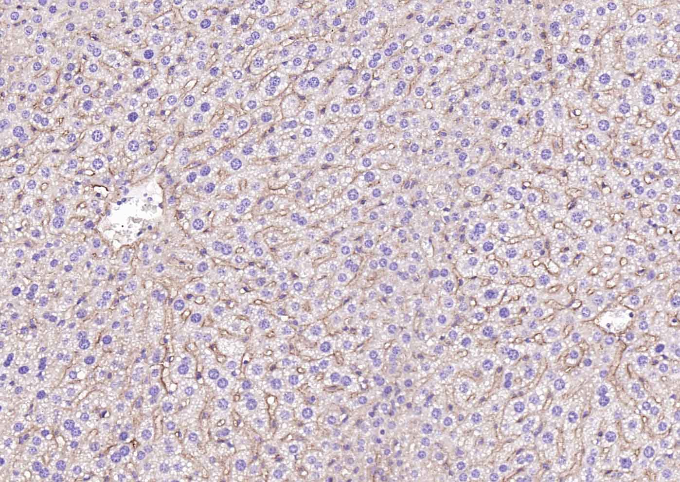
Paraformaldehyde-fixed, paraffin embedded (mouse liver); Antigen retrieval by boiling in sodium citrate buffer (pH6.0) for 15min; Block endogenous peroxidase by 3% hydrogen peroxide for 20 minutes; Blocking buffer (normal goat serum) at 37°C for 30min; Incubation with (SCARB1/Scavenger Receptor BI) Monoclonal Antibody, Unconjugated (bsm-52283R) at 1:2000 overnight at 4°C, followed by operating according to SP Kit(Rabbit) (sp-0023) instructionsand DAB staining.
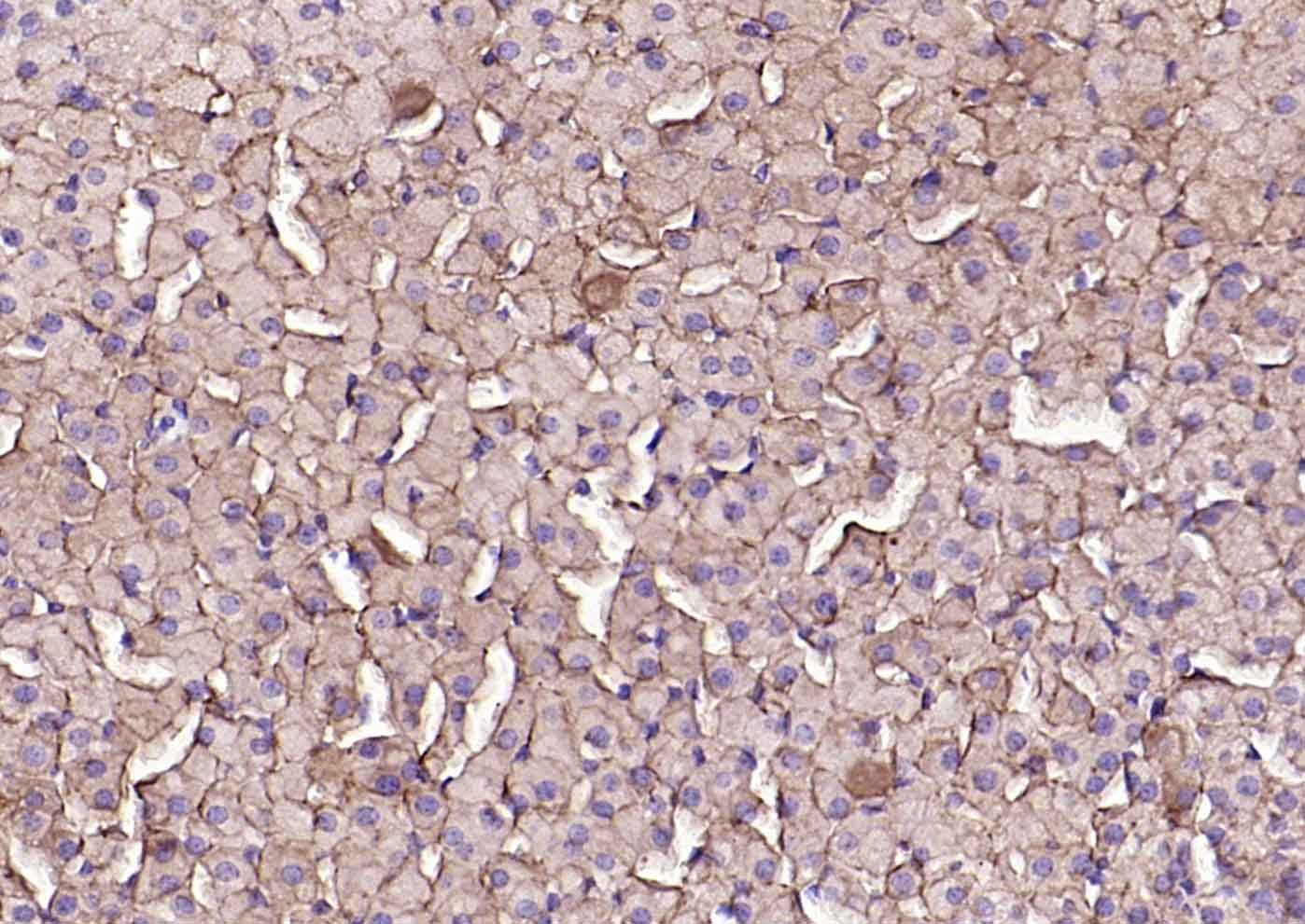
Paraformaldehyde-fixed, paraffin embedded (mouse adrenal gland); Antigen retrieval by boiling in sodium citrate buffer (pH6.0) for 15min; Block endogenous peroxidase by 3% hydrogen peroxide for 20 minutes; Blocking buffer (normal goat serum) at 37°C for 30min; Incubation with (SCARB1/Scavenger Receptor BI) Monoclonal Antibody, Unconjugated (bsm-52283R) at 1:2000 overnight at 4°C, followed by operating according to SP Kit(Rabbit) (sp-0023) instructionsand DAB staining.

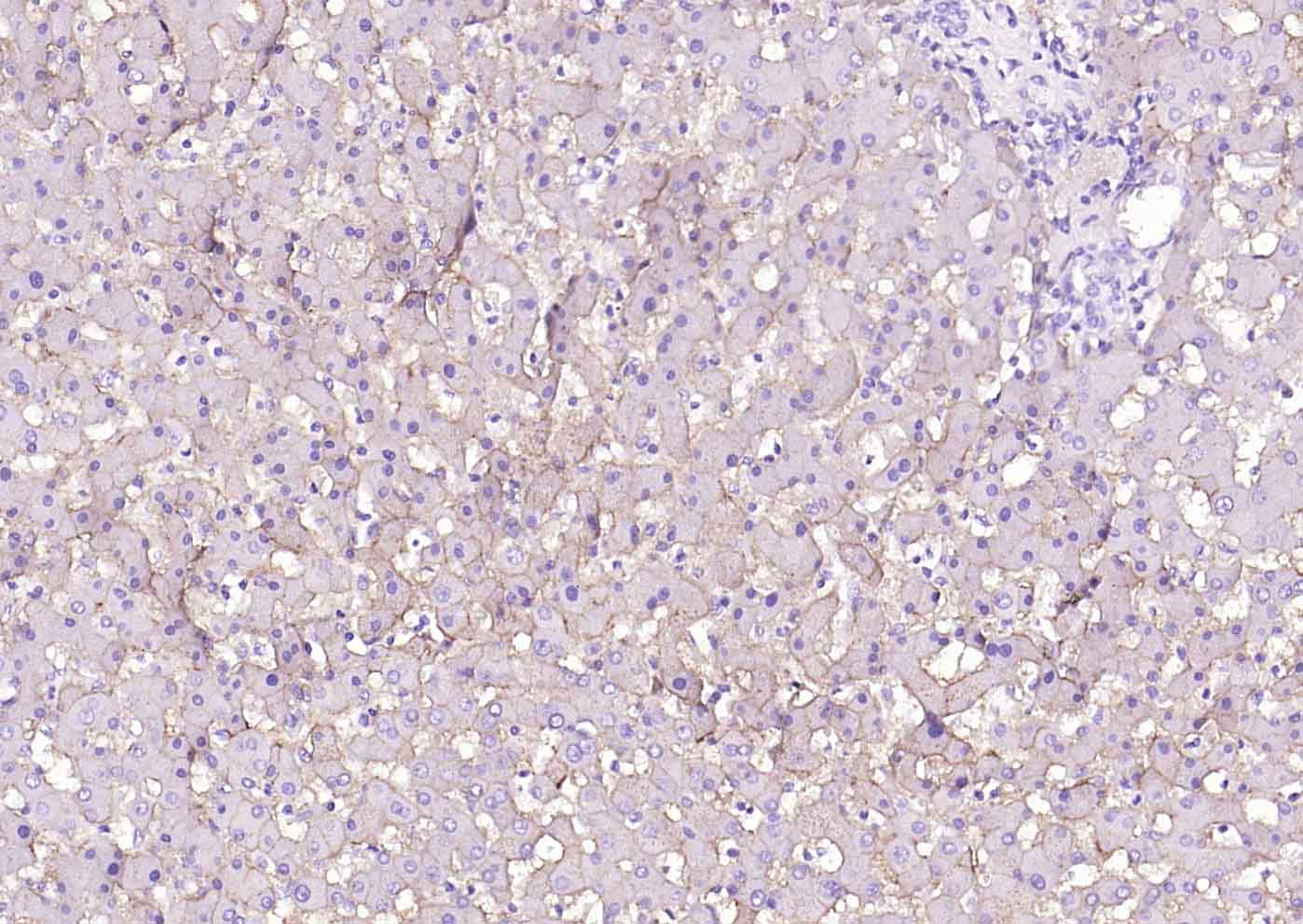
Paraformaldehyde-fixed, paraffin embedded (human adrenal gland); Antigen retrieval by boiling in sodium citrate buffer (pH6.0) for 15min; Block endogenous peroxidase by 3% hydrogen peroxide for 20 minutes; Blocking buffer (normal goat serum) at 37°C for 30min; Incubation with (SCARB1/Scavenger Receptor BI) Monoclonal Antibody, Unconjugated (bsm-52283R) at 1:2000 overnight at 4°C, followed by operating according to SP Kit(Rabbit) (sp-0023) instructionsand DAB staining.
Paraformaldehyde-fixed, paraffin embedded (human liver); Antigen retrieval by boiling in sodium citrate buffer (pH6.0) for 15min; Block endogenous peroxidase by 3% hydrogen peroxide for 20 minutes; Blocking buffer (normal goat serum) at 37°C for 30min; Incubation with (SCARB1/Scavenger Receptor BI) Monoclonal Antibody, Unconjugated (bsm-52283R) at 1:2000 overnight at 4°C, followed by operating according to SP Kit(Rabbit) (sp-0023) instructionsand DAB staining.
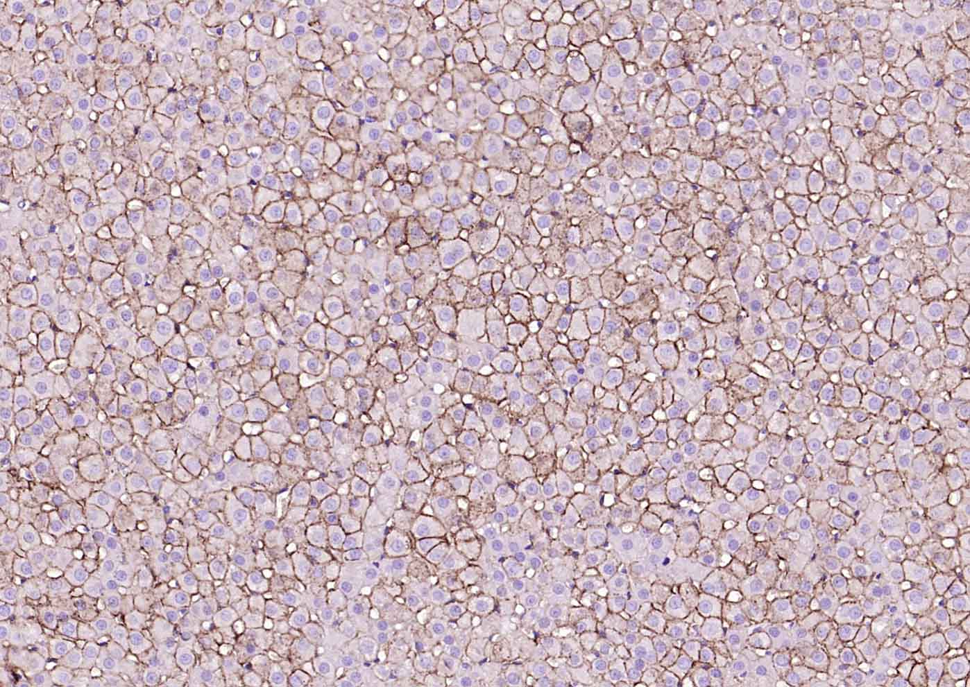
Paraformaldehyde-fixed, paraffin embedded (rat adrenal gland); Antigen retrieval by boiling in sodium citrate buffer (pH6.0) for 15min; Block endogenous peroxidase by 3% hydrogen peroxide for 20 minutes; Blocking buffer (normal goat serum) at 37°C for 30min; Incubation with (SCARB1/Scavenger Receptor BI) Monoclonal Antibody, Unconjugated (bsm-52283R) at 1:2000 overnight at 4°C, followed by operating according to SP Kit(Rabbit) (sp-0023) instructionsand DAB staining.
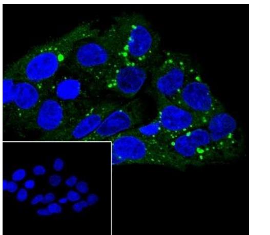
Cell line: HepG2
Fixation: 4% Paraformaldehyde
Permeabilization: 0.1% TritonX-100
Primary Ab dilution: 1:50
Primary Ab incubation condition: 4°C
overnight
Secondary Ab: Goat Anti-Rabbit IgG
Nuclear counter stain: DAPI (Blue)
Comment: Color green is the positive signal for
bsm-52283R
|
| 1、抗体溶解方法 | |
| 2、抗体修复方式 | |
| 3、常用试剂的配制 | |
| 4、免疫组化操作步骤 | |
| 5、免疫组化问题解答 | |
| 6、Western Blotting 操作步骤 | |
| 7、Western Blotting 问题解答 | |
| 8、关于肽链的设计 | |
| 9、多肽的溶解与保存 | |
| 10、酶标抗体效价测定程序 | |


