
| 产品编号 | bsm-54283R |
| 英文名称 | Furin Recombinant Rabbit mAb |
| 中文名称 | 弗林蛋白酶重组兔单抗 |
| 别 名 | FUR; PACE; PCSK3; SPC1; 9130404I01Rik; FURIN_HUMAN; FURIN; Dibasic-processing enzyme; Paired basic amino acid residue-cleaving enzyme (PACE); 3.4.21.75; FURIN_MOUSE; Prohormone convertase 3; FURIN_RAT; |
| 研究领域 | 细胞生物 信号转导 泛素 |
| 抗体来源 | Rabbit |
| 克隆类型 | Recombinant |
| 克 隆 号 | |
| 交叉反应 | Human,Mouse (predicted: Rat) |
| 产品应用 | WB=1:500-200,IHC-P=1:100-500,IHC-F=1:400-800,IF=1:100-500
not yet tested in other applications. optimal dilutions/concentrations should be determined by the end user. |
| 理论分子量 | 74 kDa |
| 检测分子量 | 87 |
| 细胞定位 | 细胞浆 细胞膜 细胞外基质 |
| 性 状 | Liquid |
| 浓 度 | 1mg/ml |
| 免 疫 原 | KLH conjugated synthetic peptide derived from human Furin |
| 亚 型 | IgG |
| 纯化方法 | affinity purified by Protein A |
| 缓 冲 液 | 0.01M TBS (pH7.4) with 1% BSA, 0.02% Proclin300 and 50% Glycerol. |
| 保存条件 | Shipped at 4℃. Store at -20℃ for one year. Avoid repeated freeze/thaw cycles. |
| 注意事项 | This product as supplied is intended for research use only, not for use in human, therapeutic or diagnostic applications. |
| PubMed | PubMed |
| 产品介绍 |
Furin is a calcium-dependent serine endoprotease that belongs to the subtilisin-like proprotein convertase family. The members of this family process latent precursor proteins into their biologically active products. Furin cleaves at paired basic amino acid processing sites within proparathyroid hormone, transforming growth factor β 1 precursor, proalbumin, pro-β-secretase, membrane type-1 matrix metalloproteinase, β subunit of pro-nerve growth factor and von Willebrand factor. Furin can directly cleave proMMP-2 within the ttrans-Golgi network leading to an inactive form of matrix metalloproteinase-2 (MMP-2). Furin is synthesized as an inactive zymogen that may minimize the occurrence of premature enzymatic activity that would lead to alternative protein activation or degradation. The inhibitory mechanism is based on the presence of an inactivating prosegment at the NH2 terminal of the Furin. After initial autocatalytic cleavage, the prosegment remains tightly associated until it reaches the trans-Golgi network where the dissociation of the prosegment and activation of furin occurs. Function: Furin is likely to represent the ubiquitous endoprotease activity within constitutive secretory pathways and capable of cleavage at the RX(K/R)R consensus motif. Subunit: Interacts with FLNA (By similarity). Binds to PACS1 which mediates TGN localization and connection to clathrin adapters. Subcellular Location: Golgi apparatus > trans-Golgi network membrane. Cell membrane. Shuttles between the trans-Golgi network and the cell surface. Propeptide cleavage is a prerequisite for exit of furin molecules out of the endoplasmic reticulum (ER). A second cleavage within the propeptide occurs in the trans Golgi network (TGN), followed by the release of the propeptide and the activation of furin. Tissue Specificity: Seems to be expressed ubiquitously. Post-translational modifications: The inhibition peptide, which plays the role of an intramolecular chaperone, is autocatalytically removed in the endoplasmic reticulum (ER) and remains non-covalently bound to furin as a potent autoinhibitor. Following transport to the trans Golgi, a second cleavage within the inhibition propeptide results in propeptide dissociation and furin activation. Similarity: Belongs to the peptidase S8 family. Furin subfamily. Contains 1 homo B/P domain. SWISS: P09958 Gene ID: 5045 Database links: Entrez Gene: 5045 Human Entrez Gene: 18550 Mouse SwissProt: P09958 Human SwissProt: P23188 Mouse 在真核生物细胞中,许多具有生物活性的多肽和蛋白是在其分泌过程中由前体蛋白经内切蛋白酶切割后激活形成的.弗林蛋白酶(Furin)就是这个内切蛋白酶家族重要成员之一,它可以识别剪切多种蛋白质,如生长医子、血清蛋白、基质金属蛋白酶、受体、病毒囊膜蛋白和细菌外毒素等.近年来Furin得到了迅速而广泛的研究,本文简介了它的表达与加工运输、生物学功能、与病毒侵染的关系,以及它的抑制剂. |
| 产品图片 |
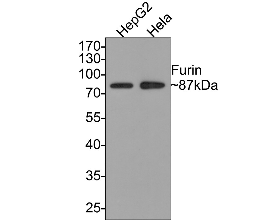
Western blot analysis of Furin on different lysates with Rabbit anti-Furin antibody (bsm-54283R) at 1/500 dilution.
Lane 1: HepG2 cell lysate
Lane 2: Hela cell lysate
Lysates/proteins at 10 µg/Lane.
Predicted band size: 87 kDa
Observed band size: 87 kDa
Exposure time: 1 minute;
10% SDS-PAGE gel.
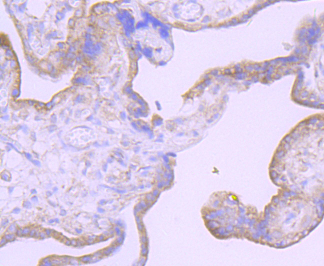
Immunohistochemical analysis of paraffin-embedded human placenta tissue using anti-Furin antibody. The section was pre-treated using heat mediated antigen retrieval with Tris-EDTA buffer (pH 8.0-8.4) for 20 minutes.The tissues were blocked in 5% BSA for 30 minutes at room temperature, washed with ddH2O and PBS, and then probed with the primary antibody (ET7107-37, 1/50) for 30 minutes at room temperature. The detection was performed using an HRP conjugated compact polymer system. DAB was used as the chromogen. Tissues were counterstained with hematoxylin and mounted with DPX.

Immunohistochemical analysis of paraffin-embedded human colon tissue using anti-Furin antibody. The section was pre-treated using heat mediated antigen retrieval with Tris-EDTA buffer (pH 8.0-8.4) for 20 minutes.The tissues were blocked in 5% BSA for 30 minutes at room temperature, washed with ddH2O and PBS, and then probed with the primary antibody (bsm-54283R, 1/50) for 30 minutes at room temperature. The detection was performed using an HRP conjugated compact polymer system. DAB was used as the chromogen. Tissues were counterstained with hematoxylin and mounted with DPX.

Immunohistochemical analysis of paraffin-embedded mouse brain tissue using anti-Furin antibody. The section was pre-treated using heat mediated antigen retrieval with Tris-EDTA buffer (pH 8.0-8.4) for 20 minutes.The tissues were blocked in 5% BSA for 30 minutes at room temperature, washed with ddH2O and PBS, and then probed with the primary antibody (bsm-54283R, 1/50) for 30 minutes at room temperature. The detection was performed using an HRP conjugated compact polymer system. DAB was used as the chromogen. Tissues were counterstained with hematoxylin and mounted with DPX.
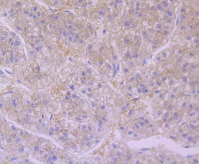
Immunohistochemical analysis of paraffin-embedded human liver tissue using anti-Furin antibody. The section was pre-treated using heat mediated antigen retrieval with Tris-EDTA buffer (pH 8.0-8.4) for 20 minutes.The tissues were blocked in 5% BSA for 30 minutes at room temperature, washed with ddH2O and PBS, and then probed with the primary antibody (bsm-54283R, 1/50) for 30 minutes at room temperature. The detection was performed using an HRP conjugated compact polymer system. DAB was used as the chromogen. Tissues were counterstained with hematoxylin and mounted with DPX.
|
| 1、抗体溶解方法 | |
| 2、抗体修复方式 | |
| 3、常用试剂的配制 | |
| 4、免疫组化操作步骤 | |
| 5、免疫组化问题解答 | |
| 6、Western Blotting 操作步骤 | |
| 7、Western Blotting 问题解答 | |
| 8、关于肽链的设计 | |
| 9、多肽的溶解与保存 | |
| 10、酶标抗体效价测定程序 | |


