
| 产品编号 | bsm-54152R |
| 英文名称 | MMP3 Recombinant Rabbit mAb |
| 中文名称 | 基质金属蛋白酶3重组兔单抗 |
| 别 名 | CHDS6; MMP-3; SL-1; STMY; STMY1; STR1; EMS-2; SLN-1; SLN1; STR-1; MMP3_HUMAN; MMP3; Matrix metalloproteinase-3 (MMP-3); Transin-1; 3.4.24.17; MMP3_MOUSE; MMP3_RAT; PTR1 protein; |
| 研究领域 | 肿瘤 |
| 抗体来源 | Rabbit |
| 克隆类型 | Recombinant |
| 克 隆 号 | 8B6 |
| 交叉反应 | Human,Mouse,Rat |
| 产品应用 | WB=1:500-2000,IHC-P=1:100-500,IHC-F=1:100-500,IF=1:100-500
not yet tested in other applications. optimal dilutions/concentrations should be determined by the end user. |
| 理论分子量 | 54 kDa |
| 检测分子量 | 54 |
| 细胞定位 | 细胞外基质 分泌型蛋白 |
| 性 状 | Liquid |
| 浓 度 | 1mg/ml |
| 免 疫 原 | A synthesized peptide derived from human MMP3: 443-477 |
| 亚 型 | IgG |
| 纯化方法 | affinity purified by Protein A |
| 缓 冲 液 | 0.01M TBS (pH7.4) with 1% BSA, 0.02% Proclin300 and 50% Glycerol. |
| 保存条件 | Shipped at 4℃. Store at -20℃ for one year. Avoid repeated freeze/thaw cycles. |
| 注意事项 | This product as supplied is intended for research use only, not for use in human, therapeutic or diagnostic applications. |
| PubMed | PubMed |
| 产品介绍 |
Proteins of the matrix metalloproteinase (MMP) family are involved in the breakdown of extracellular matrix in normal physiological processes, such as embryonic development, reproduction, and tissue remodeling, as well as in disease processes, such as arthritis and metastasis. Most MMP's are secreted as inactive proproteins which are activated when cleaved by extracellular proteinases. This gene encodes an enzyme which degrades fibronectin, laminin, collagens III, IV, IX, and X, and cartilage proteoglycans. The enzyme is thought to be involved in wound repair, progression of atherosclerosis, and tumor initiation. The gene is part of a cluster of MMP genes which localize to chromosome 11q22.3. [provided by RefSeq, Jul 2008]. Function: Can degrade fibronectin, laminin, gelatins of type I, III, IV, and V; collagens III, IV, X, and IX, and cartilage proteoglycans. Activates procollagenase. Subcellular Location: Secreted, extracellular space, extracellular matrix (Probable). Similarity: Belongs to the peptidase M10A family. Contains 4 hemopexin-like domains. SWISS: P08254 Gene ID: 4314 合成与降解(Synthesis and Degradation) 基质金属蛋白酶(matrix metalloproteinases, MMPs)是一族依赖锌离子而降解各种细胞外基质的蛋白酶,亦称IV型胶原酶或称明胶酶A,其主要功能为降解IV型胶原,因而它在肿瘤细胞突破基底膜屏障和浸润转移中起重要作用。 MMP3目前主要用于各种恶性肿瘤(如乳腺癌、胃肠道癌、卵巢癌、膀胱癌等)中的基底膜检测与肿瘤转移浸润的研究。 |
| 产品图片 |
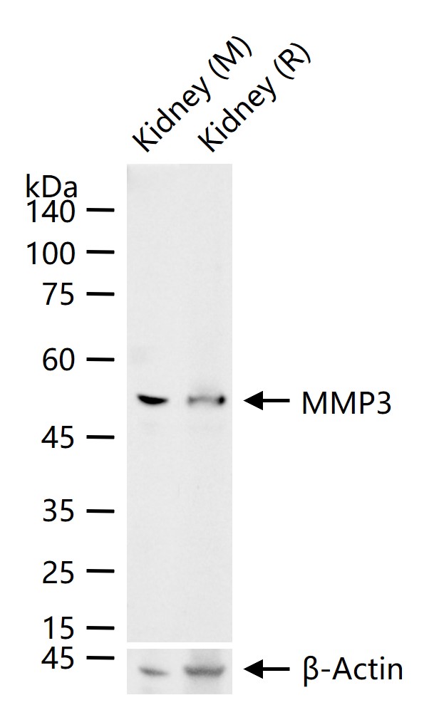
25 ug total protein per lane of various lysates (see on figure) probed with MMP3 monoclonal antibody, unconjugated (bsm-54152R) at 1:1000 dilution and 4°C overnight incubation. Followed by conjugated secondary antibody incubation at r.t. for 60 min.
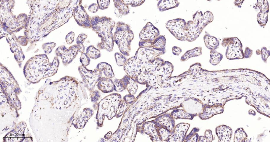
Paraformaldehyde-fixed, paraffin embedded Human Placenta; Antigen retrieval by boiling in sodium citrate buffer (pH6.0) for 15 min; The section was incubated with MMP3 Monoclonal Antibody, Unconjugated (bsm-54152R) at 1:200 overnight at 4°C, followed by conjugation to the bs-0295G-HRP and DAB (C-0010) staining.
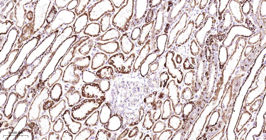
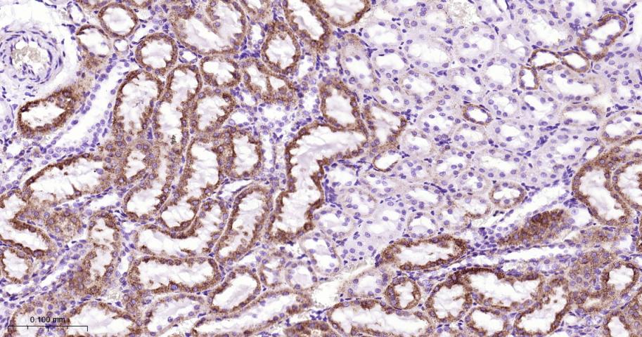
Paraformaldehyde-fixed, paraffin embedded Human Kidney; Antigen retrieval by boiling in sodium citrate buffer (pH6.0) for 15 min; The section was incubated with MMP3 Monoclonal Antibody, Unconjugated (bsm-54152R) at 1:200 overnight at 4°C, followed by conjugation to the bs-0295G-HRP and DAB (C-0010) staining.
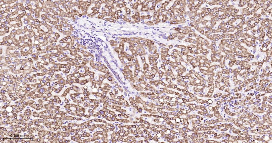
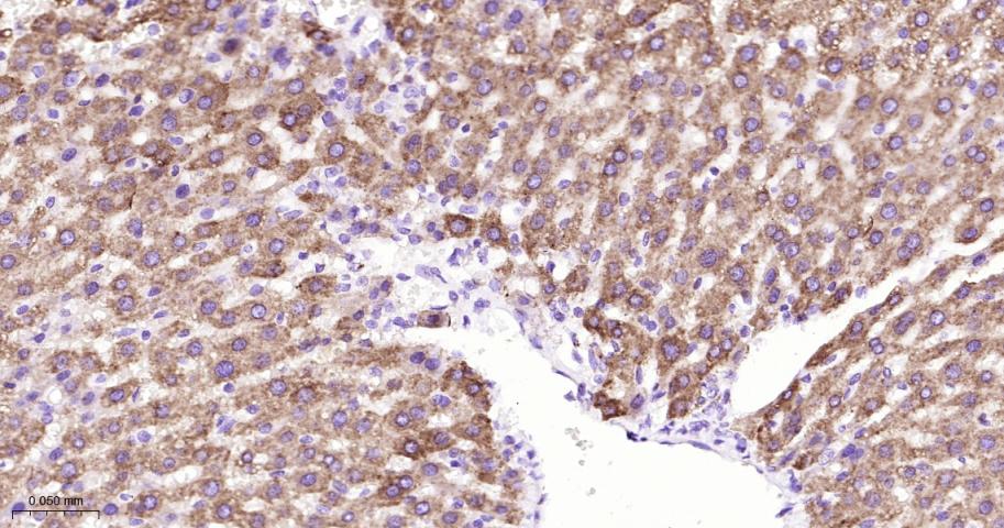
Paraformaldehyde-fixed, paraffin embedded Human Liver; Antigen retrieval by boiling in sodium citrate buffer (pH6.0) for 15 min; The section was incubated with MMP3 Monoclonal Antibody, Unconjugated (bsm-54152R) at 1:200 overnight at 4°C, followed by conjugation to the bs-0295G-HRP and DAB (C-0010) staining.
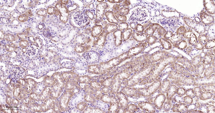
Paraformaldehyde-fixed, paraffin embedded Rat Kidney; Antigen retrieval by boiling in sodium citrate buffer (pH6.0) for 15 min; The section was incubated with MMP3 Monoclonal Antibody, Unconjugated (bsm-54152R) at 1:200 overnight at 4°C, followed by conjugation to the bs-0295G-HRP and DAB (C-0010) staining.
Paraformaldehyde-fixed, paraffin embedded Mouse Kidney; Antigen retrieval by boiling in sodium citrate buffer (pH6.0) for 15 min; The section was incubated with MMP3 Monoclonal Antibody, Unconjugated (bsm-54152R) at 1:200 overnight at 4°C, followed by conjugation to the bs-0295G-HRP and DAB (C-0010) staining.
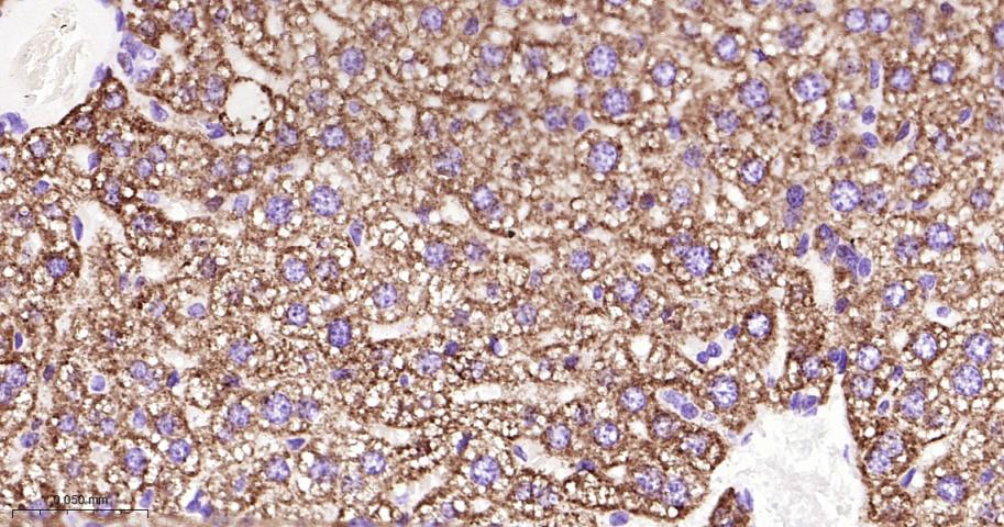
Paraformaldehyde-fixed, paraffin embedded Rat Liver; Antigen retrieval by boiling in sodium citrate buffer (pH6.0) for 15 min; The section was incubated with MMP3 Monoclonal Antibody, Unconjugated (bsm-54152R) at 1:200 overnight at 4°C, followed by conjugation to the bs-0295G-HRP and DAB (C-0010) staining.
Paraformaldehyde-fixed, paraffin embedded Mouse Liver; Antigen retrieval by boiling in sodium citrate buffer (pH6.0) for 15 min; The section was incubated with MMP3 Monoclonal Antibody, Unconjugated (bsm-54152R) at 1:200 overnight at 4°C, followed by conjugation to the bs-0295G-HRP and DAB (C-0010) staining.
|
| 1、抗体溶解方法 | |
| 2、抗体修复方式 | |
| 3、常用试剂的配制 | |
| 4、免疫组化操作步骤 | |
| 5、免疫组化问题解答 | |
| 6、Western Blotting 操作步骤 | |
| 7、Western Blotting 问题解答 | |
| 8、关于肽链的设计 | |
| 9、多肽的溶解与保存 | |
| 10、酶标抗体效价测定程序 | |


