
| 产品编号 | bsm-52323R |
| 英文名称 | Tubulin beta-III Recombinant Rabbit mAb |
| 中文名称 | 微管蛋白β3重组兔单抗 |
| 别 名 | CDCBM; CDCBM1; CFEOM3; CFEOM3A; FEOM3; TUBB4; beta-4; 3200002H15Rik; M(beta)3; M(beta)6; TBB3_HUMAN; TUBB3; Tubulin beta-4 chain; Tubulin beta-III; TBB3_MOUSE; TBB3_RAT; Neuron-specific class III beta-tubulin; tubulin beta 3 class III; tubulin, beta 3; fibrosis of extraocular muscles, congenital, 3; class III beta-tubulin |
 |
Specific References (2) | bsm-52323R has been referenced in 2 publications.
[IF=6.064] Le Wang. et al. Enteric nervous system damage caused by abnormal intestinal butyrate metabolism may lead to functional constipation. FRONT MICROBIOL. 2023; 14: 1117905 ICC ; Mouse.
[IF=5.195] Lu Hong. et al. Investigation of Naoluoxintong on the neural stem cells by facilitating proliferation and differentiation in vitro and on protecting neurons by up-regulating the expression of nestin in MCAO rats. J ETHNOPHARMACOL. 2022 Sep;:115684 WB ; Rat.
|
| 研究领域 | 细胞生物 神经生物学 细胞骨架 |
| 抗体来源 | Rabbit |
| 克隆类型 | Recombinant |
| 克 隆 号 | 3B7 |
| 交叉反应 | Human,Mouse,Rat |
| 产品应用 | WB=1:2000-20000,IHC-P=1:100-500,IHC-F=1:100-500,IF=1:100-500,Flow-Cyt=1:50-100,ICC/IF=1:100-500
not yet tested in other applications. optimal dilutions/concentrations should be determined by the end user. |
| 理论分子量 | 50kDa |
| 检测分子量 | 50 |
| 细胞定位 | 细胞浆 |
| 性 状 | Liquid |
| 浓 度 | 1mg/ml |
| 免 疫 原 | A synthesized peptide derived from human beta III Tubulin: 400-450/450 |
| 亚 型 | IgG |
| 纯化方法 | affinity purified by Protein A |
| 缓 冲 液 | 0.01M TBS (pH7.4) with 1% BSA, 0.02% Proclin300 and 50% Glycerol. |
| 保存条件 | Shipped at 4℃. Store at -20℃ for one year. Avoid repeated freeze/thaw cycles. |
| 注意事项 | This product as supplied is intended for research use only, not for use in human, therapeutic or diagnostic applications. |
| PubMed | PubMed |
| 产品介绍 |
Tubulin is the major constituent of microtubules. It binds two moles of GTP, one at an exchangeable site on the beta chain and one at a non-exchangeable site on the alpha-chain. TUBB3 plays a critical role in proper axon guidance and mantainance. Function: Tubulin is the major constituent of microtubules. It binds two moles of GTP, one at an exchangeable site on the beta chain and one at a non-exchangeable site on the alpha-chain. TUBB3 plays a critical role in proper axon guidance and mantainance. Subcellular Location: Cytoplasm, cytoskeleton. Tissue Specificity: Expression is primarily restricted to central and peripheral nervous system. Post-translational modifications: Some glutamate residues at the C-terminus are polyglutamylated. This modification occurs exclusively on glutamate residues and results in polyglutamate chains on the gamma-carboxyl group. Also monoglycylated but not polyglycylated due to the absence of functional TTLL10 in human. Monoglycylation is mainly limited to tubulin incorporated into axonemes (cilia and flagella) whereas glutamylation is prevalent in neuronal cells, centrioles, axonemes, and the mitotic spindle. Both modifications can coexist on the same protein on adjacent residues, and lowering glycylation levels increases polyglutamylation, and reciprocally. The precise function of such modifications is still unclear but they regulate the assembly and dynamics of axonemal microtubules. DISEASE: Defects in TUBB3 are the cause of congenital fibrosis of extraocular muscles type 3A (CFEOM3A) [MIM:600638]. A congenital ocular motility disorder marked by restrictive ophthalmoplegia affecting extraocular muscles innervated by the oculomotor and/or trochlear nerves. It is clinically characterized by anchoring of the eyes in downward gaze, ptosis, and backward tilt of the head. Congenital fibrosis of extraocular muscles type 3 presents as a non-progressive, autosomal dominant disorder with variable expression. Patients may be bilaterally or unilaterally affected, and their oculo-motility defects range from complete ophthalmoplegia (with the eyes fixed in a hypo- and exotropic position), to mild asymptomatic restrictions of ocular movement. Ptosis, refractive error, amblyopia, and compensatory head positions are associated with the more severe forms of the disorder. In some cases the ocular phenotype is accompanied by additional features including developmental delay, corpus callosum agenesis, basal ganglia dysmorphism, facial weakness, polyneuropathy. Similarity: Belongs to the tubulin family. SWISS: Q13509 Gene ID: 10381 Database links: Entrez Gene: 10381 Human Entrez Gene: 431043 Chicken Entrez Gene: 22152 Mouse Omim: 602661 Human SwissProt: Q13509 Human SwissProt: Q9ERD7 Mouse Unigene: 511743 Human Unigene: 40068 Mouse Unigene: 43958 Rat |
| 产品图片 |
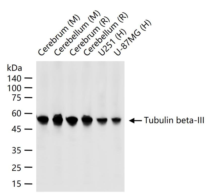
25 ug total protein per lane of various lysates (see on figure) probed with Tubulin beta-III monoclonal antibody, unconjugated (bsm-52323R) at 1:5000 dilution and 4°C overnight incubation. Followed by conjugated secondary antibody incubation at r.t. for 60 min.
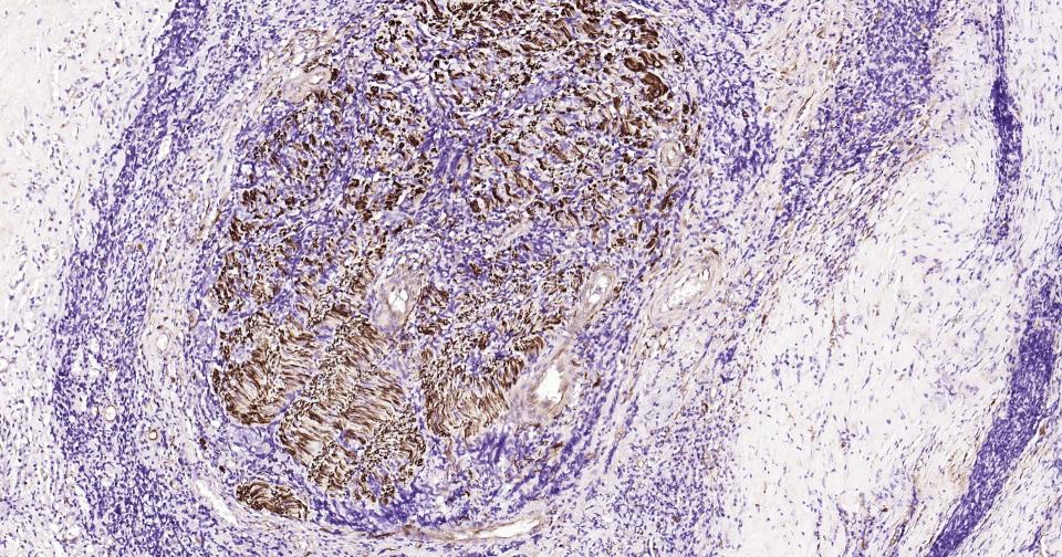
Paraformaldehyde-fixed, paraffin embedded Human Thyroid Tumor; Antigen retrieval by boiling in sodium citrate buffer (pH6.0) for 15 min; Antibody incubation with Tubulin beta-III Monoclonal Antibody, Unconjugated(bsm-52323R) at 1:200 overnight at 4°C, followed by conjugation to the SP Kit (Rabbit, SP-0023)and DAB (C-0010) staining.
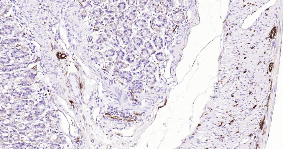
Paraformaldehyde-fixed, paraffin embedded Mouse Stomach; Antigen retrieval by boiling in sodium citrate buffer (pH6.0) for 15 min; Antibody incubation with Tubulin beta-III Monoclonal Antibody, Unconjugated(bsm-52323R) at 1:200 overnight at 4°C, followed by conjugation to the SP Kit (Rabbit, SP-0023)and DAB (C-0010) staining.
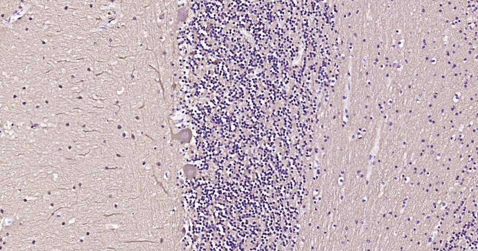
Paraformaldehyde-fixed, paraffin embedded Human Cerebellum; Antigen retrieval by boiling in sodium citrate buffer (pH6.0) for 15 min; Antibody incubation with Tubulin beta-III Monoclonal Antibody, Unconjugated(bsm-52323R) at 1:200 overnight at 4°C, followed by conjugation to the SP Kit (Rabbit, SP-0023)and DAB (C-0010) staining.
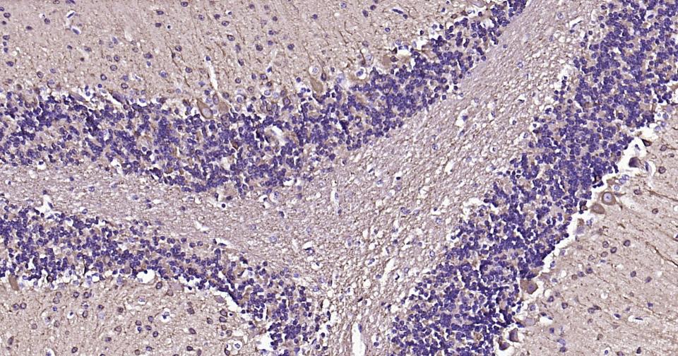
Paraformaldehyde-fixed, paraffin embedded Mouse Cerebellum; Antigen retrieval by boiling in sodium citrate buffer (pH6.0) for 15 min; Antibody incubation with Tubulin beta-III Monoclonal Antibody, Unconjugated(bsm-52323R) at 1:200 overnight at 4°C, followed by conjugation to the SP Kit (Rabbit, SP-0023)and DAB (C-0010) staining.
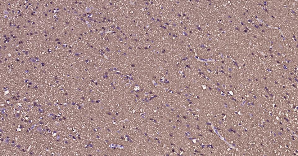
Paraformaldehyde-fixed, paraffin embedded Human Cerebrum; Antigen retrieval by boiling in sodium citrate buffer (pH6.0) for 15 min; Antibody incubation with Tubulin beta-III Monoclonal Antibody, Unconjugated(bsm-52323R) at 1:200 overnight at 4°C, followed by conjugation to the SP Kit (Rabbit, SP-0023)and DAB (C-0010) staining.
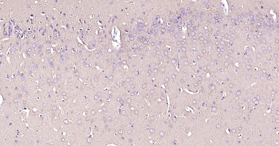
Paraformaldehyde-fixed, paraffin embedded Rat Cerebrum; Antigen retrieval by boiling in sodium citrate buffer (pH6.0) for 15 min; Antibody incubation with Tubulin beta-III Monoclonal Antibody, Unconjugated(bsm-52323R) at 1:200 overnight at 4°C, followed by conjugation to the SP Kit (Rabbit, SP-0023)and DAB (C-0010) staining.
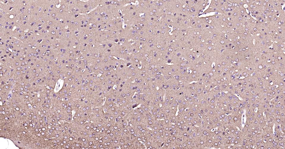
Paraformaldehyde-fixed, paraffin embedded Mouse Cerebrum; Antigen retrieval by boiling in sodium citrate buffer (pH6.0) for 15 min; Antibody incubation with Tubulin beta-III Monoclonal Antibody, Unconjugated(bsm-52323R) at 1:200 overnight at 4°C, followed by conjugation to the SP Kit (Rabbit, SP-0023)and DAB (C-0010) staining.
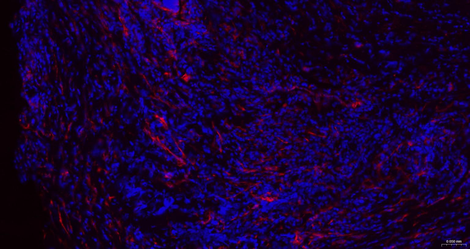
Paraformaldehyde-fixed, paraffin embedded Human Thyroid tumor; Antigen retrieval by boiling in sodium citrate buffer (pH6.0) for 15 min; The section was incubated with Tubulin beta-III Monoclonal Antibody, Unconjugated (bsm-52323R) at 1:200 overnight at 4°C. Followed by conjugated Goat Anti-Rabbit IgG antibody (Red, bs-0295G-BF594), DAPI (blue, C02-04002) was used to stain the cell nuclei.
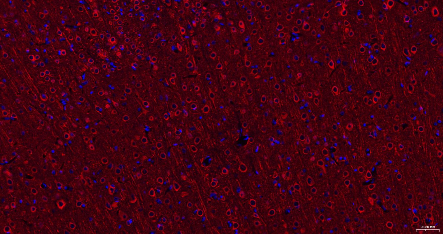
Paraformaldehyde-fixed, paraffin embedded Rat Cerebrum; Antigen retrieval by boiling in sodium citrate buffer (pH6.0) for 15 min; The section was incubated with Tubulin beta-III Monoclonal Antibody, Unconjugated (bsm-52323R) at 1:200 overnight at 4°C. Followed by conjugated Goat Anti-Rabbit IgG antibody (Red, bs-0295G-BF594), DAPI (blue, C02-04002) was used to stain the cell nuclei.
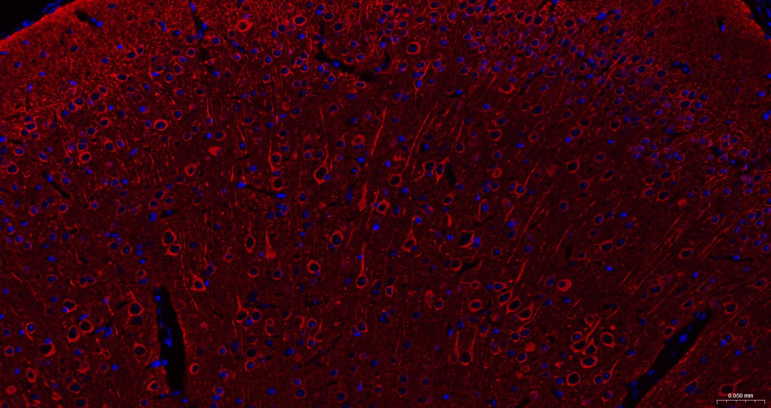
Paraformaldehyde-fixed, paraffin embedded Mouse Cerebrum; Antigen retrieval by boiling in sodium citrate buffer (pH6.0) for 15 min; The section was incubated with Tubulin beta-III Monoclonal Antibody, Unconjugated (bsm-52323R) at 1:200 overnight at 4°C. Followed by conjugated Goat Anti-Rabbit IgG antibody (Red, bs-0295G-BF594), DAPI (blue, C02-04002) was used to stain the cell nuclei.
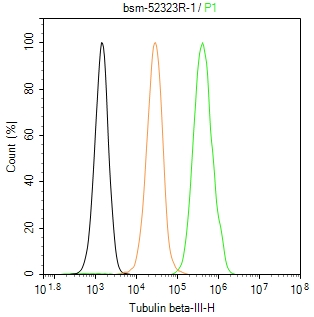
The U-87MG (H) cells were fixed with 4% PFA (10 min at r.t.) and then permeabilized with 90% ice-cold methanol for 20 min at -20℃,the cells then were incubated in 5%BSA to block non-specific protein-protein interactions (30 min at r.t.), followed by secondary antibody incubation for 40 min at room temperature. Primary Antibody (green):Rabbit Anti-Tubulin beta-III antibody (bsm-52323R,1:100); Isotype Control (orange): Rabbit IgG (bs-0295P). Blank control (black): PBS. Acquisition of 20,000 events was performed.
|
| 1、抗体溶解方法 | |
| 2、抗体修复方式 | |
| 3、常用试剂的配制 | |
| 4、免疫组化操作步骤 | |
| 5、免疫组化问题解答 | |
| 6、Western Blotting 操作步骤 | |
| 7、Western Blotting 问题解答 | |
| 8、关于肽链的设计 | |
| 9、多肽的溶解与保存 | |
| 10、酶标抗体效价测定程序 | |


