
| 产品编号 | bsm-54212R |
| 英文名称 | villin Recombinant Rabbit mAb |
| 中文名称 | 绒毛蛋白重组兔单抗 |
| 别 名 | D2S1471; VIL; VILI_HUMAN; VIL1; VILI_MOUSE; villin 1 |
 |
Specific References (1) | bsm-54212R has been referenced in 1 publications.
[IF=14.957] Cynthia Bülck. et al. Proteolytic processing of galectin-3 by meprin metalloproteases is crucial for host-microbiome homeostasis. SCI ADV. 2023 Mar;9(13) IF ; Mouse.
|
| 研究领域 | 肿瘤 免疫学 神经生物学 内分泌病 |
| 抗体来源 | Rabbit |
| 克隆类型 | Recombinant |
| 克 隆 号 | 6G2 |
| 交叉反应 | Human,Mouse,Rat |
| 产品应用 | WB=1:500-2000,IHC-P=1:200-1000,IHC-F=1:200-1000,IF=1:200-1000
not yet tested in other applications. optimal dilutions/concentrations should be determined by the end user. |
| 理论分子量 | 93kDa |
| 检测分子量 | 100 |
| 细胞定位 | 细胞浆 |
| 性 状 | Liquid |
| 浓 度 | 1mg/ml |
| 免 疫 原 | Recombinant human villin protein |
| 亚 型 | IgG |
| 纯化方法 | affinity purified by Protein A |
| 缓 冲 液 | 0.01M TBS (pH7.4) with 1% BSA, 0.02% Proclin300 and 50% Glycerol. |
| 保存条件 | Shipped at 4℃. Store at -20℃ for one year. Avoid repeated freeze/thaw cycles. |
| 注意事项 | This product as supplied is intended for research use only, not for use in human, therapeutic or diagnostic applications. |
| PubMed | PubMed |
| 产品介绍 |
Villin can cap, nucleate, sever and bundle actin in a calcium and phosphoinositide regulated manner. It is associated with the microvillar actin core bundle of intestinal and renal brush border implicated in adsorption. Villin is composed of six repeats, each containing 150 residues that together constitute the core domain followed by the carboxyl terminal headpiece domain of 87 residues. The core domain retains the calcium dependent capping nucleating and severing activity, whereas the headpiece domain contributes towards actin filament bundling and binding F actin, independently of Calcium. Function: Epithelial cell-specific Ca(2+)-regulated actin-modifying protein that modulates the reorganization of microvillar actin filaments. Plays a role in the actin nucleation, actin filament bundle assembly, actin filament capping and severing. Binds phosphatidylinositol 4,5-bisphosphate (PIP2) and lysophosphatidic acid (LPA); binds LPA with higher affinity than PIP2. Binding to LPA increases its phosphorylation by SRC and inhibits all actin-modifying activities. Binding to PIP2 inhibits actin-capping and -severing activities but enhances actin-bundling activity. Regulates the intestinal epithelial cell morphology, cell invasion, cell migration and apoptosis. Protects against apoptosis induced by dextran sodium sulfate (DSS) in the gastrointestinal epithelium. Appears to regulate cell death by maintaining mitochondrial integrity. Enhances hepatocyte growth factor (HGF)-induced epithelial cell motility, chemotaxis and wound repair. Upon S.flexneri cell infection, its actin-severing activity enhances actin-based motility of the bacteria and plays a role during the dissemination. Subunit: Monomer. Homodimer; homodimerization is necessary for actin-bundling. Associates with F-actin; phosphorylation at tyrosines residues decreases the association with F-actin. Interacts (phosphorylated at C-terminus tyrosine phosphorylation sites) with PLCG1 (via the SH2 domains). Interacts (phosphorylated form) with PLCG1; the interaction is enhanced by hepatocyte growth factor (HGF) (By similarity). Subcellular Location: Cytoplasm, cytoskeleton. Cell projection, lamellipodium. Cell projection, ruffle. Cell projection, microvillus. Cell projection, filopodium tip (By similarity). Cell projection, filopodium (By similarity). Note=Relocalized in the tip of cellular protrusions and filipodial extensions upon infection with S.flexneri in primary intestinal epithelial cells (IEC) and in the tail-like structures forming the actin comets of S.flexneri. Redistributed to the leading edge of hepatocyte growth factor (HGF)-induced lamellipodia (By similarity). Rapidly redistributed to ruffles and lamellipodia structures in response to autotaxin, lysophosphatidic acid (LPA) and epidermal growth factor (EGF) treatment. Tissue Specificity: Specifically expressed in epithelial cells. Major component of microvilli of intestinal epithelial cells and kidney proximal tubule cells. Expressed in canalicular microvilli of hepatocytes (at protein level). Post-translational modifications: Tyrosine phosphorylation is induced by epidermal growth factor (EGF) and stimulates cell migration (By similarity). Phosphorylated on tyrosine residues by SRC. The unphosphorylated form increases the initial rate of actin-nucleating activity, whereas the tyrosine-phosphorlyated form inhibits actin-nucleating activity, enhances actin-bundling activity and enhances actin-severing activity by reducing high Ca(2+) requirements. The tyrosine-phosphorlyated form does not regulate actin-capping activity. Tyrosine phosphorylation is essential for cell migration: tyrosine phosphorylation sites in the N-terminus half regulate actin reorganization and cell morphology, whereas tyrosine phosphorylation sites in the C-terminus half regulate cell migration via interaction with PLCG1. DISEASE: Note=Biliary atresia is a chronic and progressive cholestatic liver disease of chilhood characterized by an abnormal villin gene expression and severe malformation of canalicular microvillus structure. Similarity: Belongs to the villin/gelsolin family. Contains 6 gelsolin-like repeats. Contains 1 HP (headpiece) domain SWISS: P09327 Gene ID: 7429 Database links: Entrez Gene: 7429 Human Entrez Gene: 22349 Mouse Omim: 193040 Human SwissProt: P09327 Human SwissProt: Q62468 Mouse Unigene: 654595 Human Unigene: 471601 Mouse |
| 产品图片 |
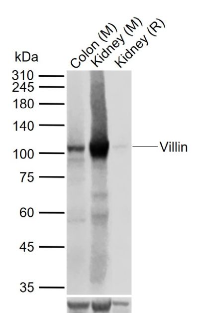
Sample:
Lane 1: Mouse Colon tissue lysates
Lane 2: Mouse Kidney tissue lysates
Lane 3: Rat Kidney tissue lysates
Primary: Anti-Villin (bsm-60340R) at 1/1000 dilution
Secondary: IRDye800CW Goat Anti-Rabbit IgG at 1/20000 dilution
Predicted band size: 93 kDa
Observed band size: 100 kDa
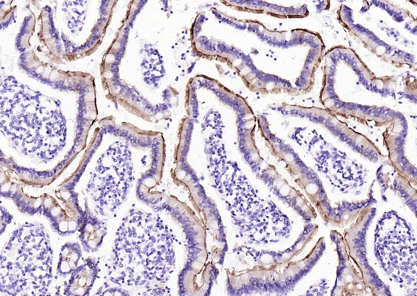
Paraformaldehyde-fixed, paraffin embedded (Human duodenum); Antigen retrieval by boiling in sodium citrate buffer (pH6.0) for 15min; Block endogenous peroxidase by 3% hydrogen peroxide for 20 minutes; Blocking buffer (normal goat serum) at 37°C for 30min; Incubation with (villin) Monoclonal Antibody, Unconjugated (bsm-54212R) at 1:200 overnight at 4°C, followed by operating according to SP Kit(Rabbit) (sp-0023) instructionsand DAB staining.
Paraformaldehyde-fixed, paraffin embedded (rat intestine); Antigen retrieval by boiling in sodium citrate buffer (pH6.0) for 15min; Block endogenous peroxidase by 3% hydrogen peroxide for 20 minutes; Blocking buffer (normal goat serum) at 37°C for 30min; Incubation with (villin) Monoclonal Antibody, Unconjugated (bsm-54212R) at 1:200 overnight at 4°C, followed by operating according to SP Kit(Rabbit) (sp-0023) instructionsand DAB staining.

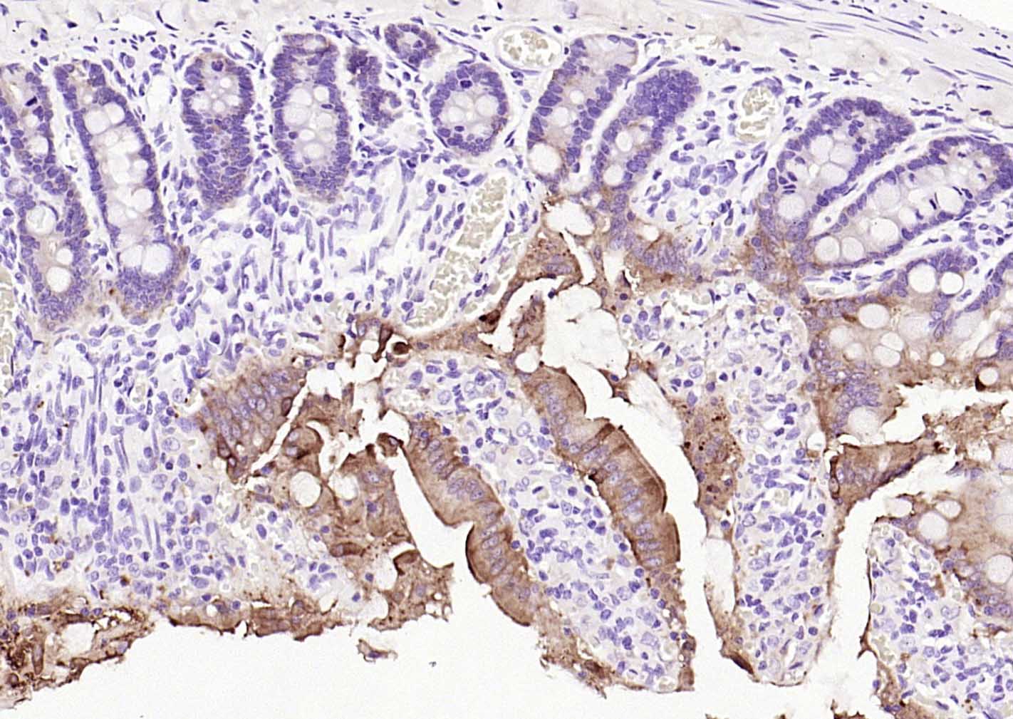
Paraformaldehyde-fixed, paraffin embedded (rat colon); Antigen retrieval by boiling in sodium citrate buffer (pH6.0) for 15min; Block endogenous peroxidase by 3% hydrogen peroxide for 20 minutes; Blocking buffer (normal goat serum) at 37°C for 30min; Incubation with (villin) Monoclonal Antibody, Unconjugated (bsm-54212R) at 1:200 overnight at 4°C, followed by operating according to SP Kit(Rabbit) (sp-0023) instructionsand DAB staining.
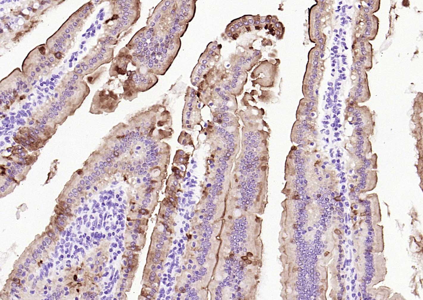
Paraformaldehyde-fixed, paraffin embedded (Mouse small intestine); Antigen retrieval by boiling in sodium citrate buffer (pH6.0) for 15min; Block endogenous peroxidase by 3% hydrogen peroxide for 20 minutes; Blocking buffer (normal goat serum) at 37°C for 30min; Incubation with (villin) Monoclonal Antibody, Unconjugated (bsm-54212R) at 1:200 overnight at 4°C, followed by operating according to SP Kit(Rabbit) (sp-0023) instructionsand DAB staining.
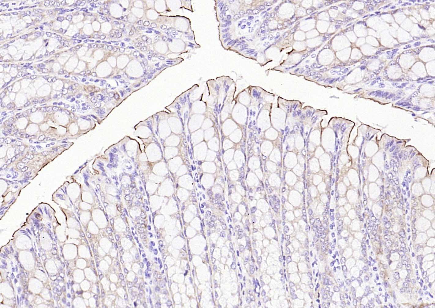
Paraformaldehyde-fixed, paraffin embedded (mouse colon); Antigen retrieval by boiling in sodium citrate buffer (pH6.0) for 15min; Block endogenous peroxidase by 3% hydrogen peroxide for 20 minutes; Blocking buffer (normal goat serum) at 37°C for 30min; Incubation with (villin) Monoclonal Antibody, Unconjugated (bsm-54212R) at 1:200 overnight at 4°C, followed by operating according to SP Kit(Rabbit) (sp-0023) instructionsand DAB staining.
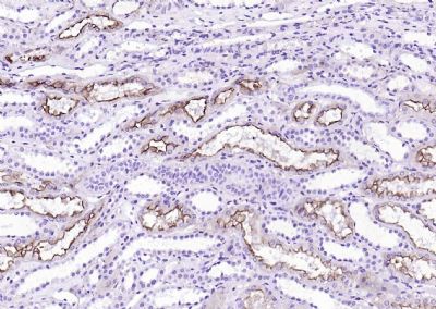
Paraformaldehyde-fixed, paraffin embedded (Human kidney); Antigen retrieval by boiling in sodium citrate buffer (pH6.0) for 15min; Block endogenous peroxidase by 3% hydrogen peroxide for 20 minutes; Blocking buffer (normal goat serum) at 37°C for 30min; Antibody incubation with (Villin) Monoclonal Antibody, Unconjugated (bsm-60340R) at 1:200 overnight at 4°C, followed by operating according to SP Kit(Rabbit) (sp-0023) instructionsand DAB staining.
|
| 1、抗体溶解方法 | |
| 2、抗体修复方式 | |
| 3、常用试剂的配制 | |
| 4、免疫组化操作步骤 | |
| 5、免疫组化问题解答 | |
| 6、Western Blotting 操作步骤 | |
| 7、Western Blotting 问题解答 | |
| 8、关于肽链的设计 | |
| 9、多肽的溶解与保存 | |
| 10、酶标抗体效价测定程序 | |


