扫码关注公众号           扫码咨询技术支持           扫码咨询技术服务
  
客服热线:400-901-9800  客服QQ:4009019800  技术答疑  技术支持  质量反馈  人才招聘  关于我们  联系我们
产品中心-北京博奥森生物技术有限公司
首页 > 产品中心 > 一抗 > 产品信息
Rabbit Anti-STAT2  antibody (bsm-52234R)
~~~促销代码KT202409~~~
订购热线:400-901-9800
订购邮箱:sales@bioss.com.cn
订购QQ:  400-901-9800
技术支持:techsupport@bioss.com.cn
说明书: 50ul  100ul  
50ul/1580.00元
100ul/2500.00元
大包装/询价

产品编号 bsm-52234R
英文名称 Rabbit Anti-STAT2  antibody
中文名称 信号转导和转录激活因子2重组兔单抗
别    名 signal transducers and activators of transduction2; Homo sapiens interferon alpha induced transcriptional activator; interferon alpha induced transcriptional activator; ISGF3; P113; signal transducer and activator of transcription 2 113kD; STAT113; STAT2_HUMAN; p113.   
研究领域 细胞生物  神经生物学  
抗体来源 Rabbit
克隆类型 Recombinant
克 隆 号 4B1
交叉反应 Human
产品应用 WB=1:500-1000,IHC-P=1:100-500,IHC-F=1:400-800,ICC/IF=1:50-200,IF=1:50-200
not yet tested in other applications.
optimal dilutions/concentrations should be determined by the end user.
理论分子量 97kDa
细胞定位 细胞核 细胞浆 
性    状 Liquid
浓    度 1mg/ml
免 疫 原 Recombinant human STAT2 protein (1-100aa) 
亚    型 IgG
纯化方法 affinity purified by Protein A
缓 冲 液 0.01M TBS(pH7.4) with 1% BSA, 0.03% Proclin300 and 50% Glycerol.
保存条件 Shipped at 4℃. Store at -20 °C for one year. Avoid repeated freeze/thaw cycles.
注意事项 This product as supplied is intended for research use only, not for use in human, therapeutic or diagnostic applications.
PubMed PubMed
产品介绍 The protein encoded by this gene is a member of the STAT protein family. In response to cytokines and growth factors, STAT family members are phosphorylated by the receptor associated kinases, and then form homo- or heterodimers that translocate to the cell nucleus where they act as transcription activators. In response to interferon (IFN), this protein forms a complex with STAT1 and IFN regulatory factor family protein p48 (ISGF3G), in which this protein acts as a transactivator, but lacks the ability to bind DNA directly. Transcription adaptor P300/CBP (EP300/CREBBP) has been shown to interact specifically with this protein, which is thought to be involved in the process of blocking IFN-alpha response by adenovirus. Multiple transcript variants encoding different isoforms have been found for this gene. [provided by RefSeq, Mar 2010].

Function:
Signal transducer and activator of transcription that mediates signaling by type I IFNs (IFN-alpha and IFN-beta). Following type I IFN binding to cell surface receptors, Jak kinases (TYK2 and JAK1) are activated, leading to tyrosine phosphorylation of STAT1 and STAT2. The phosphorylated STATs dimerize, associate with ISGF3G/IRF-9 to form a complex termed ISGF3 transcription factor, that enters the nucleus. ISGF3 binds to the IFN stimulated response element (ISRE) to activate the transcription of interferon stimulated genes, which drive the cell in an antiviral state.

Subunit:
Interacts with ISGF3G/IRF-9 in the cytoplasm. Heterodimer with STAT1 upon IFN-alpha/beta induced phosphorylation. Interacts with CRSP2 and CRSP6. Interacts with Simian virus 5 protein V and rabies virus phosphoprotein (By similarity). Can form a homodimer upon IFN-alpha induced phosphorylation. Interacts with IFNAR1; the interaction requires the phosphorylation of IFNAR1 at 'Tyr-466'. Interacts with IFNAR2. Interacts with dengue virus NS5; this interaction inhibits the phosphorylation of STAT2, and, when all viral proteins are present (polyprotein), targets STAT2 for degradation. Interacts with human cytomegalovirus/HHV-5 protein UL123; this interaction promotes viral growth.

Subcellular Location:
Cytoplasm. Nucleus. Note=Translocated into the nucleus upon activation by IFN-alpha/beta.

Post-translational modifications:
Tyrosine phosphorylated in response to IFN-alpha.

Similarity:
Belongs to the transcription factor STAT family.
Contains 1 SH2 domain.

SWISS:
P52630

Gene ID:
6773

Database links:

Entrez Gene: 6773 Human

Entrez Gene: 20847 Mouse

Entrez Gene: 288774 Rat

Omim: 600556 Human

SwissProt: P52630 Human

SwissProt: Q9WVL2 Mouse

Unigene: 530595 Human

Unigene: 293120 Mouse

Unigene: 471333 Mouse

Unigene: 24237 Rat


产品图片
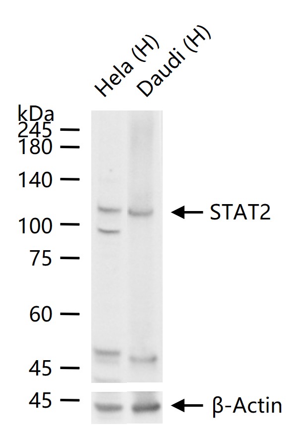
25 ug total protein per lane of various lysates (see on figure) probed with STAT2 monoclonal antibody, unconjugated (bsm-52234R) at 1:1000 dilution and 4°C overnight incubation. Followed by conjugated secondary antibody incubation at r.t. for 60 min.
Immunohistochemical analysis of paraffin-embedded human colon cancer tissue with Rabbit anti-STAT2 antibody (bsm-52234R) at 1/50 dilution. The section was pre-treated using heat mediated antigen retrieval with Tris-EDTA buffer (pH 9.0) for 20 minutes. The tissues were blocked in 1% BSA for 20 minutes at room temperature, washed with ddH2O and PBS, and then probed with the primary antibody (bsm-52234R) at 1/400 dilution for 1 hour at room temperature. The detection was performed using an HRP conjugated compact polymer system. DAB was used as the chromogen. Tissues were counterstained with hematoxylin and mounted with DPX.
Immunohistochemical analysis of paraffin-embedded mouse skin cancer tissue with Rabbit anti-STAT2 antibody (bsm-52234R) at 1/50 dilution. The section was pre-treated using heat mediated antigen retrieval with Tris-EDTA buffer (pH 9.0) for 20 minutes. The tissues were blocked in 1% BSA for 20 minutes at room temperature, washed with ddH2O and PBS, and then probed with the primary antibody (bsm-52234R) at 1/400 dilution for 1 hour at room temperature. The detection was performed using an HRP conjugated compact polymer system. DAB was used as the chromogen. Tissues were counterstained with hematoxylin and mounted with DPX.
Immunohistochemical analysis of paraffin-embedded human lung cancer tissue with Rabbit anti-STAT2 antibody (bsm-52234R) at 1/50 dilution. The section was pre-treated using heat mediated antigen retrieval with Tris-EDTA buffer (pH 9.0) for 20 minutes. The tissues were blocked in 1% BSA for 20 minutes at room temperature, washed with ddH2O and PBS, and then probed with the primary antibody (bsm-52234R) at 1/400 dilution for 1 hour at room temperature. The detection was performed using an HRP conjugated compact polymer system. DAB was used as the chromogen. Tissues were counterstained with hematoxylin and mounted with DPX.
Immunocytochemistry analysis of SW480 cells labeling STAT2 with Rabbit anti-STAT2 antibody (bsm-52234R) at 1/50 dilution. Cells were fixed in 4% paraformaldehyde for 10 minutes at 37 ℃, permeabilized with 0.05% Triton X-100 in PBS for 20 minutes, and then blocked with 2% negative goat serum for 30 minutes at room temperature. Cells were then incubated with Rabbit anti-STAT2 antibody (bsm-52234R) at 1/200 dilution in 2% negative goat serum overnight at 4 ℃. Goat Anti-Rabbit IgG H&L (iFluor™ 488, HA1121) was used as the secondary antibody at 1/1,000 dilution. PBS instead of the primary antibody was used as the secondary antibody only control. Nuclear DNA was labelled in blue with DAPI.
Immunocytochemistry analysis of A549 cells labeling STAT2 with Rabbit anti-STAT2 antibody (bsm-52234R) at 1/50 dilution. Cells were fixed in 4% paraformaldehyde for 10 minutes at 37 ℃, permeabilized with 0.05% Triton X-100 in PBS for 20 minutes, and then blocked with 2% negative goat serum for 30 minutes at room temperature. Cells were then incubated with Rabbit anti-STAT2 antibody (bsm-52234R) at 1/200 dilution in 2% negative goat serum overnight at 4 ℃. Goat Anti-Rabbit IgG H&L (iFluor™ 488, HA1121) was used as the secondary antibody at 1/1,000 dilution. PBS instead of the primary antibody was used as the secondary antibody only control. Nuclear DNA was labelled in blue with DAPI.
Immunocytochemistry analysis of A431 cells labeling STAT2 with Rabbit anti-STAT2 antibody (bsm-52234R) at 1/50 dilution. Cells were fixed in 4% paraformaldehyde for 10 minutes at 37 ℃, permeabilized with 0.05% Triton X-100 in PBS for 20 minutes, and then blocked with 2% negative goat serum for 30 minutes at room temperature. Cells were then incubated with Rabbit anti-STAT2 antibody (bsm-52234R) at 1/200 dilution in 2% negative goat serum overnight at 4 ℃. Goat Anti-Rabbit IgG H&L (iFluor™ 488) was used as the secondary antibody at 1/1,000 dilution. PBS instead of the primary antibody was used as the secondary antibody only control. Nuclear DNA was labelled in blue with DAPI.
版权所有 2004-2026 www.bioss.com.cn 北京博奥森生物技术有限公司
通过国际质量管理体系ISO 9001:2015 GB/T 19001-2016    证书编号: 00124Q34771R2M/1100
通过国际医疗器械-质量管理体系ISO 13485:2016 GB/T 42061-2022    证书编号: CQC24QY10047R0M/1100
京ICP备05066980号-1         京公网安备110107000727号