
| 产品编号 | bsm-52052R |
| 英文名称 | CK10/Cytokeratin 10 Recombinant Rabbit mAb |
| 中文名称 | 细胞角蛋白10重组兔单抗 |
| 别 名 | BCIE; BIE; CK10; EHK; EHK2; EHK2A; EHK2B; IHL; K10; KPP; D130054E02Rik; K1C1; Krt-1.10; Krt1-10; Ka10; K1C10_HUMAN; KRT10; Cytokeratin-10 (CK-10); Keratin-10 (K10); K1C10_MOUSE; 56 kDa cytokeratin; Keratin, type I cytoskeletal 59 kDa; K1C10_RAT; Type I keratin Ka10; keratin 10; keratosis palmaris et plantaris; keratin 10, type I; cytokeratin 10; epidermolytic hyperkeratosis |
| 研究领域 | 信号转导 细胞骨架 细胞外基质 |
| 抗体来源 | Rabbit |
| 克隆类型 | Recombinant |
| 克 隆 号 | 1D8 |
| 交叉反应 | Human,Mouse,Rat |
| 产品应用 | WB=1:1000-10000,IHC-P=1:1000-5000,IHC-F=1:1000-5000,IF=1:1000-5000
not yet tested in other applications. optimal dilutions/concentrations should be determined by the end user. |
| 理论分子量 | 59kDa |
| 检测分子量 | 59 |
| 细胞定位 | 细胞浆 |
| 性 状 | Liquid |
| 浓 度 | 1mg/ml |
| 免 疫 原 | A synthesized peptide derived from human Cytokeratin 10: 550-584 |
| 亚 型 | IgG |
| 纯化方法 | affinity purified by Protein A |
| 缓 冲 液 | 0.01M TBS (pH7.4) with 1% BSA, 0.02% Proclin300 and 50% Glycerol. |
| 保存条件 | Shipped at 4℃. Store at -20℃ for one year. Avoid repeated freeze/thaw cycles. |
| 注意事项 | This product as supplied is intended for research use only, not for use in human, therapeutic or diagnostic applications. |
| PubMed | PubMed |
| 产品介绍 |
Cytokeratins comprise a diverse group of intermediate filament proteins (IFPs) that are expressed as pairs in both keratinized and non-keratinized epithelial tissue. Cytokeratins play a critical role in differentiation and tissue specialization and function to maintain the overall structural integrity of epithelial cells. Cytokeratins have been found to be useful markers of tissue differentiation which is directly applicable to the characterization of malignant tumors. Cytokeratins 10 and 13 are present in the cytoskeletal region of a subset of squamous cell carcinomas. Cytokeratin 10 is a heterotetramer of two type I and two type II keratins, is generally associated with keratin 1, and is seen in all suprabasal cell layers including stratum corneum. Subunit: Belongs to the intermediate filament family. Subcellular Location: Cytoplasm. Tissue Specificity: Seen in all suprabasal cell layers including stratum corneum. DISEASE: Defects in KRT10 are a cause of bullous congenital ichthyosiform erythroderma (BCIE) [MIM:113800]; also known as epidermolytic hyperkeratosis (EHK) or bullous erythroderma ichthyosiformis congenita of Brocq. BCIE is an autosomal dominant skin disorder characterized by widespread blistering and an ichthyotic erythroderma at birth that persist into adulthood. Histologically there is a diffuse epidermolytic degeneration in the lower spinous layer of the epidermis. Within a few weeks from birth, erythroderma and blister formation diminish and hyperkeratoses develop. Defects in KRT10 are a cause of ichthyosis annular epidermolytic (AEI) [MIM:607602]; also known as cyclic ichthyosis with epidermolytic hyperkeratosis. AEI is a skin disorder resembling bullous congenital ichthyosiform erythroderma. Affected individuals present with bullous ichthyosis in early childhood and hyperkeratotic lichenified plaques in the flexural areas and extensor surfaces at later ages. The feature that distinguishes AEI from BCIE is dramatic episodes of flares of annular polycyclic plaques with scale, which coalesce to involve most of the body surface and can persist for several weeks or even months. Similarity: Belongs to the intermediate filament family. SWISS: P13645 Gene ID: 3858 Database links: Entrez Gene: 3858 Human Entrez Gene: 16661 Mouse Omim: 148080 Human SwissProt: P13645 Human SwissProt: P02535 Mouse Unigene: 99936 Human Unigene: 22662 Mouse |
| 产品图片 |
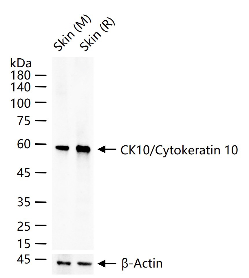
25 ug total protein per lane of various lysates (see on figure) probed with CK10/Cytokeratin 10 monoclonal antibody, unconjugated (bsm-52052R) at 1:1000 dilution and 4°C overnight incubation. Followed by conjugated secondary antibody incubation at r.t. for 60 min.
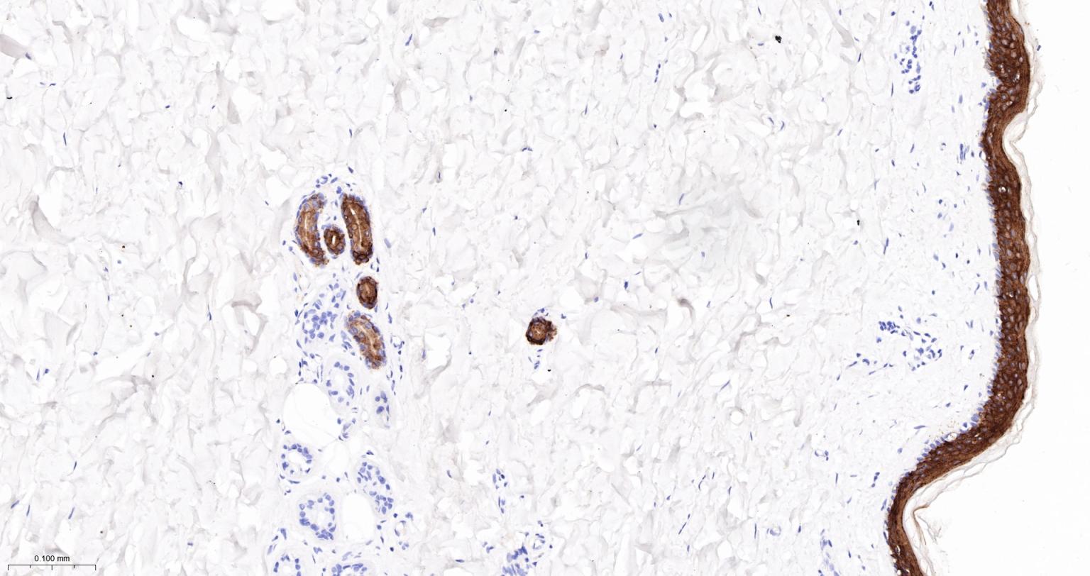
Paraformaldehyde-fixed, paraffin embedded Human Skin; Antigen retrieval by boiling in sodium citrate buffer (pH6.0) for 15 min; Antibody incubation with CK10/Cytokeratin 10 Monoclonal Antibody, Unconjugated(bsm-52052R) at 1:1000 overnight at 4°C, followed by conjugation to the bs-0295G-HRP and DAB (C-0010) staining.
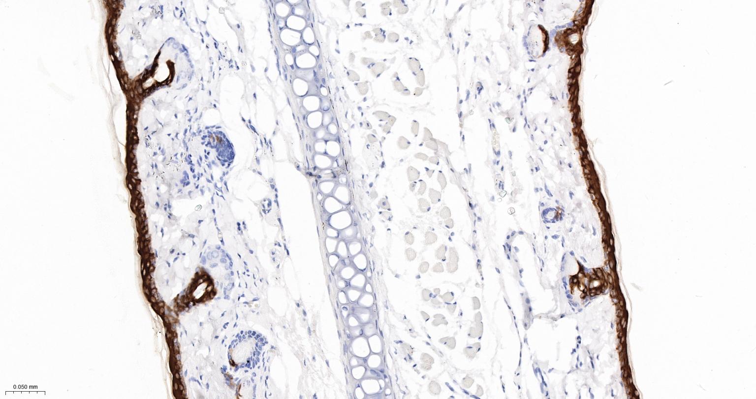
Paraformaldehyde-fixed, paraffin embedded Rat Skin; Antigen retrieval by boiling in sodium citrate buffer (pH6.0) for 15 min; Antibody incubation with CK10/Cytokeratin 10 Monoclonal Antibody, Unconjugated(bsm-52052R) at 1:1000 overnight at 4°C, followed by conjugation to the bs-0295G-HRP and DAB (C-0010) staining.
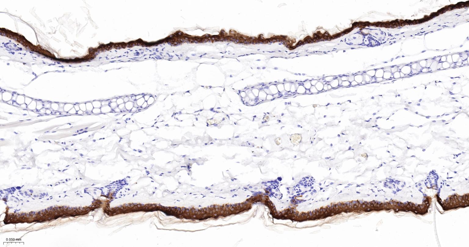
Paraformaldehyde-fixed, paraffin embedded Mouse Skin; Antigen retrieval by boiling in sodium citrate buffer (pH6.0) for 15 min; Antibody incubation with CK10/Cytokeratin 10 Monoclonal Antibody, Unconjugated(bsm-52052R) at 1:1000 overnight at 4°C, followed by conjugation to the bs-0295G-HRP and DAB (C-0010) staining.
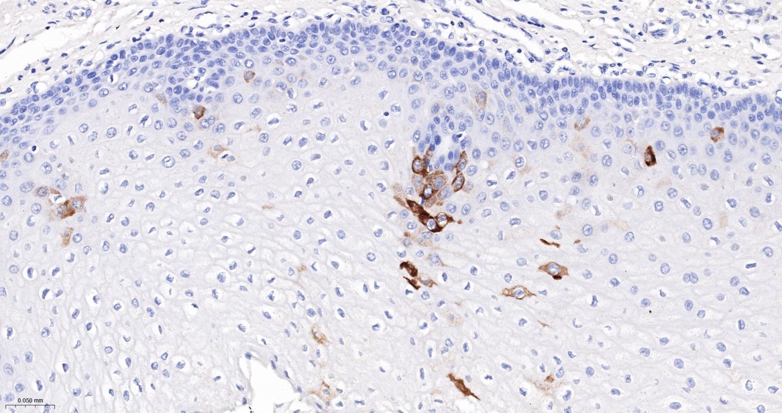
Paraformaldehyde-fixed, paraffin embedded Human Esophagus; Antigen retrieval by boiling in sodium citrate buffer (pH6.0) for 15 min; Antibody incubation with CK10/Cytokeratin 10 Monoclonal Antibody, Unconjugated(bsm-52052R) at 1:1000 overnight at 4°C, followed by conjugation to the bs-0295G-HRP and DAB (C-0010) staining.
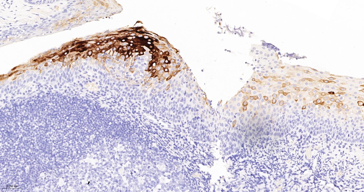
Paraformaldehyde-fixed, paraffin embedded Human Tonsil; Antigen retrieval by boiling in sodium citrate buffer (pH6.0) for 15 min; Antibody incubation with CK10/Cytokeratin 10 Monoclonal Antibody, Unconjugated(bsm-52052R) at 1:1000 overnight at 4°C, followed by conjugation to the bs-0295G-HRP and DAB (C-0010) staining.
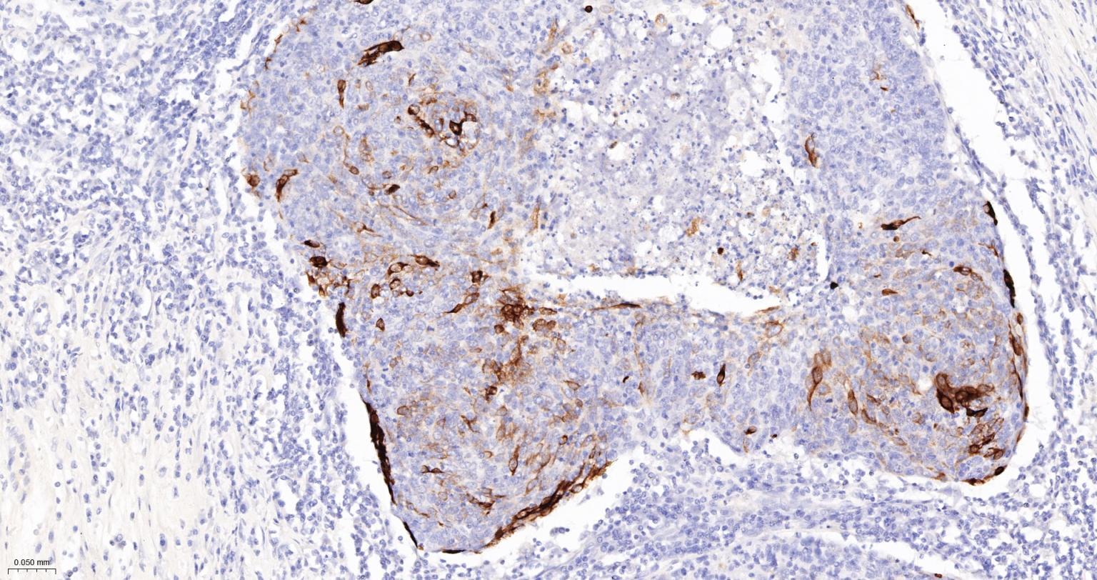
Paraformaldehyde-fixed, paraffin embedded Human Skin Cancer; Antigen retrieval by boiling in sodium citrate buffer (pH6.0) for 15 min; Antibody incubation with CK10/Cytokeratin 10 Monoclonal Antibody, Unconjugated(bsm-52052R) at 1:1000 overnight at 4°C, followed by conjugation to the bs-0295G-HRP and DAB (C-0010) staining.
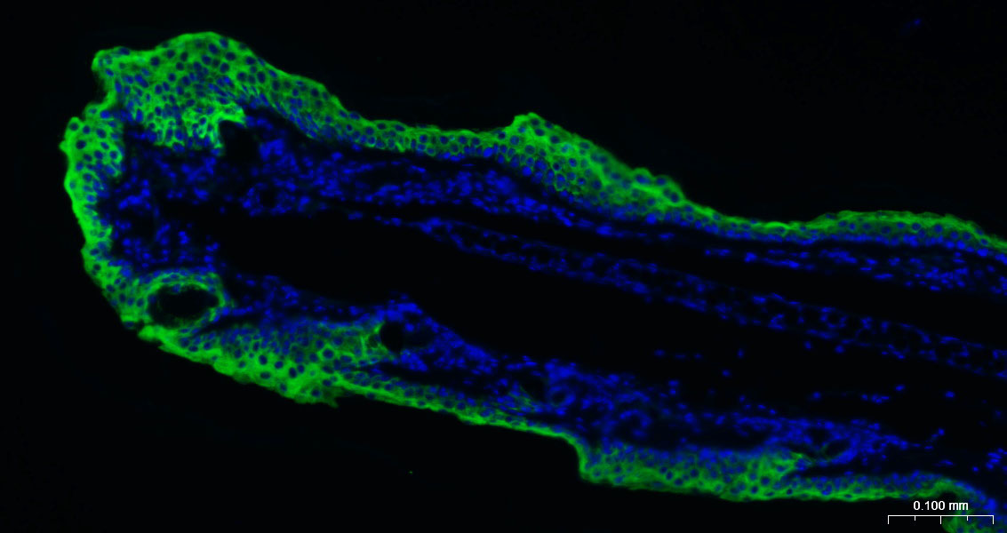
Paraformaldehyde-fixed, paraffin embedded Mouse Skin; Antigen retrieval by boiling in sodium citrate buffer (pH6.0) for 15 min; Antibody incubation with CK10/Cytokeratin 10 Monoclonal Antibody, Unconjugated (bsm-52052R) at 1:200 overnight at 4°C. Followed by conjugated Goat Anti-Rabbit IgG antibody (green, bs-0295G-BF488), DAPI (blue, C02-04002) was used to stain the cell nuclei.
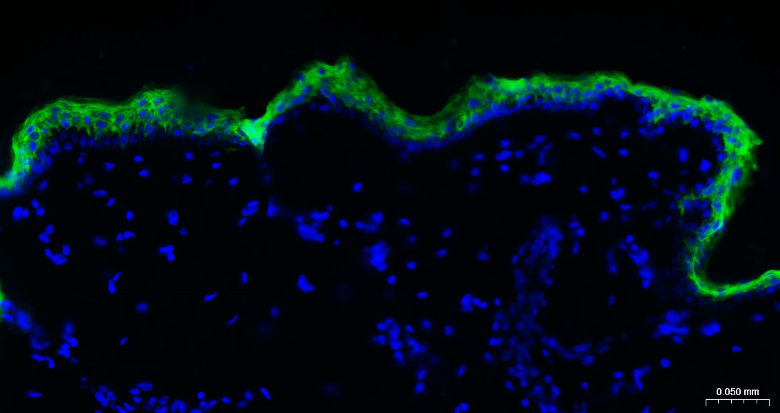
Paraformaldehyde-fixed, paraffin embedded Human Skin; Antigen retrieval by boiling in sodium citrate buffer (pH6.0) for 15 min; Antibody incubation with CK10/Cytokeratin 10 Monoclonal Antibody, Unconjugated (bsm-52052R) at 1:200 overnight at 4°C. Followed by conjugated Goat Anti-Rabbit IgG antibody (green, bs-0295G-BF488), DAPI (blue, C02-04002) was used to stain the cell nuclei.
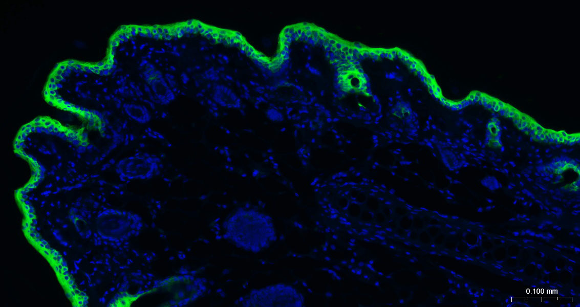
Paraformaldehyde-fixed, paraffin embedded Rat Skin; Antigen retrieval by boiling in sodium citrate buffer (pH6.0) for 15 min; Antibody incubation with CK10/Cytokeratin 10 Monoclonal Antibody, Unconjugated (bsm-52052R) at 1:200 overnight at 4°C. Followed by conjugated Goat Anti-Rabbit IgG antibody (green, bs-0295G-BF488), DAPI (blue, C02-04002) was used to stain the cell nuclei.
|
| 1、抗体溶解方法 | |
| 2、抗体修复方式 | |
| 3、常用试剂的配制 | |
| 4、免疫组化操作步骤 | |
| 5、免疫组化问题解答 | |
| 6、Western Blotting 操作步骤 | |
| 7、Western Blotting 问题解答 | |
| 8、关于肽链的设计 | |
| 9、多肽的溶解与保存 | |
| 10、酶标抗体效价测定程序 | |


