

| 产品编号 | bsm-33309M |
| 英文名称 | LC3A Mouse mAb |
| 中文名称 | 自噬微管相关蛋白轻链3单克隆抗体 |
| 别 名 | ATG8E; LC3; LC3A; MAP1ALC3; MAP1BLC3; 1010001H21Rik; 4922501H04Rik; LC3-I; LC3-II; MLP3A_HUMAN; MAP1LC3A; Autophagy-related protein LC3 A; Autophagy-related ubiquitin-like modifier LC3 A; MAP1 light chain 3-like protein 1; Microtubule-associated proteins |
 |
Specific References (1) | bsm-33309M has been referenced in 1 publications.
[IF=2.1] Rong Hu. et al. Apatinib sensitizes chemoresistant NSCLC cells to doxetaxel via regulating autophagy and enhances the therapeutic efficacy in advanced and refractory/recurrent NSCLC. Mol Med Rep. 2020 Nov;22(5):3935-3943 WB,IHC ; Human.
|
| 研究领域 | 肿瘤 细胞生物 免疫学 细胞凋亡 细胞自噬 |
| 抗体来源 | Mouse |
| 克隆类型 | Monoclonal |
| 克 隆 号 | 7G10 |
| 交叉反应 | Human,Mouse,Rat |
| 产品应用 | WB=1:500-1000,IHC-P=1:100-500,IHC-F=1:100-500,IF=1:100-500
not yet tested in other applications. optimal dilutions/concentrations should be determined by the end user. |
| 理论分子量 | 14/16 kDa |
| 检测分子量 | 15 |
| 细胞定位 | 细胞浆 细胞膜 |
| 性 状 | Liquid |
| 浓 度 | 1mg/ml |
| 免 疫 原 | KLH conjugated synthetic peptide derived from human LC3A: 1-100/121 |
| 亚 型 | IgG |
| 纯化方法 | affinity purified by Protein G |
| 缓 冲 液 | 0.01M TBS (pH7.4) with 1% BSA, 0.02% Proclin300 and 50% Glycerol. |
| 保存条件 | Shipped at 4℃. Store at -20℃ for one year. Avoid repeated freeze/thaw cycles. |
| 注意事项 | This product as supplied is intended for research use only, not for use in human, therapeutic or diagnostic applications. |
| PubMed | PubMed |
| 产品介绍 |
Microtubule-associated MAPILC3A constitutes nearly half of the mass of all the microtubule associated proteins that copurify with brain microtubules. MAP1LC3A is one of three human orthologs of the rat Map1LC3, (named MAP1LC3A, MAP1LC3B, and MAP1LC3C). The three isoforms of human MAP1LC3 exhibit distinct expression patterns in different human tissues and also differ in their post-translation modifications. MAP1LC3A and MAP1LC3C are produced by the proteolytic cleavage after the conserved C-terminal Gly residue; MAP1LC3B does not undergo C-terminal cleavage and exists in a single modified form. Function: Probably involved in formation of autophagosomal vacuoles (autophagosomes). Subunit: 3 different light chains, LC1, LC2 and LC3, can associate with MAP1A and MAP1B proteins. Interacts with SQSTM1. Interacts with TP53INP1 and TP53INP2. Subcellular Location: Cytoplasm, cytoskeleton. Endomembrane system; Lipid-anchor. Cytoplasmic vesicle, autophagosome membrane; Lipid-anchor. Cytoplasmic vesicle, autophagosome. Note=LC3-II binds to the autophagic membranes. Tissue Specificity: Most abundant in heart, brain, liver, skeletal muscle and testis but absent in thymus and peripheral blood leukocytes. Post-translational modifications: The precursor molecule is cleaved by APG4B/ATG4B to form the cytosolic form, LC3-I. This is activated by APG7L/ATG7, transferred to ATG3 and conjugated to phospholipid to form the membrane-bound form, LC3-II. Similarity: Detects a band of approximately 16 kDa (predicted molecular weight: 14 kDa). SWISS: Q9H492 Gene ID: 84557 Database links: Entrez Gene: 84557 Human Entrez Gene: 66734 Mouse Omim: 601242 Human SwissProt: Q9H492 Human SwissProt: Q91VR7 Mouse Unigene: 632273 Human Unigene: 196239 Mouse Unigene: 3135 Rat |
| 产品图片 |

Sample:
Lane 1: Mouse Cerebrum tissue lysates
Lane 2: Rat Cerebrum tissue lysates
Lane 3: Rat Cerebellum tissue lysates
Primary: Anti-LC3A (bsm-33309M) at 1/1000 dilution
Secondary: IRDye800CW Goat Anti-Mouse IgG at 1/20000 dilution
Predicted band size: 14/16 kDa
Observed band size: 15 kDa

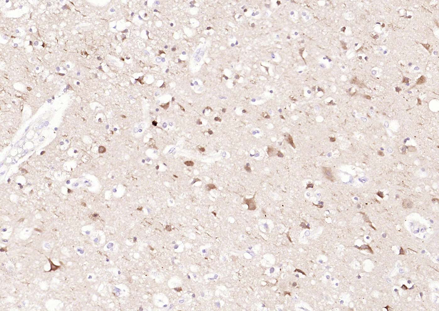
Paraformaldehyde-fixed, paraffin embedded (Rat brain); Antigen retrieval by boiling in sodium citrate buffer (pH6.0) for 15min; Block endogenous peroxidase by 3% hydrogen peroxide for 20 minutes; Blocking buffer (normal goat serum) at 37°C for 30min; Antibody incubation with (LC3A) Monoclonal Antibody, Unconjugated (bsm-33309M) at 1:400 overnight at 4°C, followed by a conjugated secondary (sp-0023) for 20 minutes and DAB staining.
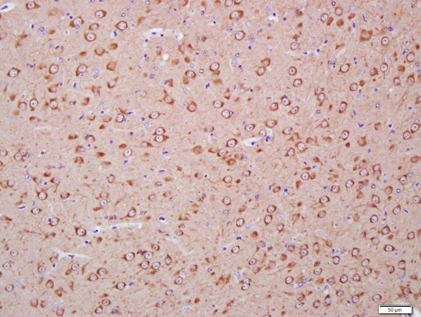
Paraformaldehyde-fixed, paraffin embedded (Mouse brain); Antigen retrieval by boiling in sodium citrate buffer (pH6.0) for 15min; Block endogenous peroxidase by 3% hydrogen peroxide for 20 minutes; Blocking buffer (normal goat serum) at 37°C for 30min; Antibody incubation with (LC3A) Monoclonal Antibody, Unconjugated (bsm-33309M) at 1:400 overnight at 4°C, followed by a conjugated secondary (sp-0023) for 20 minutes and DAB staining.
Paraformaldehyde-fixed, paraffin embedded (human brain); Antigen retrieval by boiling in sodium citrate buffer (pH6.0) for 15min; Block endogenous peroxidase by 3% hydrogen peroxide for 20 minutes; Blocking buffer (normal goat serum) at 37°C for 30min; Incubation with (LC3A ) Monoclonal Antibody, Unconjugated (bsm-33309M) at 1:200 overnight at 4°C, followed by operating according to SP Kit(Mouse)(sp-0024) instructionsand DAB staining.
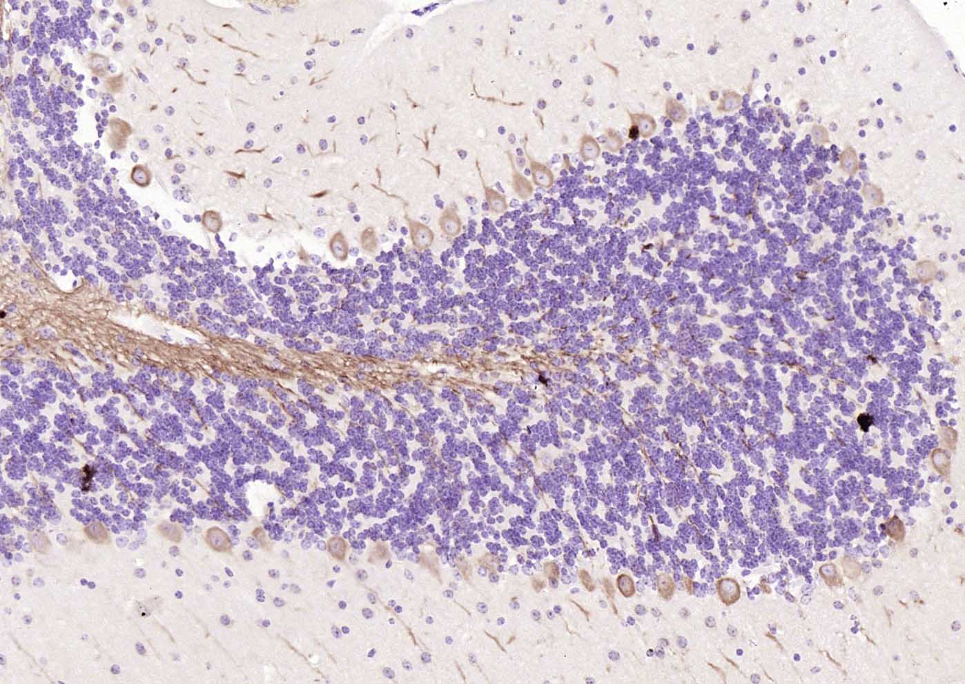
Paraformaldehyde-fixed, paraffin embedded (mouse cerebellum); Antigen retrieval by boiling in sodium citrate buffer (pH6.0) for 15min; Block endogenous peroxidase by 3% hydrogen peroxide for 20 minutes; Blocking buffer (normal goat serum) at 37°C for 30min; Incubation with (LC3A ) Monoclonal Antibody, Unconjugated (bsm-33309M) at 1:200 overnight at 4°C, followed by operating according to SP Kit(Mouse)(sp-0024) instructionsand DAB staining.

Paraformaldehyde-fixed, paraffin embedded (human cerebellum); Antigen retrieval by boiling in sodium citrate buffer (pH6.0) for 15min; Block endogenous peroxidase by 3% hydrogen peroxide for 20 minutes; Blocking buffer (normal goat serum) at 37°C for 30min; Incubation with (LC3A ) Monoclonal Antibody, Unconjugated (bsm-33309M) at 1:200 overnight at 4°C, followed by operating according to SP Kit(Mouse)(sp-0024) instructionsand DAB staining.
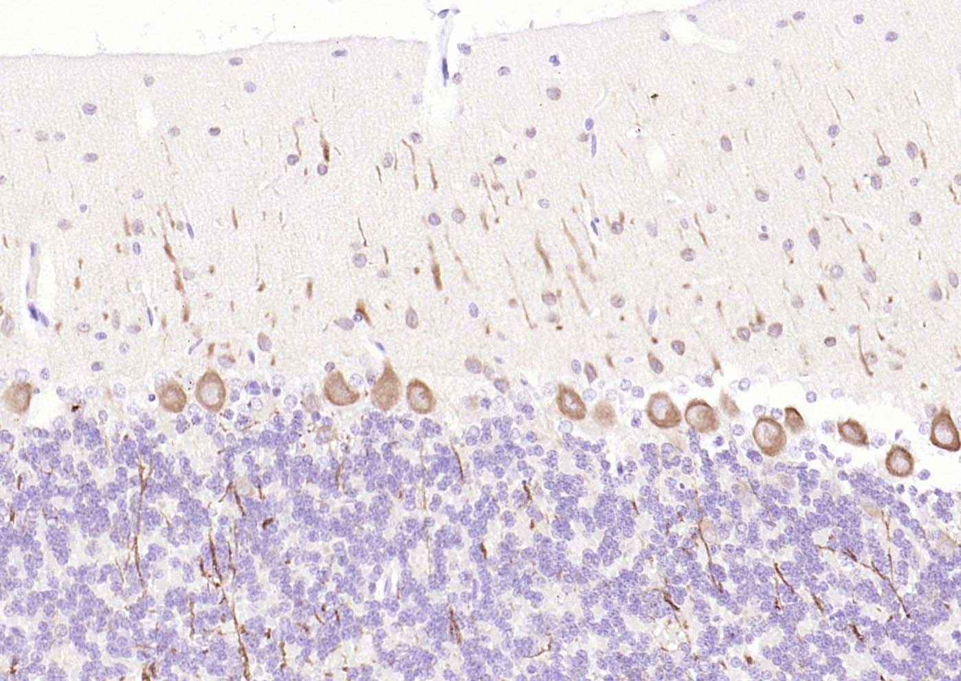
Paraformaldehyde-fixed, paraffin embedded (rat cerebellum); Antigen retrieval by boiling in sodium citrate buffer (pH6.0) for 15min; Block endogenous peroxidase by 3% hydrogen peroxide for 20 minutes; Blocking buffer (normal goat serum) at 37°C for 30min; Incubation with (LC3A ) Monoclonal Antibody, Unconjugated (bsm-33309M) at 1:200 overnight at 4°C, followed by operating according to SP Kit(Mouse)(sp-0024) instructionsand DAB staining.

Paraformaldehyde-fixed, paraffin embedded (Mouse brain); Antigen retrieval by boiling in sodium citrate buffer (pH6.0) for 15min; Block endogenous peroxidase by 3% hydrogen peroxide for 20 minutes; Blocking buffer (normal goat serum) at 37°C for 30min; Antibody incubation with (LC3A) Monoclonal Antibody, Unconjugated (bsm-33309M) at 1:400 overnight at 4°C, followed by a conjugated Goat Anti-Mouse IgG antibody (bs-0296G-FITC) for 90 minutes, and DAPI for nuclei staining.
|
| 1、抗体溶解方法 | |
| 2、抗体修复方式 | |
| 3、常用试剂的配制 | |
| 4、免疫组化操作步骤 | |
| 5、免疫组化问题解答 | |
| 6、Western Blotting 操作步骤 | |
| 7、Western Blotting 问题解答 | |
| 8、关于肽链的设计 | |
| 9、多肽的溶解与保存 | |
| 10、酶标抗体效价测定程序 | |


