

| 产品编号 | bsm-33219M |
| 英文名称 | PIK3R1 Mouse mAb |
| 中文名称 | 磷脂酰肌醇激酶单克隆抗体 |
| 别 名 | AGM7; GRB1; IMD36; p85; p85-ALPHA; p85alpha; PI3K; p50alpha; p55alpha; PI3KA; P85A_HUMAN; PIK3R1; PI3-kinase regulatory subunit alpha; PI3K regulatory subunit alpha; PtdIns-3-kinase regulatory subunit alpha; Phosphatidylinositol 3-kinase 85 kDa regulatory |
 |
Specific References (7) | bsm-33219M has been referenced in 7 publications.
|
| 研究领域 | 肿瘤 细胞生物 免疫学 信号转导 激酶和磷酸酶 |
| 抗体来源 | Mouse |
| 克隆类型 | Monoclonal |
| 克 隆 号 | 5C11 |
| 交叉反应 | Human,Mouse,Rat (predicted: Pig) |
| 产品应用 | WB=1:500-2000,IHC-P=1:100-500,IHC-F=1:100-500,IF=1:100-500,ICC/IF=1:100-500
not yet tested in other applications. optimal dilutions/concentrations should be determined by the end user. |
| 理论分子量 | 80 kDa |
| 检测分子量 | 80 |
| 细胞定位 | 细胞核 细胞浆 细胞膜 |
| 性 状 | Liquid |
| 浓 度 | 1mg/ml |
| 免 疫 原 | Recombinant mouse PI3K p85 Protein: 1-110/724 |
| 亚 型 | IgG |
| 纯化方法 | affinity purified by Protein G |
| 缓 冲 液 | 0.01M TBS (pH7.4) with 1% BSA, 0.02% Proclin300 and 50% Glycerol. |
| 保存条件 | Shipped at 4℃. Store at -20℃ for one year. Avoid repeated freeze/thaw cycles. |
| 注意事项 | This product as supplied is intended for research use only, not for use in human, therapeutic or diagnostic applications. |
| PubMed | PubMed |
| 产品介绍 |
The enzyme phosphatidylinositol 3 kinase (PI3 kinase) is a lipid kinase that generates phosphatidylinositol 3, 4, 5-triphosphate in response to receptor activation in many signal transduction pathways. Class IA PI3Ks exist as a heterodimer of a catalytic 110 kDa (p110) and a regulatory p85 subunit (e.g. p85 alpha). p85 alpha is an adaptor molecule that regulates the activity of the catalytic p110 subunit by binding to phosphorylated receptor tyrosine kinases (RTKs) through its SH2 domain and mediating the interaction between p110 and the plasma membrane. p85 alpha is necessary for insulin-stimulated increase in glucose uptake and glycogen synthesis in insulin-sensitive tissues. Function: Binds to activated (phosphorylated) protein-Tyr kinases, through its SH2 domain, and acts as an adapter, mediating the association of the p110 catalytic unit to the plasma membrane. Necessary for the insulin-stimulated increase in glucose uptake and glycogen synthesis in insulin-sensitive tissues. Subunit: Heterodimer of a regulatory subunit PIK3R1 and a p110 catalytic subunit (PIK3CA, PIK3CB or PIK3CD). Interacts with FER. Interacts (via SH2 domain) with TEK/TIE2 (tyrosine phosphorylated). Interacts with PTK2/FAK1 (By similarity). Interacts with phosphorylated TOM1L1. Interacts with phosphorylated LIME1 upon TCR and/or BCR activation. Interacts with SOCS7. Interacts with RUFY3. Interacts (via SH2 domain) with CSF1R (tyrosine phosphorylated). Interacts with LYN (via SH3 domain); this enhances enzyme activity. Interacts with phosphorylated LAT, LAX1 and TRAT1 upon TCR activation. Interacts with CBLB. Interacts with HIV-1 Nef to activate the Nef associated p21-activated kinase (PAK). This interaction depends on the C-terminus of both proteins and leads to increased production of HIV. Interacts with HCV NS5A. The SH2 domains interact with the YTHM motif of phosphorylated INSR in vitro. Also interacts with tyrosine-phosphorylated IGF1R in vitro. Interacts with CD28 and CD3Z upon T-cell activation. Interacts with IRS1 and phosphorylated IRS4, as well as with NISCH and HCST. Interacts with FASLG, KIT and BCR. Interacts with AXL, FGFR1, FGFR2, FGFR3 and FGFR4 (phosphorylated). Interacts with FGR and HCK. Interacts with PDGFRA (tyrosine phosphorylated) and PDGFRB (tyrosine phosphorylated). Interacts with ERBB4 (phosphorylated). Interacts with NTRK1 (phosphorylated upon ligand-binding). Tissue Specificity: Isoform 2 is expressed in skeletal muscle and brain, and at lower levels in kidney and cardiac muscle. Isoform 2 and isoform 4 are present in skeletal muscle (at protein level). Post-translational modifications: Polyubiquitinated in T-cells by CBLB; which does not promote proteasomal degradation but impairs association with CD28 and CD3Z upon T-cell activation. Phosphorylated. Tyrosine phosphorylated in response to signaling by FGFR1, FGFR2, FGFR3 and FGFR4. Phosphorylated by CSF1R. Phosphorylated by ERBB4. Phosphorylated on tyrosine residues by TEK/TIE2. Dephosphorylated by PTPRJ. Phosphorylated by PIK3CA at Ser-608; phosphorylation is stimulated by insulin and PDGF. The relevance of phosphorylation by PIK3CA is however unclear. Phosphorylated in response to KIT and KITLG/SCF. Phosphorylated by FGR. Similarity: Belongs to the PI3K p85 subunit family. Contains 1 Rho-GAP domain. Contains 2 SH2 domains. Contains 1 SH3 domain. SWISS: P26450 Gene ID: 18708 Database links: Entrez Gene: 5295 Human Entrez Gene: 18708 Mouse Omim: 171833 Human SwissProt: P27986 Human SwissProt: P26450 Mouse Unigene: 132225 Human |
| 产品图片 |
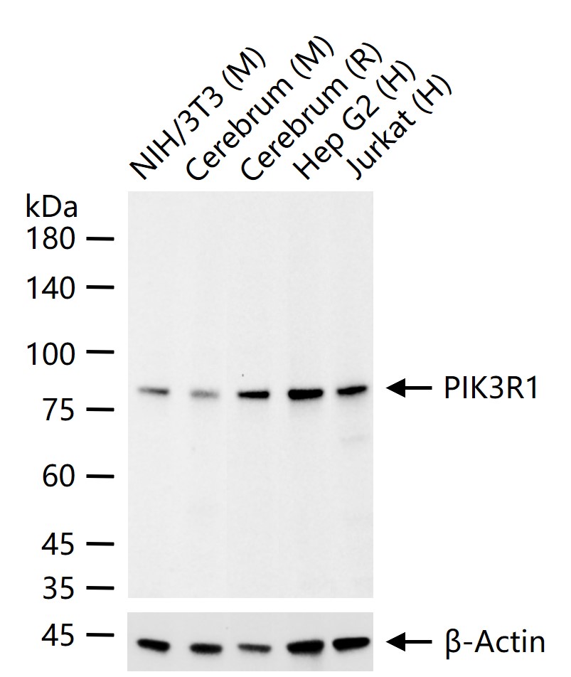
25 ug total protein per lane of various lysates (see on figure) probed with PIK3R1 monoclonal antibody, unconjugated (bsm-33219M) at 1:1000 dilution and 4°C overnight incubation. Followed by conjugated secondary antibody incubation at r.t. for 60 min.
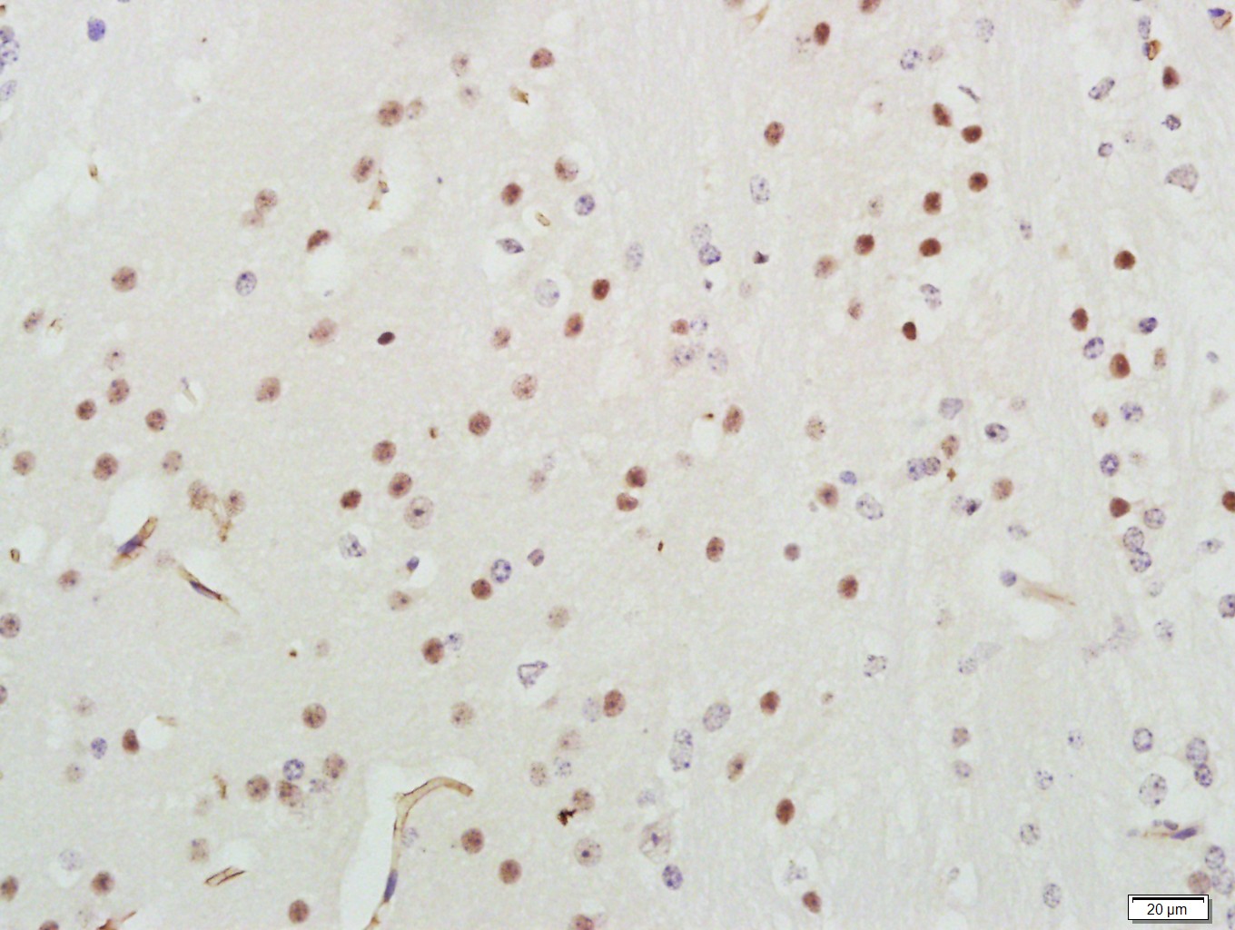
Paraformaldehyde-fixed, paraffin embedded (Mouse brain); Antigen retrieval by boiling in sodium citrate buffer (pH6.0) for 15min; Block endogenous peroxidase by 3% hydrogen peroxide for 20 minutes; Blocking buffer (normal goat serum) at 37°C for 30min; Antibody incubation with (PI3K p85) Monoclonal Antibody, Unconjugated (bsm-33219M) at 1:400 overnight at 4°C, followed by a conjugated secondary (sp-0023) for 20 minutes and DAB staining.
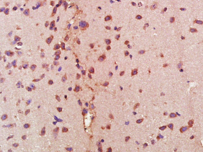
Paraformaldehyde-fixed, paraffin embedded (Mouse brain); Antigen retrieval by boiling in sodium citrate buffer (pH6.0) for 15min; Block endogenous peroxidase by 3% hydrogen peroxide for 20 minutes; Blocking buffer (normal goat serum) at 37°C for 30min; Antibody incubation with (PI 3 Kinase p85 alpha) Monoclonal Antibody, Unconjugated (bsm-33219M) at 1:400 overnight at 4°C, followed by operating according to SP Kit(Mouse) (sp-0024) instructionsand DAB staining.
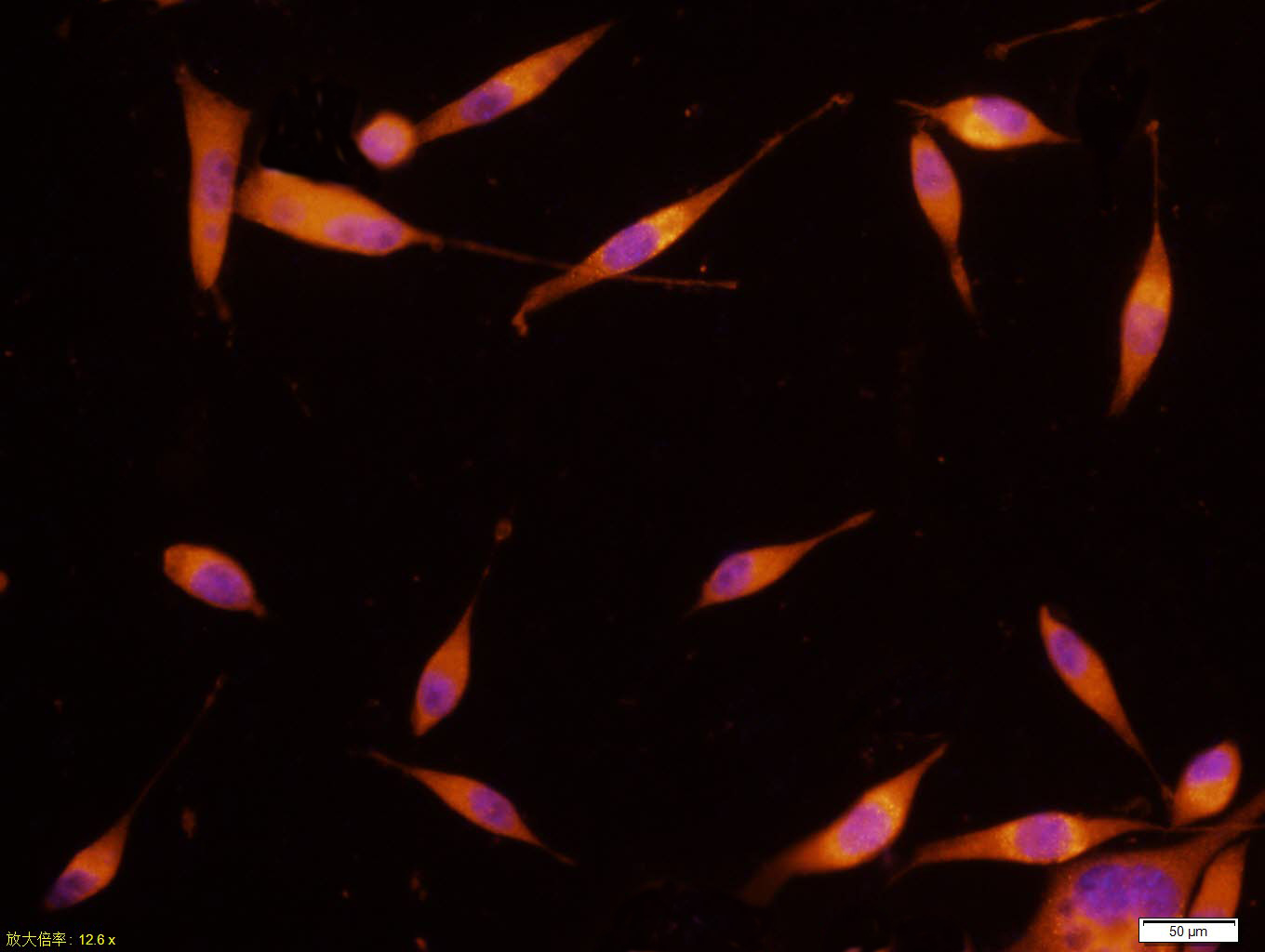
SH-SY5Y cell; 4% Paraformaldehyde-fixed; Triton X-100 at room temperature for 20 min; Blocking buffer (normal goat serum, C-0005) at 37°C for 20 min; Antibody incubation with (PI 3 Kinase p85 alpha) monoclonal Antibody, Unconjugated (bsm-33219M) 1:100, 90 minutes at 37°C; followed by a conjugated Goat Anti-Mouse IgG antibody at 37°C for 90 minutes, DAPI (blue, C02-04002) was used to stain the cell nuclei.
|
| 1、抗体溶解方法 | |
| 2、抗体修复方式 | |
| 3、常用试剂的配制 | |
| 4、免疫组化操作步骤 | |
| 5、免疫组化问题解答 | |
| 6、Western Blotting 操作步骤 | |
| 7、Western Blotting 问题解答 | |
| 8、关于肽链的设计 | |
| 9、多肽的溶解与保存 | |
| 10、酶标抗体效价测定程序 | |


