

| 产品编号 | bsm-10825M |
| 英文名称 | CD31 Mouse mAb |
| 中文名称 | 血小板内皮细胞黏附分子1单克隆抗体 |
| 别 名 | CD31; CD31/EndoCAM; GPIIA'; PECA1; PECAM-1; endoCAM; Pecam; PECA1_HUMAN; PECAM1; PECA1_MOUSE; platelet and endothelial cell adhesion molecule 1; platelet/endothelial cell adhesion molecule 1; CD31 antigen |
 |
Specific References (2) | bsm-10825M has been referenced in 2 publications.
[IF=11.322] Jinxiu Yu. et al. Promoting osseointegration of titanium by pH-responsive releasing of H2S and optimizing polarization time for macrophages. COMPOS PART B-ENG. 2023 Mar;253:110554 IHC,IF ; Rat.
[IF=10.041] Bingcheng Yi. et al. Lysine-doped polydopamine coating enhances antithrombogenicity and endothelialization of an electrospun aligned fibrous vascular graft. Appl Mater Today. 2021 Dec;25:101198 IF ; Human.
|
| 研究领域 | 肿瘤 心血管 细胞生物 免疫学 干细胞 细胞粘附分子 血管内皮细胞 内皮细胞 |
| 抗体来源 | Mouse |
| 克隆类型 | Monoclonal |
| 克 隆 号 | 2D4 |
| 交叉反应 | Human |
| 产品应用 | WB=1:1000-10000,IHC-P=1:200-2000,IHC-F=1:200-2000,IF=1:200-2000,Flow-Cyt=1ug/Test,ICC/IF=1:200-800
not yet tested in other applications. optimal dilutions/concentrations should be determined by the end user. |
| 理论分子量 | 78kDa |
| 检测分子量 | 130 |
| 细胞定位 | 细胞膜 |
| 性 状 | Liquid |
| 浓 度 | 1mg/ml |
| 免 疫 原 | Recombinant human CD31 28-601/727aa (C-6x His-Tag) <Extracellular> |
| 亚 型 | IgG1 |
| 纯化方法 | affinity purified by Protein A |
| 缓 冲 液 | 0.01M TBS (pH7.4) with 1% BSA, 0.02% Proclin300 and 50% Glycerol. |
| 保存条件 | Shipped at 4℃. Store at -20℃ for one year. Avoid repeated freeze/thaw cycles. |
| 注意事项 | This product as supplied is intended for research use only, not for use in human, therapeutic or diagnostic applications. |
| PubMed | PubMed |
| 产品介绍 |
This protein is a cell adhesion molecule expressed on platelets and at endothelial cell intercellular junctions. Type I membrane protein. SIZE: 738 amino acids; 82536 Da. SUBCELLULAR LOCATION: Membrane; Single-pass type I membrane protein. TISSUE SPECIFICITY: Long isoform predominates all tissues examined, isoform Delta12 was detected only in trachea and isoform Delta14-15 only in lung, isoform Delta14 was detected in all tissues examined with the strongest expression in heart. PTM: Phosphorylated on Ser and Tyr residues after cellular activation. SIMILARITY: Contains 6 Ig-like C2-type (immunoglobulin-like) domains. CD31, also known as platelet endothelial cell adhesion molecule 1 (PECAM1), is a type I integral membrane glycoprotein and a member of the immunoglobulin superfamily of cell surface receptors. It is found on the surface of platelets, monocytes, neutrophils, and some types of T-cells, and makes up a large portion of endothelial cell intercellular junctions. CD31 is implicated in several functions, including transendothelial migration of leukocytes, angiogenesis, and integrin activation. Tyr-690 plays a critical role in leukocyte transendothelial migration (TEM) and is required for efficient trafficking of CD31 to and from the lateral border recycling compartment (LBRC) and is also essential for the LBRC membrane to be targeted around migrating leukocytes. CD31 prevents phagocyte ingestion of closely apposed viable cells by transmitting 'detachment' signals, and changes function on apoptosis, promoting tethering of dying cells to phagocytes (the encounter of a viable cell with a phagocyte via the homophilic interaction of CD31 on both cell surfaces leads to the viable cell's active repulsion from the phagocyte. During apoptosis, the inside-out signaling of CD31 is somehow disabled so that the apoptotic cell does not actively reject the phagocyte anymore. The lack of this repulsion signal together with the interaction of the eat-me signals and their respective receptors causes the attachment of the apoptotic cell to the phagocyte, thus triggering the process of engulfment). CD31 has been used to measure angiogenesis in association with tumor recurrence. Other studies have also indicated that CD31 and CD34 can be used as markers for myeloid progenitor cells and recognize different subsets of myeloid leukemia infiltrates (granular sarcomas). Function: Induces susceptibility to atherosclerosis (By similarity). Cell adhesion molecule which is required for leukocyte transendothelial migration (TEM) under most inflammatory conditions. Tyr-690 plays a critical role in TEM and is required for efficient trafficking of PECAM1 to and from the lateral border recycling compartment (LBRC) and is also essential for the LBRC membrane to be targeted around migrating leukocytes. Prevents phagocyte ingestion of closely apposed viable cells by transmitting 'detachment' signals, and changes function on apoptosis, promoting tethering of dying cells to phagocytes (the encounter of a viable cell with a phagocyte via the homophilic interaction of PECAM1 on both cell surfaces leads to the viable cell's active repulsion from the phagocyte. During apoptosis, the inside-out signaling of PECAM1 is somehow disabled so that the apoptotic cell does not actively reject the phagocyte anymore. The lack of this repulsion signal together with the interaction of the eat-me signals and their respective receptors causes the attachment of the apoptotic cell to the phagocyte, thus triggering the process of engulfment). Isoform Delta15 is unable to protect against apoptosis. Modulates BDKRB2 activation. Regulates bradykinin- and hyperosmotic shock-induced ERK1/2 activation in human umbilical cord vein cells (HUVEC). Subunit: Interacts with PTPN11; Tyr-713 is critical for PTPN11 recruitment. Forms a complex with BDKRB2 and GNAQ. Interacts with BDKRB2 and GNAQ. Subcellular Location: Isoform Long: Membrane; Single-pass type I membrane protein. Cell junction. Note=Localizes to the lateral border recycling compartment (LBRC) and recycles from the LBRC to the junction in resting endothelial cells. Isoform Delta15: Cell junction. Note=Localizes to the lateral border recycling compartment (LBRC) and recycles from the LBRC to the junction in resting endothelial cells. Tissue Specificity: Expressed on platelets and leukocytes and is primarily concentrated at the borders between endothelial cells. Isoform Long predominates in all tissues examined. Isoform Delta12 is detected only in trachea. Isoform Delta14-15 is only detected in lung. Isoform Delta14 is detected in all tissues examined with the strongest expression in heart. Isoform Delta15 is expressed in brain, testis, ovary, cell surface of platelets, human umbilical vein endothelial cells (HUVECs), Jurkat T-cell leukemia, human erythroleukemia (HEL) and U937 histiocytic lymphoma cell lines (at protein level). Post-translational modifications: Phosphorylated on Ser and Tyr residues after cellular activation. Phosphorylated on tyrosine residues by FER and FES in response to FCER1 activation. In endothelial cells Fyn mediates mechanical-force (stretch or pull) induced tyrosine phosphorylation. Similarity: Contains 6 Ig-like C2-type (immunoglobulin-like) domains. SWISS: P16284 Gene ID: 5175 Database links: Entrez Gene: 5175 Human Entrez Gene: 18613 Mouse Omim: 173445 Human SwissProt: P16284 Human SwissProt: Q08481 Mouse Unigene: 376675 Human Unigene: 514412 Human Unigene: 343951 Mouse 血小板内皮细胞黏附分子-1 ( Platelet/ endothelial cell adhesion molecule, PECAM-1)在血小板、内皮细胞、单核细胞、嗜中性细胞及某些T细胞亚群上表达的质膜糖蛋白,属于免疫球蛋白超基因家族成员,在细胞外结构域中有6个C2亚类免疫球蛋白样保守性同原单位。在炎症应答中起作用。 |
| 产品图片 |
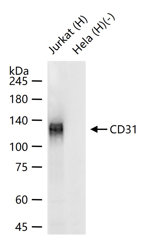
25 ug total protein per lane of various lysates (see on figure) probed with CD31 monoclonal antibody, unconjugated (bsm-10825M) at 1:1000 dilution and 4°C overnight incubation. Followed by conjugated secondary antibody incubation at r.t. for 60 min.
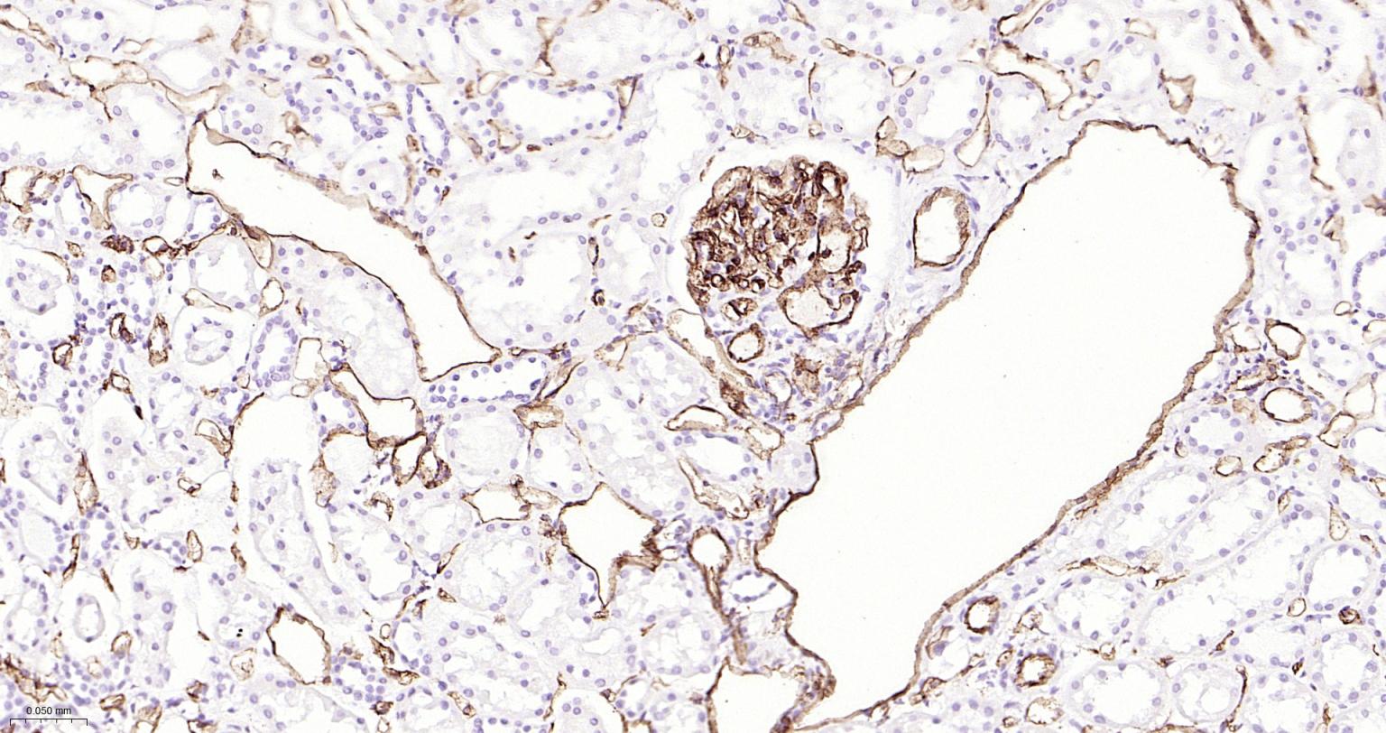
Paraformaldehyde-fixed, paraffin embedded Human Kidney; Antigen retrieval by boiling in sodium citrate buffer (pH6.0) for 15 min; The section was incubated with CD31 Monoclonal Antibody, Unconjugated (ascites of bsm-10825M) at 1:1500 overnight at 4°C, followed by conjugation to the bs-40296G-HRP and DAB (C-0010) staining.
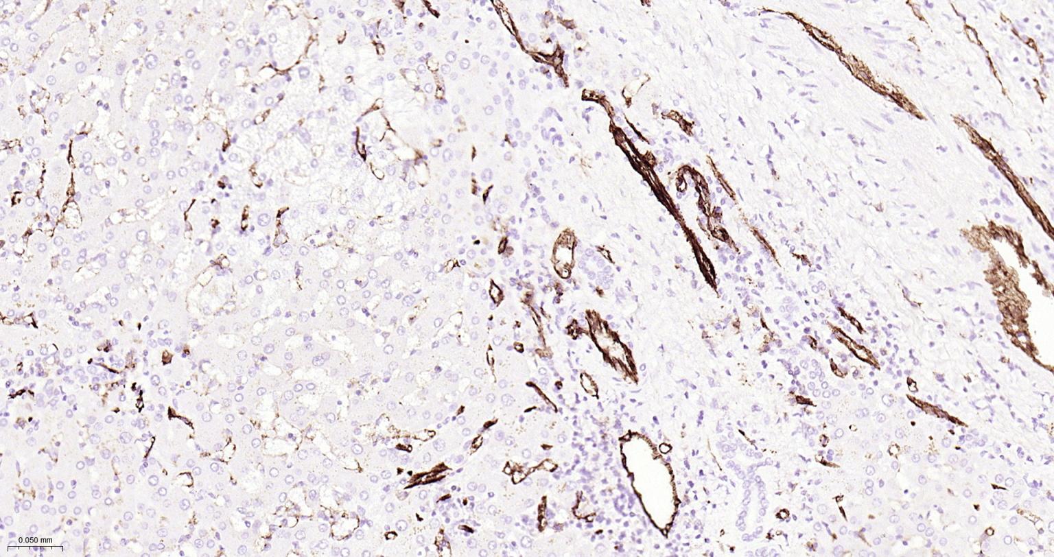
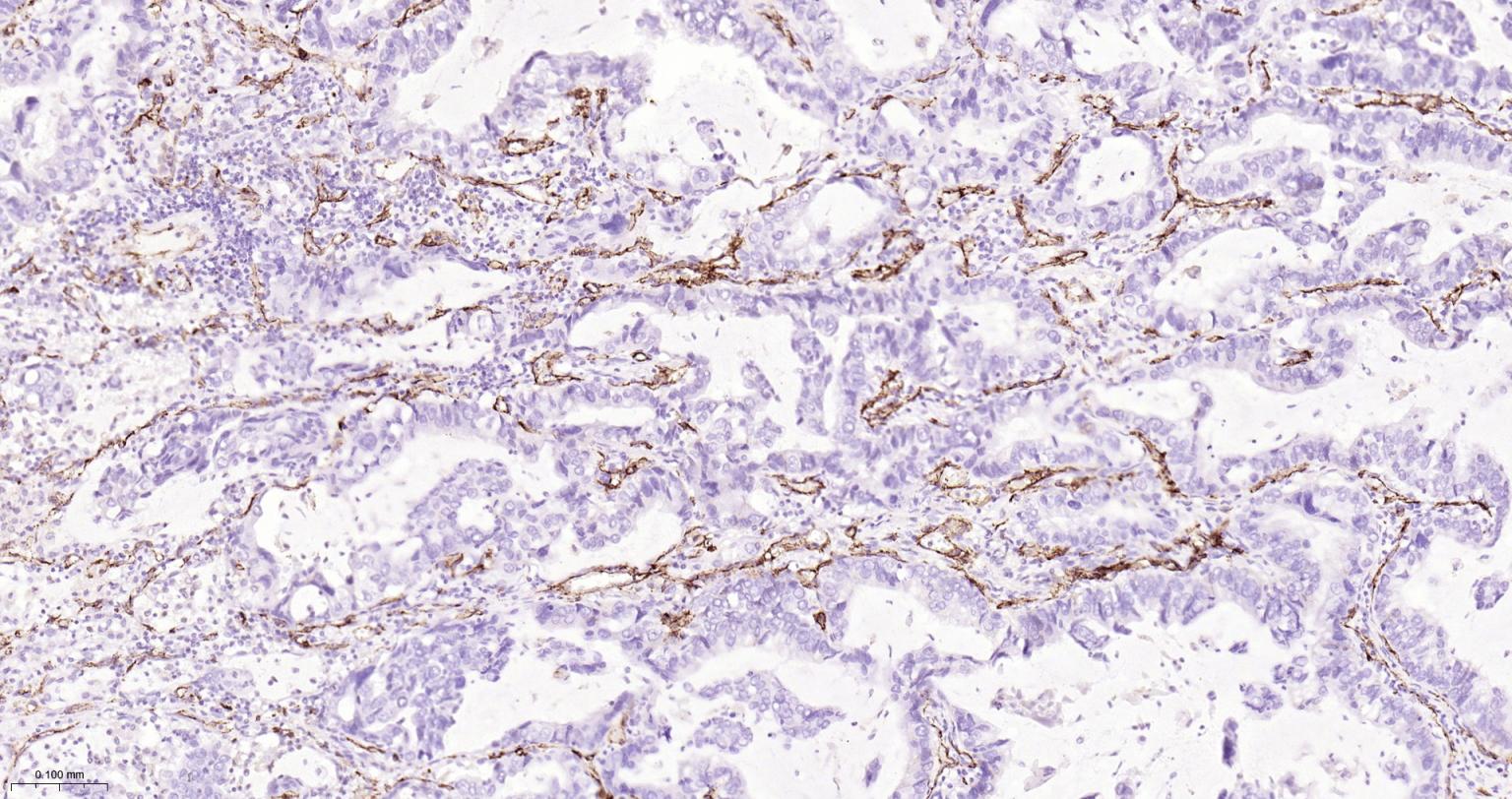
Paraformaldehyde-fixed, paraffin embedded Human Liver; Antigen retrieval by boiling in sodium citrate buffer (pH6.0) for 15 min; The section was incubated with CD31 Monoclonal Antibody, Unconjugated (ascites of bsm-10825M) at 1:1500 overnight at 4°C, followed by conjugation to the bs-40296G-HRP and DAB (C-0010) staining.
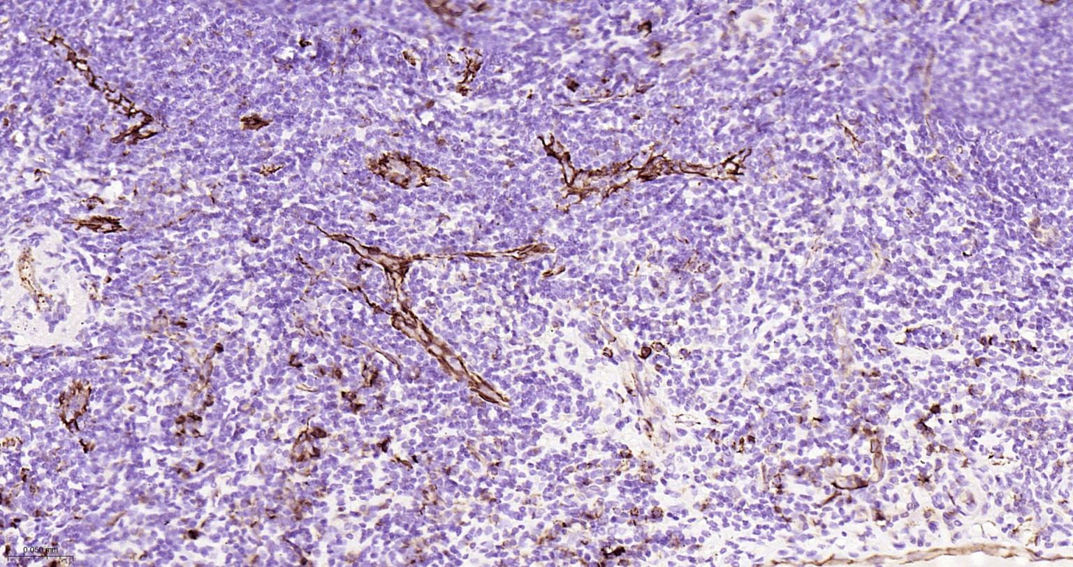
Paraformaldehyde-fixed, paraffin embedded Human Tonsil; Antigen retrieval by boiling in sodium citrate buffer (pH6.0) for 15 min; The section was incubated with CD31 Monoclonal Antibody, Unconjugated (ascites of bsm-10825M) at 1:1500 overnight at 4°C, followed by conjugation to the bs-40296G-HRP and DAB (C-0010) staining.
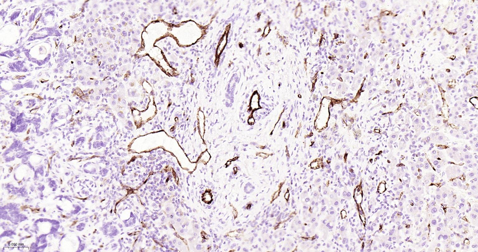
Paraformaldehyde-fixed, paraffin embedded Human Liver Cancer; Antigen retrieval by boiling in sodium citrate buffer (pH6.0) for 15 min; The section was incubated with CD31 Monoclonal Antibody, Unconjugated (ascites of bsm-10825M) at 1:1500 overnight at 4°C, followed by conjugation to the bs-40296G-HRP and DAB (C-0010) staining.
Paraformaldehyde-fixed, paraffin embedded Human Lung Cancer; Antigen retrieval by boiling in sodium citrate buffer (pH6.0) for 15 min; The section was incubated with CD31 Monoclonal Antibody, Unconjugated (ascites of bsm-10825M) at 1:1500 overnight at 4°C, followed by conjugation to the bs-40296G-HRP and DAB (C-0010) staining.
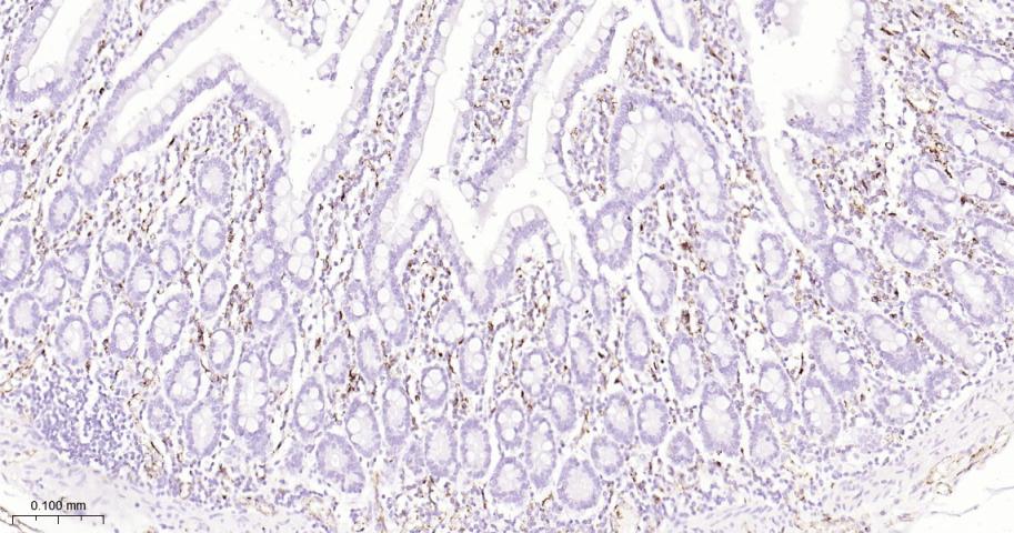
Paraformaldehyde-fixed, paraffin embedded Human Duodenum; Antigen retrieval by boiling in sodium citrate buffer (pH6.0) for 15 min; The section was incubated with CD31 Monoclonal Antibody, Unconjugated (ascites of bsm-10825M) at 1:1500 overnight at 4°C, followed by conjugation to the bs-40296G-HRP and DAB (C-0010) staining.
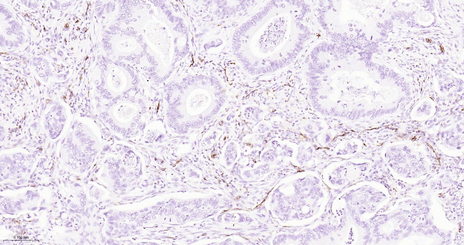
Paraformaldehyde-fixed, paraffin embedded Human Colon Cancer; Antigen retrieval by boiling in sodium citrate buffer (pH6.0) for 15 min; The section was incubated with CD31 Monoclonal Antibody, Unconjugated (ascites of bsm-10825M) at 1:1500 overnight at 4°C, followed by conjugation to the bs-40296G-HRP and DAB (C-0010) staining.
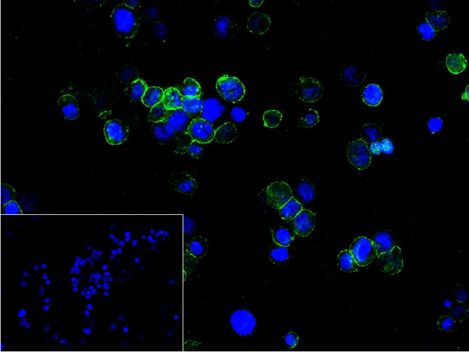
4% Paraformaldehyde-fixed THP-1 (H) cell; Antibody incubation with (CD31) monoclonal Antibody, unconjugated (bsm-10825M) 1:200, 90 min at 37°C; followed by conjugated Goat Anti-Mouse IgG antibody (green, bs-60296G-BF488) at 37°C for 90 min, DAPI (blue, C02-04002) was used to stain the cell nuclei. PBS instead of the primary antibody was used as the blank control.
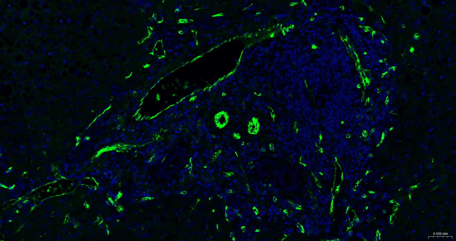
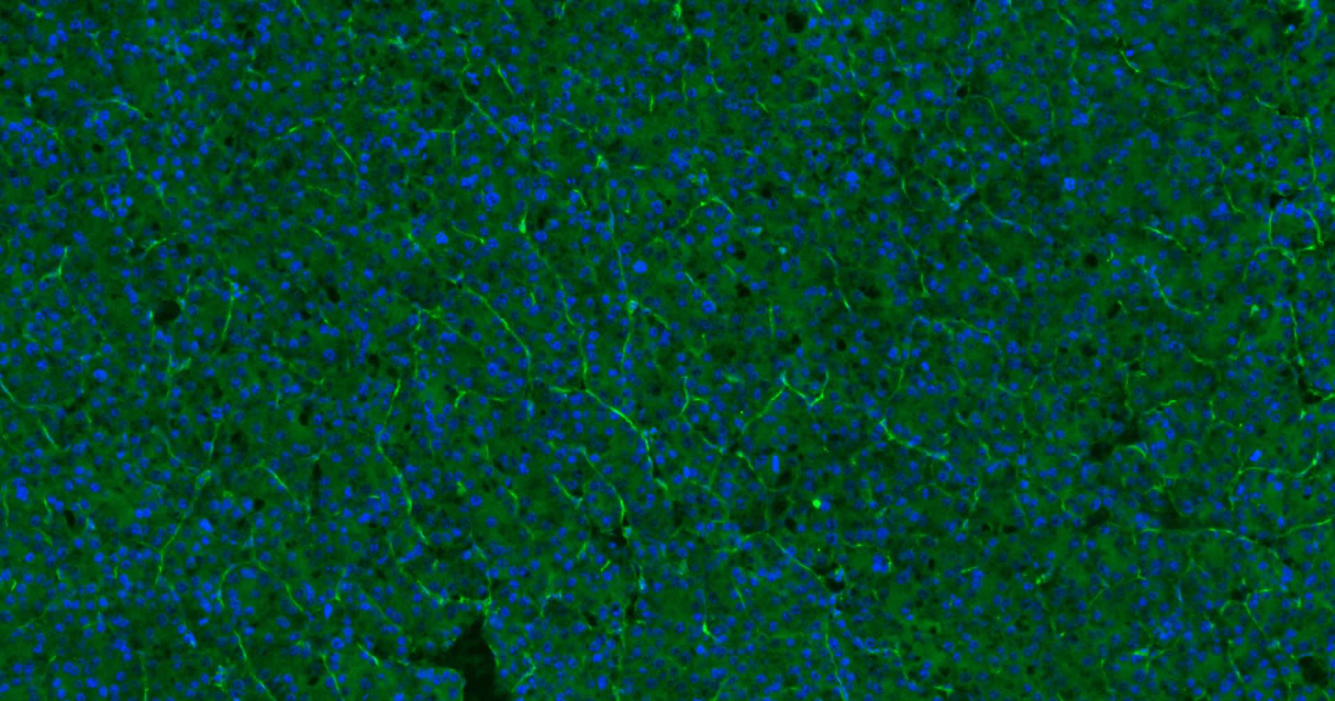
Paraformaldehyde-fixed, paraffin embedded Human Liver; Antigen retrieval by boiling in sodium citrate buffer (pH6.0) for 15 min; Antibody incubation with CD31 Monoclonal Antibody, Unconjugated (bsm-10825M) at 1:200 overnight at 4°C. Followed by conjugated Goat Anti-Rabbit IgG antibody (green, bs-0296G-BF488), DAPI (blue, C02-04002) was used to stain the cell nuclei.
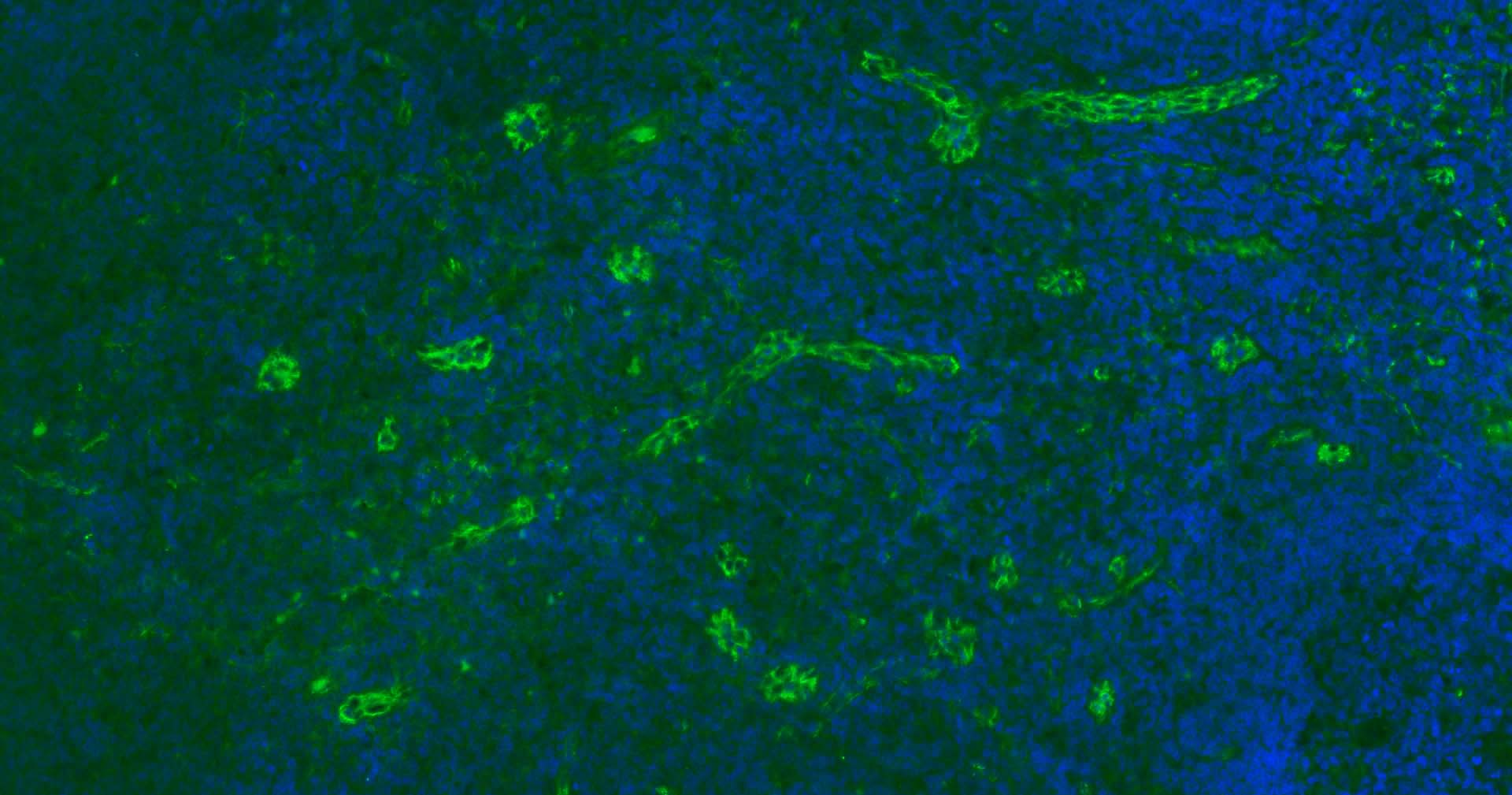
Paraformaldehyde-fixed, paraffin embedded Human Tonsil; Antigen retrieval by boiling in sodium citrate buffer (pH6.0) for 15 min; Antibody incubation with CD31 Monoclonal Antibody, Unconjugated (bsm-10825M) at 1:200 overnight at 4°C. Followed by conjugated Goat Anti-Rabbit IgG antibody (green, bs-0296G-BF488), DAPI (blue, C02-04002) was used to stain the cell nuclei.
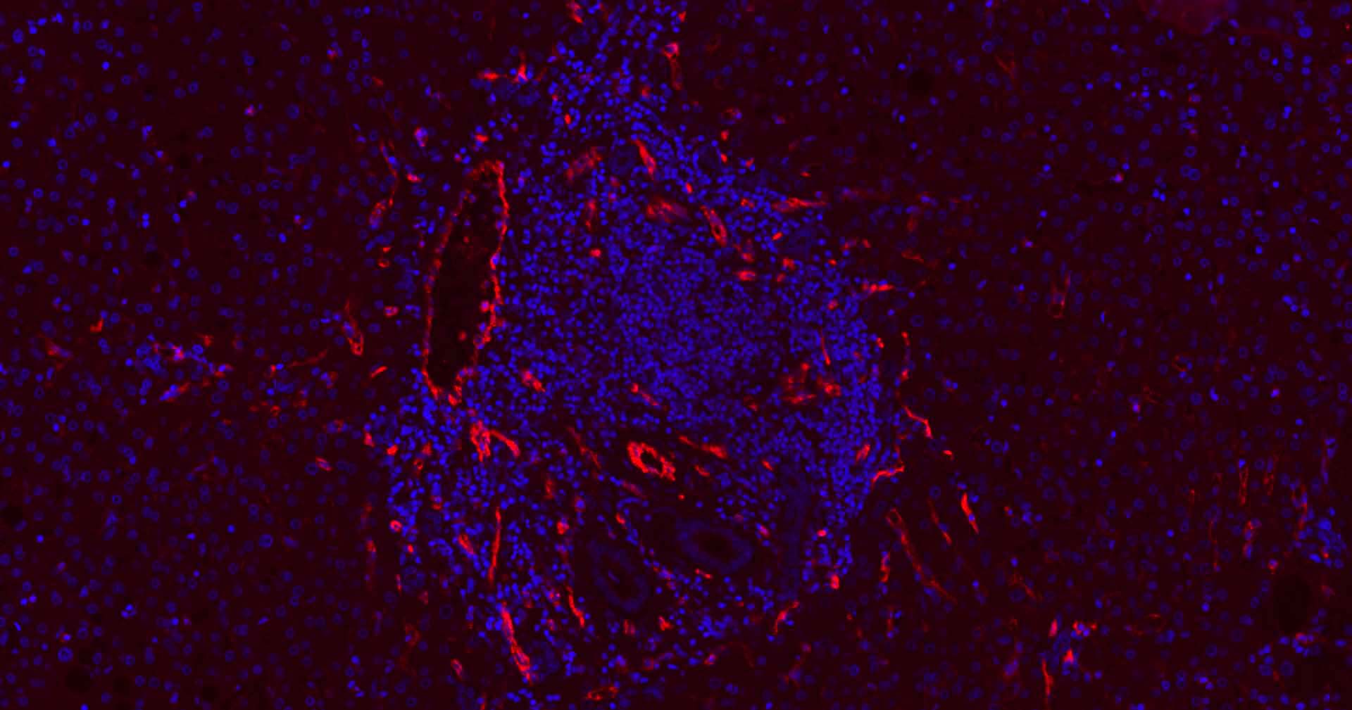
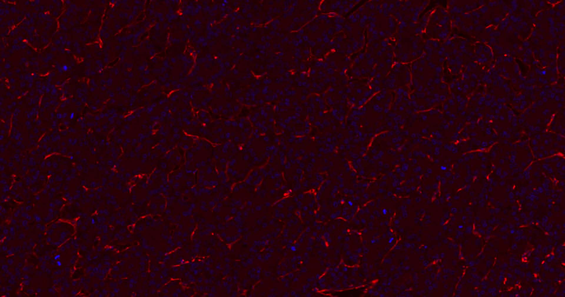
Paraformaldehyde-fixed, paraffin embedded Human Liver ; Antigen retrieval by boiling in sodium citrate buffer (pH6.0) for 15 min; Antibody incubation with CD31 Monoclonal Antibody, Unconjugated (bsm-10825M) at 1:200 overnight at 4°C. Followed by conjugated Goat Anti-Rabbit IgG antibody (Red, bs-0296G-BF594), DAPI (blue, C02-04002) was used to stain the cell nuclei.
Paraformaldehyde-fixed, paraffin embedded Human Colon Cancer; Antigen retrieval by boiling in sodium citrate buffer (pH6.0) for 15 min; Antibody incubation with CD31 Monoclonal Antibody, Unconjugated (bsm-10825M) at 1:200 overnight at 4°C. Followed by conjugated Goat Anti-Rabbit IgG antibody (Red, bs-0296G-BF594), DAPI (blue, C02-04002) was used to stain the cell nuclei.
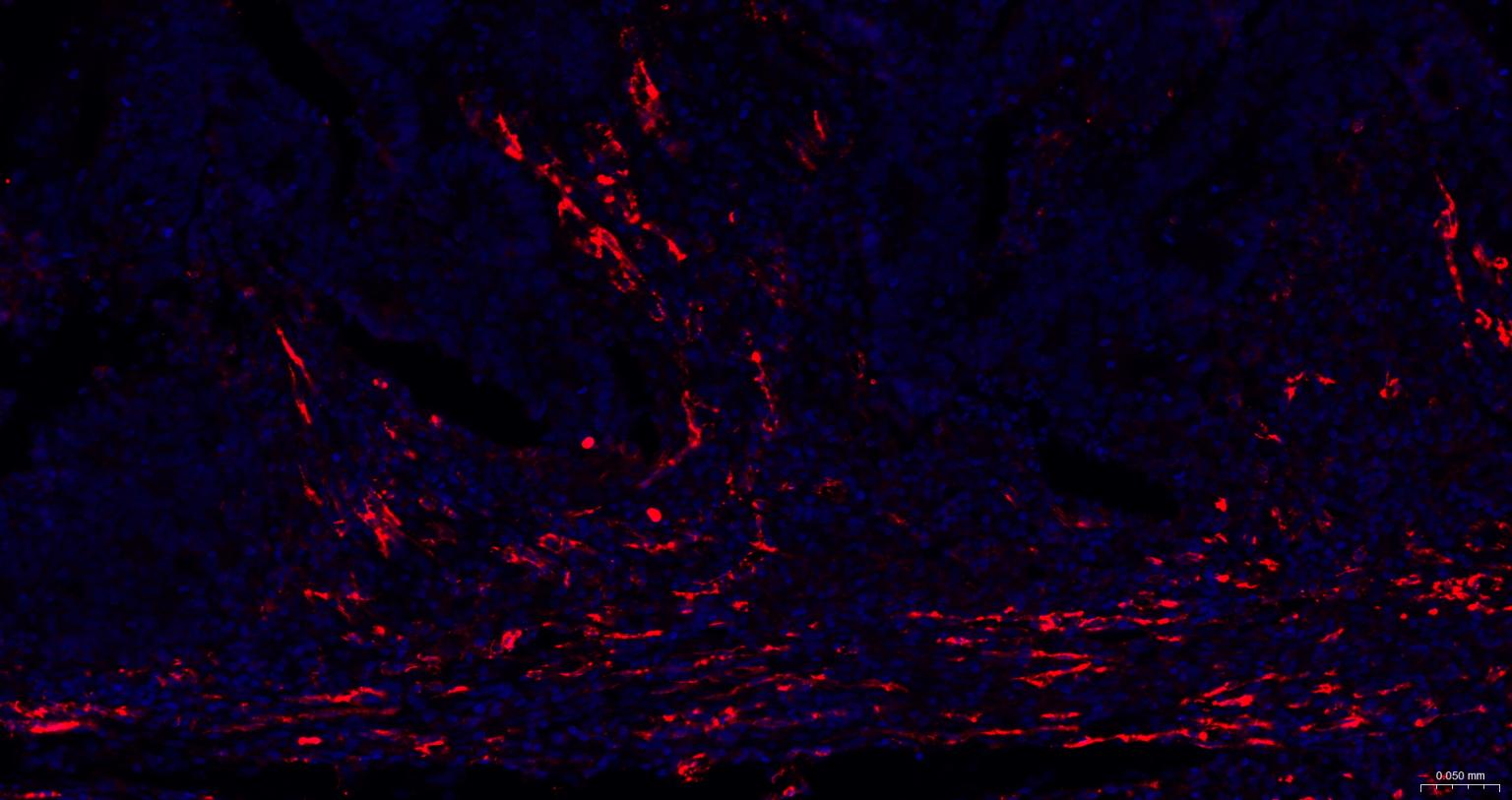
Paraformaldehyde-fixed, paraffin embedded Human Liver Cancer; Antigen retrieval by boiling in sodium citrate buffer (pH6.0) for 15 min; Antibody incubation with CD31 Monoclonal Antibody, Unconjugated (bsm-10825M) at 1:200 overnight at 4°C. Followed by conjugated Goat Anti-Rabbit IgG antibody (Red, bs-0296G-BF594), DAPI (blue, C02-04002) was used to stain the cell nuclei.
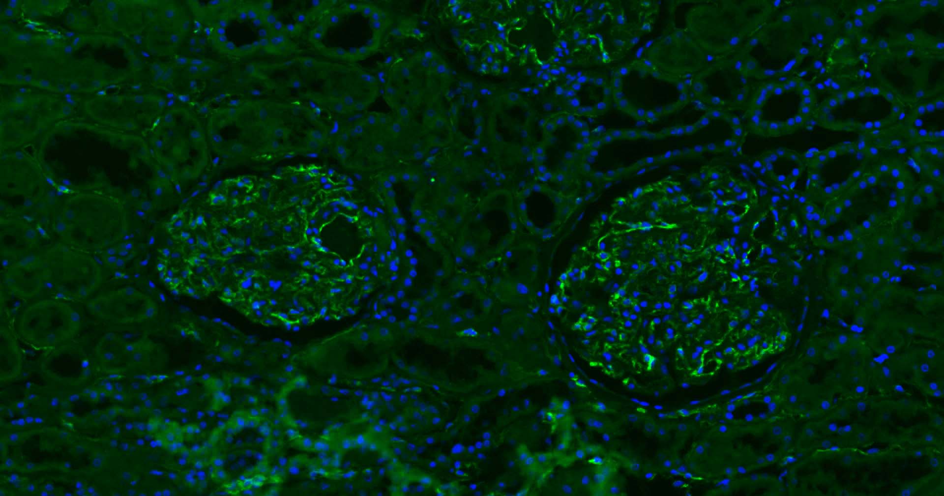
Paraformaldehyde-fixed, paraffin embedded Human Kidney; Antigen retrieval by boiling in sodium citrate buffer (pH6.0) for 15 min; Antibody incubation with CD31 Monoclonal Antibody, Unconjugated (bsm-10825M) at 1:200 overnight at 4°C. Followed by conjugated Goat Anti-Rabbit IgG antibody (green, bs-0296G-BF488), DAPI (blue, C02-04002) was used to stain the cell nuclei.
Paraformaldehyde-fixed, paraffin embedded Human Colon Cancer; Antigen retrieval by boiling in sodium citrate buffer (pH6.0) for 15 min; Antibody incubation with CD31 Monoclonal Antibody, Unconjugated (bsm-10825M) at 1:200 overnight at 4°C. Followed by conjugated Goat Anti-Rabbit IgG antibody (green, bs-0296G-BF488), DAPI (blue, C02-04002) was used to stain the cell nuclei.
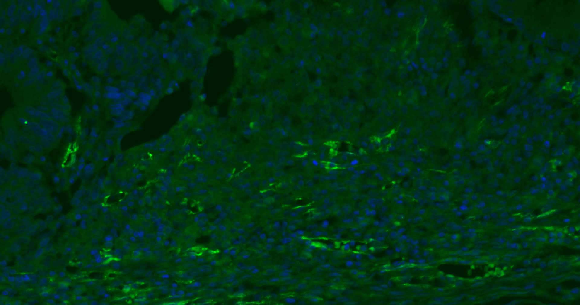
Paraformaldehyde-fixed, paraffin embedded Human Liver Cancer; Antigen retrieval by boiling in sodium citrate buffer (pH6.0) for 15 min; Antibody incubation with CD31 Monoclonal Antibody, Unconjugated (bsm-10825M) at 1:200 overnight at 4°C. Followed by conjugated Goat Anti-Rabbit IgG antibody (green, bs-0296G-BF488), DAPI (blue, C02-04002) was used to stain the cell nuclei.
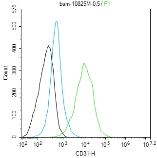
The THP-1(H) cells were incubated in 5%BSA to block non-specific protein-protein interactions (30 min at r.t.). Primary Antibody (green): Mouse Anti-CD31 antibody (bsm-10825M): 0.5 μg/10^6 cells; Secondary Antibody (white blue): Goat anti-Mouse IgG-FITC (bs-60296G-FITC): 1 μg/test. Blank control (black): PBS. Acquisition of 20,000 events was performed.
|
| 1、抗体溶解方法 | |
| 2、抗体修复方式 | |
| 3、常用试剂的配制 | |
| 4、免疫组化操作步骤 | |
| 5、免疫组化问题解答 | |
| 6、Western Blotting 操作步骤 | |
| 7、Western Blotting 问题解答 | |
| 8、关于肽链的设计 | |
| 9、多肽的溶解与保存 | |
| 10、酶标抗体效价测定程序 | |


