

| 产品编号 | bs-16681R |
| 英文名称 | phospho-HEF1 (Ser369) Rabbit pAb |
| 中文名称 | 磷酸化蛋白激酶底物相关蛋白抗体 |
| 别 名 | NEDD9 | HEF1 (phospho-S369); p-HEF1; phospho-HEF1; p-NEDD9; NEDD9 | HEF1 (phospho-Ser369); CAS-L; CAS2; CASL; CASS2; HEF1; MEF1; Nedd-9; p105; CASL_HUMAN; NEDD9; CRK-associated substrate-related protein (CAS-L | CasL); Cas scaffolding protein family membe |
| 产品类型 | 磷酸化抗体 |
| 研究领域 | 肿瘤 细胞生物 信号转导 细胞凋亡 转录调节因子 表观遗传学 |
| 抗体来源 | Rabbit |
| 克隆类型 | Polyclonal |
| 克 隆 号 | |
| 交叉反应 | Human (predicted: Sheep,Cow,Horse) |
| 产品应用 | WB=1:500-2000
not yet tested in other applications. optimal dilutions/concentrations should be determined by the end user. |
| 理论分子量 | 93 kDa |
| 检测分子量 | 105,115 |
| 细胞定位 | 细胞浆 |
| 性 状 | Liquid |
| 浓 度 | 1mg/ml |
| 免 疫 原 | KLH conjugated synthesised phosphopeptide derived from human HEF1 around the phosphorylation site of Ser369: RL(p-S)FS |
| 亚 型 | IgG |
| 纯化方法 | affinity purified by Protein A |
| 缓 冲 液 | 0.01M TBS (pH7.4) with 1% BSA, 0.02% Proclin300 and 50% Glycerol. |
| 保存条件 | Shipped at 4℃. Store at -20℃ for one year. Avoid repeated freeze/thaw cycles. |
| 注意事项 | This product as supplied is intended for research use only, not for use in human, therapeutic or diagnostic applications. |
| PubMed | PubMed |
| 产品介绍 |
The protein encoded by this gene is a member of the CRK-associated substrates family. Members of this family are adhesion docking molecules that mediate protein-protein interactions for signal transduction pathways. This protein is a focal adhesion protein that acts as a scaffold to regulate signaling complexes important in cell attachment, migration and invasion as well as apoptosis and the cell cycle. This protein has also been reported to have a role in cancer metastasis. Alternative splicing results in multiple transcript variants. [provided by RefSeq, Aug 2012] Function: Docking protein which plays a central coordinating role for tyrosine-kinase-based signaling related to cell adhesion. May function in transmitting growth control signals between focal adhesions at the cell periphery and the mitotic spindle in response to adhesion or growth factor signals initiating cell proliferation. May play an important role in integrin beta-1 or B cell antigen receptor (BCR) mediated signaling in B- and T-cells. Integrin beta-1 stimulation leads to recruitment of various proteins including CRK, NCK and SHPTP2 to the tyrosine phosphorylated form. Subcellular Location: Cytoplasm; cytoskeleton; spindle and Cytoplasm; cell cortex. Nucleus. Golgi apparatus. Cell projection; lamellipodium. Cytoplasm. Cell junction ; focal adhesion. Localizes to both the cell nucleus and the cell periphery and is differently localized in fibroblasts and epithelial cells. In fibroblasts is predominantly nuclear and in some cells is present in the Golgi apparatus. In epithelial cells localized predominantly in the cell periphery with particular concentration in lamellipodia but is also found in the nucleus. Isoforms p105 and p115 are predominantly cytoplasmic and associate with focal adhesions while p55 associates with mitotic spindle. Tissue Specificity: Widely expressed. Higher levels detected in kidney, lung, and placenta. Also detected in T-cells, B-cells and diverse cell lines. The protein has been detected in lymphocytes, in diverse cell lines, and in lung tissues. Post-translational modifications: Cell cycle-regulated processing produces four isoforms: p115, p105, p65, and p55. Isoform p115 arises from p105 phosphorylation and appears later in the cell cycle. Isoform p55 arises from p105 as a result of cleavage at a caspase cleavage-related site and it appears specifically at mitosis. The p65 isoform is poorly detected. Focal adhesion kinase 1 phosphorylates the protein at the YDYVHL motif (conserved among all cas proteins). The SRC family kinases (FYN, SRC, LCK and CRK) are recruited to the phosphorylated sites and can phosphorylate other tyrosine residues. Ligation of either integrin beta-1 or B-cell antigen receptor on tonsillar B-cells and B-cell lines promotes tyrosine phosphorylation and both integrin and BCR-mediated tyrosine phosphorylation requires an intact actin network. In fibroblasts transformation with oncogene v-ABL results in an increase in tyrosine phosphorylation. Transiently phosphorylated following CD3 cross-linking and this phosphorylated form binds to CRK and C3G. A mutant lacking the SH3 domain is phosphorylated upon CD3 cross-linking but not upon integrin beta-1 cross-linking. Tyrosine phosphorylation occurs upon stimulation of the G-protein coupled C1a calcitonin receptor in rabbit. Calcitonin-stimulated tyrosine phosphorylation is mediated by calcium- and protein kinase C-dependent mechanisms and requires the integrity of the actin cytoskeleton. Similarity: Belongs to the CAS family. Contains 1 SH3 domain. SWISS: Q14511 Gene ID: 4739 Database links: Entrez Gene: 4739 Human Entrez Gene: 18003 Mouse Omim: 602265 Human SwissProt: Q14511 Human SwissProt: O35177 Mouse Unigene: 37982 Human |
| 产品图片 |
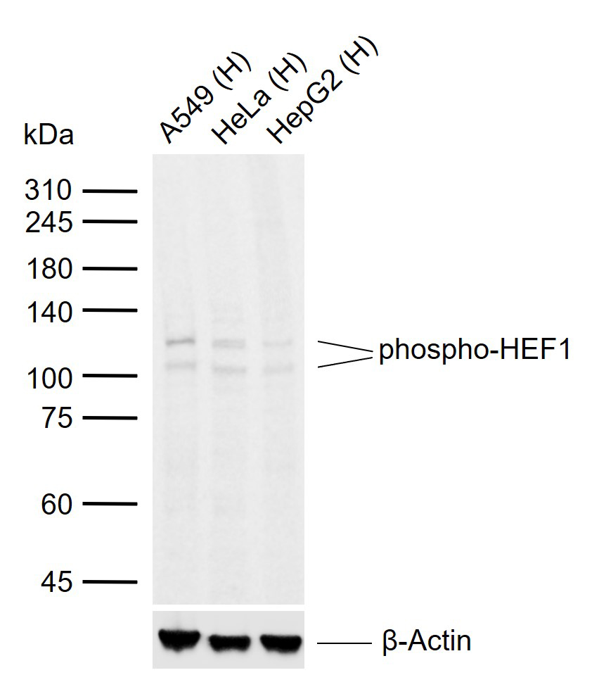
Sample:
Lane 1: Human A549 cell lysates
Lane 2: Human HeLa cell lysates
Lane 3: Human HepG2 cell lysates
Primary: Anti-phospho-HEF1 (Ser369) (bs-16681R) at 1/1000 dilution
Secondary: IRDye800CW Goat Anti-Rabbit IgG at 1/20000 dilution
Predicted band size: 93 kDa
Observed band size: 105,115 kDa
|
| 1、抗体溶解方法 | |
| 2、抗体修复方式 | |
| 3、常用试剂的配制 | |
| 4、免疫组化操作步骤 | |
| 5、免疫组化问题解答 | |
| 6、Western Blotting 操作步骤 | |
| 7、Western Blotting 问题解答 | |
| 8、关于肽链的设计 | |
| 9、多肽的溶解与保存 | |
| 10、酶标抗体效价测定程序 | |


