

| 产品编号 | bs-14548R |
| 英文名称 | eIF4A3 Rabbit pAb |
| 中文名称 | eIF4A3蛋白抗体 |
| 别 名 | IF4A3_CHICK; EIF4A3; eIF-4A-III; eIF4A-III; ATP-dependent RNA helicase DDX48; ATP-dependent RNA helicase eIF4A-3; DEAD box protein 48; Eukaryotic translation initiation factor 4A isoform 3; 3.6.4.13; DDX48; IF4A3_BOVIN; IF4A3_MACFA; IF4A3_HUMAN; Eukaryoti |
 |
Specific References (1) | bs-14548R has been referenced in 1 publications.
[IF=11.161] Zhipeng Jiang. et al. EIF4A3-induced circ_0084615 contributes to the progression of colorectal cancer via miR-599/ONECUT2 pathway. J Exp Clin Canc Res. 2021 Dec;40(1):1-15 IP ; Human.
|
| 研究领域 | 细胞生物 细胞分化 表观遗传学 |
| 抗体来源 | Rabbit |
| 克隆类型 | Polyclonal |
| 克 隆 号 | |
| 交叉反应 | Human,Mouse,Rat (predicted: Cow,Chicken,Dog,Horse) |
| 产品应用 | WB=1:500-2000,IHC-P=1:100-500,IHC-F=1:100-500,IF=1:100-500,Flow-Cyt=2ug/Test,ICC/IF=1:100-500
not yet tested in other applications. optimal dilutions/concentrations should be determined by the end user. |
| 理论分子量 | 47 kDa |
| 检测分子量 | 47 |
| 细胞定位 | 细胞核 细胞浆 |
| 性 状 | Liquid |
| 浓 度 | 1mg/ml |
| 免 疫 原 | KLH conjugated synthetic peptide derived from human eIF4A3: 351-411/411 |
| 亚 型 | IgG |
| 纯化方法 | affinity purified by Protein A |
| 缓 冲 液 | 0.01M TBS (pH7.4) with 1% BSA, 0.02% Proclin300 and 50% Glycerol. |
| 保存条件 | Shipped at 4℃. Store at -20℃ for one year. Avoid repeated freeze/thaw cycles. |
| 注意事项 | This product as supplied is intended for research use only, not for use in human, therapeutic or diagnostic applications. |
| PubMed | PubMed |
| 产品介绍 |
This gene encodes a member of the DEAD box protein family. DEAD box proteins, characterized by the conserved motif Asp-Glu-Ala-Asp (DEAD), are putative RNA helicases. They are implicated in a number of cellular processes involving alteration of RNA secondary structure, such as translation initiation, nuclear and mitochondrial splicing, and ribosome and spliceosome assembly. Based on their distribution patterns, some members of this family are believed to be involved in embryogenesis, spermatogenesis, and cellular growth and division. The protein encoded by this gene is a nuclear matrix protein. Its amino acid sequence is highly similar to the amino acid sequences of the translation initiation factors eIF4AI and eIF4AII, two other members of the DEAD box protein family. [provided by RefSeq, Jul 2008] Function: ATP-dependent RNA helicase. Component of a splicing-dependent multiprotein exon junction complex (EJC) deposited at splice junction on mRNAs. The EJC is a dynamic structure consisting of a few core proteins and several more peripheral nuclear and cytoplasmic associated factors that join the complex only transiently either during EJC assembly or during subsequent mRNA metabolism. Core components of the EJC, that remains bound to spliced mRNAs throughout all stages of mRNA metabolism, functions to mark the position of the exon-exon junction in the mature mRNA and thereby influences downstream processes of gene expression including mRNA splicing, nuclear mRNA export, subcellular mRNA localization, translation efficiency and nonsense-mediated mRNA decay (NMD). Constitutes at least part of the platform anchoring other EJC proteins to spliced mRNAs. Its RNA-dependent ATPase and RNA-helicase activities are induced by CASC3, but abolished in presence of the MAGOH/RBM8A heterodimer, thereby trapping the ATP-bound EJC core onto spliced mRNA in a stable conformation. The inhibition of ATPase activity by the MAGOH/RBM8A heterodimer increases the RNA-binding affinity of the EJC. Involved in translational enhancement of spliced mRNAs after formation of the 80S ribosome complex. Binds spliced mRNA in sequence-independent manner, 20-24 nucleotides upstream of mRNA exon-exon junctions. Shows higher affinity for single-stranded RNA in an ATP-bound core EJC complex than after the ATP is hydrolyzed. Subcellular Location: Nucleus. Nucleus speckle. Cytoplasm. Nucleocytoplasmic shuttling protein. Travels to the cytoplasm as part of the exon junction complex (EJC) bound to mRNA. Detected in dendritic layer as well as the nuclear and cytoplasmic (somatic) compartments of neurons. Colocalizes with STAU1 and FMR1 in dendrites. Tissue Specificity: Ubiquitously expressed. Similarity: Belongs to the DEAD box helicase family. eIF4A subfamily. Contains 1 helicase ATP-binding domain. Contains 1 helicase C-terminal domain. SWISS: P38919 Gene ID: 9775 Database links: Entrez Gene: 9775 Human Entrez Gene: 515145 CowEntrez Gene: 192170 Mouse Entrez Gene: 399362 Xenopus laevis Entrez Gene: 394053 Zebrafish Omim: 608546 Human SwissProt: Q5ZM36 Chicken SwissProt: Q4R3Q1 Cynomolgus Monkey SwissProt: P38919 Human SwissProt: Q91VC3 Mouse SwissProt: O42226 Xenopus laevis SwissProt: Q5U526 Xenopus laevis SwissProt: Q7ZVA6 Zebrafish Unigene: 389649 Human Unigene: 197555 Mouse Unigene: 422975 Mouse Unigene: 202617 Rat Unigene: 78203 Zebrafish |
| 产品图片 |
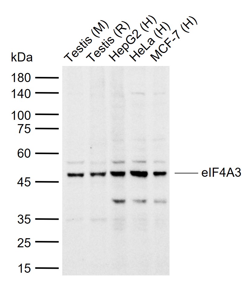
Sample:
Lane 1: Mouse Testis tissue lysates
Lane 2: Rat Testis tissue lysates
Lane 3: Human HepG2 cell lysates
Lane 4: Human HeLa cell lysates
Lane 5: Human MCF-7 cell lysates
Primary: Anti-eIF4A3 (bs-14548R) at 1/1000 dilution
Secondary: IRDye800CW Goat Anti-Rabbit IgG at 1/20000 dilution
Predicted band size: 47 kDa
Observed band size: 47 kDa

Tissue/cell: mouse embryo tissue; 4% Paraformaldehyde-fixed and paraffin-embedded;
Antigen retrieval: citrate buffer ( 0.01M, pH 6.0 ), Boiling bathing for 15min; Block endogenous peroxidase by 3% Hydrogen peroxide for 30min; Blocking buffer (normal goat serum,C-0005) at 37℃ for 20 min;
Incubation: Anti-eIF4A3 Polyclonal Antibody, Unconjugated(bs-14548R) 1:200, overnight at 4°C, followed by conjugation to the secondary antibody(SP-0023) and DAB(C-0010) staining
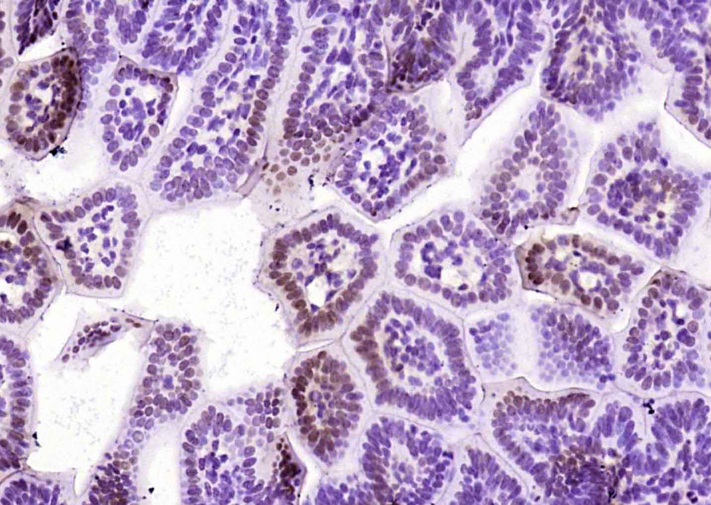
Paraformaldehyde-fixed, paraffin embedded (mouse embryos tissue); Antigen retrieval by boiling in sodium citrate buffer (pH6.0) for 15min; Block endogenous peroxidase by 3% hydrogen peroxide for 20 minutes; Blocking buffer (normal goat serum) at 37°C for 30min; Antibody incubation with (eIF4A3) Polyclonal Antibody, Unconjugated (bs-14548R) at 1:200 overnight at 4°C, followed by operating according to SP Kit(Rabbit) (sp-0023) instructionsand DAB staining.
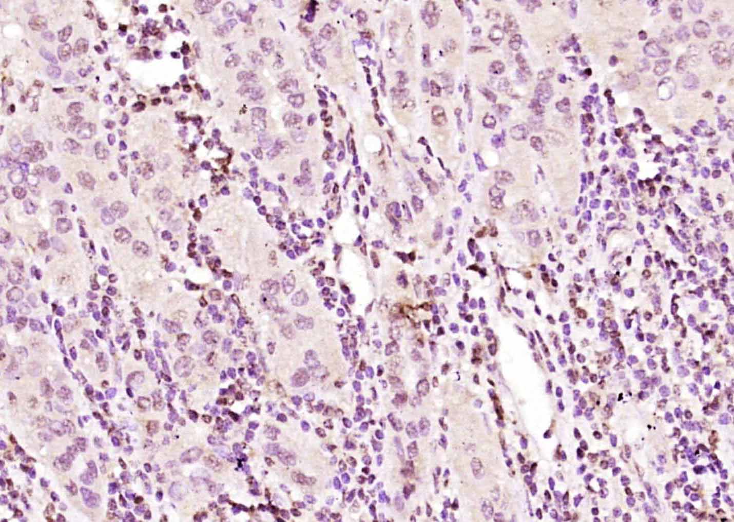
Paraformaldehyde-fixed, paraffin embedded (human liver carcinoma); Antigen retrieval by boiling in sodium citrate buffer (pH6.0) for 15min; Block endogenous peroxidase by 3% hydrogen peroxide for 20 minutes; Blocking buffer (normal goat serum) at 37°C for 30min; Antibody incubation with (eIF4A3) Polyclonal Antibody, Unconjugated (bs-14548R) at 1:200 overnight at 4°C, followed by operating according to SP Kit(Rabbit) (sp-0023) instructionsand DAB staining.
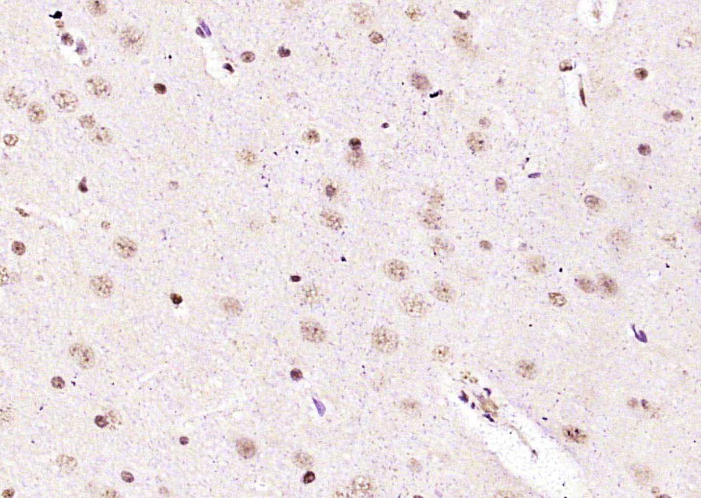
Paraformaldehyde-fixed, paraffin embedded (rat brain tissue); Antigen retrieval by boiling in sodium citrate buffer (pH6.0) for 15min; Block endogenous peroxidase by 3% hydrogen peroxide for 20 minutes; Blocking buffer (normal goat serum) at 37°C for 30min; Antibody incubation with (eIF4A3) Polyclonal Antibody, Unconjugated (bs-14548R) at 1:200 overnight at 4°C, followed by operating according to SP Kit(Rabbit) (sp-0023) instructionsand DAB staining
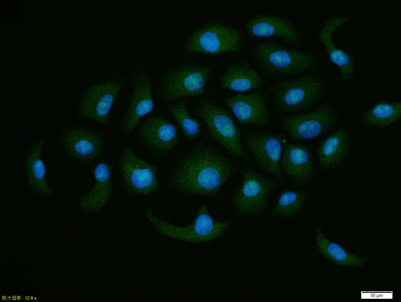
HepG2 cell; 4% Paraformaldehyde-fixed; Triton X-100 at room temperature for 20 min; Blocking buffer (normal goat serum, C-0005) at 37°C for 20 min; Antibody incubation with (eIF4A3) polyclonal Antibody, Unconjugated (bs-14548R) 1:100, 90 minutes at 37°C; followed by a conjugated Goat Anti-Rabbit IgG antibody at 37°C for 90 minutes, DAPI (blue, C02-04002) was used to stain the cell nuclei.
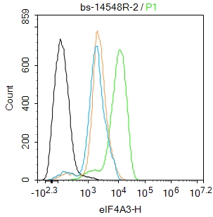
Blank control:Hela.
Primary Antibody (green line): Rabbit Anti-eIF4A3 antibody (bs-14548R)
Dilution: 2ug/Test;
Secondary Antibody : Goat anti-rabbit IgG-FITC
Dilution: 0.5ug/Test.
Protocol
The cells were fixed with 4% PFA (10min at room temperature)and then permeabilized with 90% ice-cold methanol for 20 min at -20℃.The cells were then incubated in 5%BSA to block non-specific protein-protein interactions for 30 min at room temperature .Cells stained with Primary Antibody for 30 min at room temperature. The secondary antibody used for 40 min at room temperature. Acquisition of 20,000 events was performed.
|
| 1、抗体溶解方法 | |
| 2、抗体修复方式 | |
| 3、常用试剂的配制 | |
| 4、免疫组化操作步骤 | |
| 5、免疫组化问题解答 | |
| 6、Western Blotting 操作步骤 | |
| 7、Western Blotting 问题解答 | |
| 8、关于肽链的设计 | |
| 9、多肽的溶解与保存 | |
| 10、酶标抗体效价测定程序 | |


