

| 产品编号 | bs-8533R |
| 英文名称 | [KO验证抗体] Vimentin Rabbit pAb |
| 中文名称 | 波形蛋白抗体 |
| 别 名 | VIME_HUMAN; VIM; VIME_MOUSE; VIME_RAT; |
 |
Specific References (40) | bs-8533R has been referenced in 40 publications.
|
| 研究领域 | 肿瘤 细胞生物 免疫学 信号转导 干细胞 细胞骨架 肿瘤细胞生物标志物 |
| 抗体来源 | Rabbit |
| 克隆类型 | Polyclonal |
| 克 隆 号 | |
| 交叉反应 | Human,Mouse,Rat (predicted: Rabbit,Pig,Cow,Dog,Horse) |
| 产品应用 | WB=1:1000-5000,IHC-P=1:200-1000,IHC-F=1:200-1000,IF=1:200-1000,Flow-Cyt=1μg/Test,ICC/IF=1:100-500
not yet tested in other applications. optimal dilutions/concentrations should be determined by the end user. |
| 理论分子量 | 51 kDa |
| 检测分子量 | 53 |
| 细胞定位 | 细胞浆 |
| 性 状 | Liquid |
| 浓 度 | 1mg/ml |
| 免 疫 原 | KLH conjugated synthetic peptide derived from human Vimentin: 371-466/466 |
| 亚 型 | IgG |
| 纯化方法 | affinity purified by Protein A |
| 缓 冲 液 | 0.01M TBS (pH7.4) with 1% BSA, 0.02% Proclin300 and 50% Glycerol. |
| 保存条件 | Shipped at 4℃. Store at -20℃ for one year. Avoid repeated freeze/thaw cycles. |
| 注意事项 | This product as supplied is intended for research use only, not for use in human, therapeutic or diagnostic applications. |
| PubMed | PubMed |
| 产品介绍 |
This gene encodes a member of the intermediate filament family. Intermediate filamentents, along with microtubules and actin microfilaments, make up the cytoskeleton. The protein encoded by this gene is responsible for maintaining cell shape, integrity of the cytoplasm, and stabilizing cytoskeletal interactions. It is also involved in the immune response, and controls the transport of low-density lipoprotein (LDL)-derived cholesterol from a lysosome to the site of esterification. It functions as an organizer of a number of critical proteins involved in attachment, migration, and cell signaling. Mutations in this gene causes a dominant, pulverulent cataract.[provided by RefSeq, Jun 2009] Function: Vimentins are class-III intermediate filaments found in various non-epithelial cells, especially mesenchymal cells. Vimentin is attached to the nucleus, endoplasmic reticulum, and mitochondria, either laterally or terminally. Involved with LARP6 in the stabilization of type I collagen mRNAs for CO1A1 and CO1A2. Subunit : Homopolymer assembled from elementary dimers. Interacts with HCV core protein. Interacts with LGSN and SYNM. Interacts (via rod region) with PLEC (via CH 1 domain) (By similarity). Interacts with SLC6A4. Interacts with STK33. Interacts with LARP6. Interacts with RAB8B (By similarity). Subcellular Location: Cytoplasm. Tissue Specificity: Highly expressed in fibroblasts, some expression in T- and B-lymphocytes, and little or no expression in Burkitt's lymphoma cell lines. Expressed in many hormone-independent mammary carcinoma cell lines. Post-translational modifications: Filament disassembly during mitosis is promoted by phosphorylation at Ser-55 as well as by nestin. One of the most prominent phosphoproteins in various cells of mesenchymal origin. Phosphorylation is enhanced during cell division, at which time vimentin filaments are significantly reorganized. Phosphorylation by PKN1 inhibits the formation of filaments. Phosphorylated at Ser-56 by CDK5 during neutrophil secretion in the cytoplasm. Phosphorylated by STK33. Similarity: Belongs to the intermediate filament family. SWISS: P08670 Gene ID: 7431 Database links: Entrez Gene: 7431 Human Entrez Gene: 22352 Mouse Omim: 193060 Human SwissProt: P08670 Human SwissProt: P20152 Mouse Unigene: 455493 Human |
| 产品图片 |
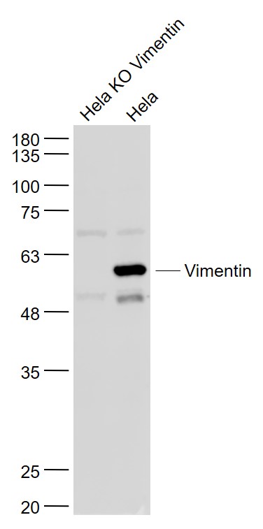
Sample:
Hela KO Vimentin (Human) Cell Lysate at 30 ug
Hela(Human) Cell Lysate at 30 ug
Primary: Anti- Vimentin (bs-8533R) at 1/1000 dilution
Secondary: IRDye800CW Goat Anti-Rabbit IgG at 1/20000 dilution
Predicted band size: 51 kD
Observed band size: 53 kD
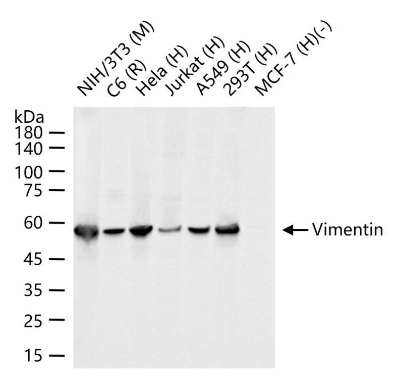
25 ug total protein per lane of various lysates (see on figure) probed with Vimentin polyclonal antibody, unconjugated (bs-8533R) at 1:1000 dilution and 4°C overnight incubation. Followed by conjugated secondary antibody incubation at r.t. for 60 min.
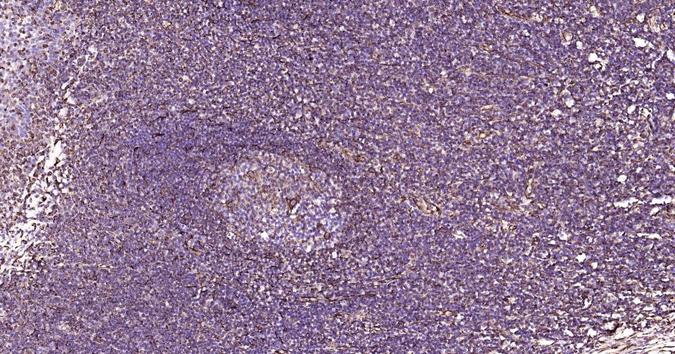
Paraformaldehyde-fixed, paraffin embedded Human Tonsil; Antigen retrieval by boiling in sodium citrate buffer (pH6.0) for 15 min; Antibody incubation with Vimentin Polyclonal Antibody, Unconjugated (bs-8533R) at 1:200 overnight at 4°C, followed by conjugation to the SP Kit (Rabbit, SP-0023) and DAB (C-0010) staining.
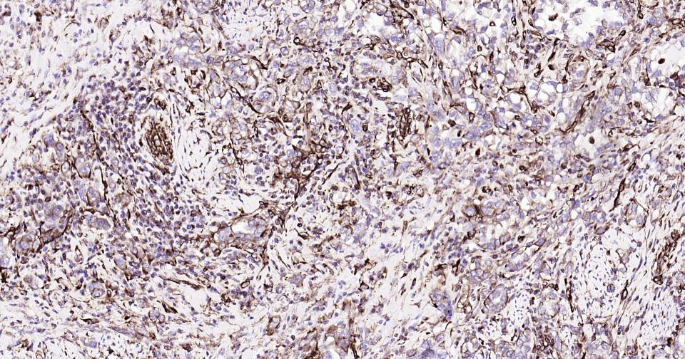
Paraformaldehyde-fixed, paraffin embedded Human Cervical Cancer; Antigen retrieval by boiling in sodium citrate buffer (pH6.0) for 15 min; Antibody incubation with Vimentin Polyclonal Antibody, Unconjugated (bs-8533R) at 1:200 overnight at 4°C, followed by conjugation to the SP Kit (Rabbit, SP-0023) and DAB (C-0010) staining.

Paraformaldehyde-fixed, paraffin embedded Human Lung Cancer; Antigen retrieval by boiling in sodium citrate buffer (pH6.0) for 15 min; Antibody incubation with Vimentin Polyclonal Antibody, Unconjugated (bs-8533R) at 1:200 overnight at 4°C, followed by conjugation to the SP Kit (Rabbit, SP-0023) and DAB (C-0010) staining.
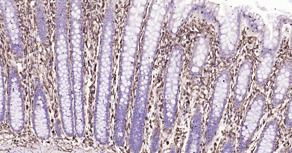
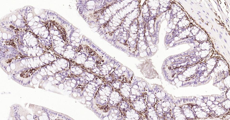
Paraformaldehyde-fixed, paraffin embedded Human Colon; Antigen retrieval by boiling in sodium citrate buffer (pH6.0) for 15 min; Antibody incubation with Vimentin Polyclonal Antibody, Unconjugated (bs-8533R) at 1:200 overnight at 4°C, followed by conjugation to the SP Kit (Rabbit, SP-0023) and DAB (C-0010) staining.
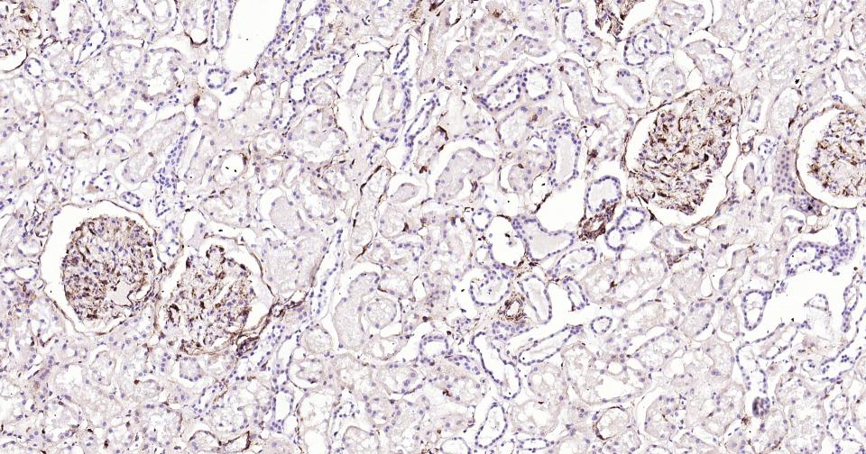
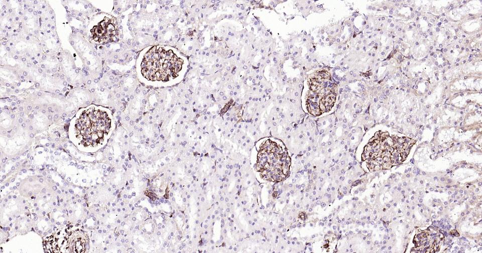
Paraformaldehyde-fixed, paraffin embedded Human Kidney; Antigen retrieval by boiling in sodium citrate buffer (pH6.0) for 15 min; Antibody incubation with Vimentin Polyclonal Antibody, Unconjugated (bs-8533R) at 1:200 overnight at 4°C, followed by conjugation to the SP Kit (Rabbit, SP-0023) and DAB (C-0010) staining.
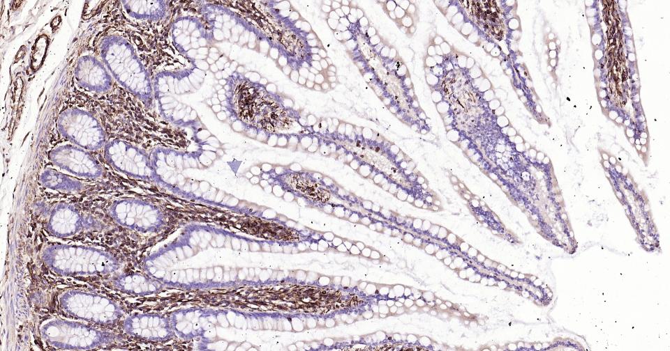
Paraformaldehyde-fixed, paraffin embedded Human Small Intestine; Antigen retrieval by boiling in sodium citrate buffer (pH6.0) for 15 min; Antibody incubation with Vimentin Polyclonal Antibody, Unconjugated (bs-8533R) at 1:200 overnight at 4°C, followed by conjugation to the SP Kit (Rabbit, SP-0023) and DAB (C-0010) staining.
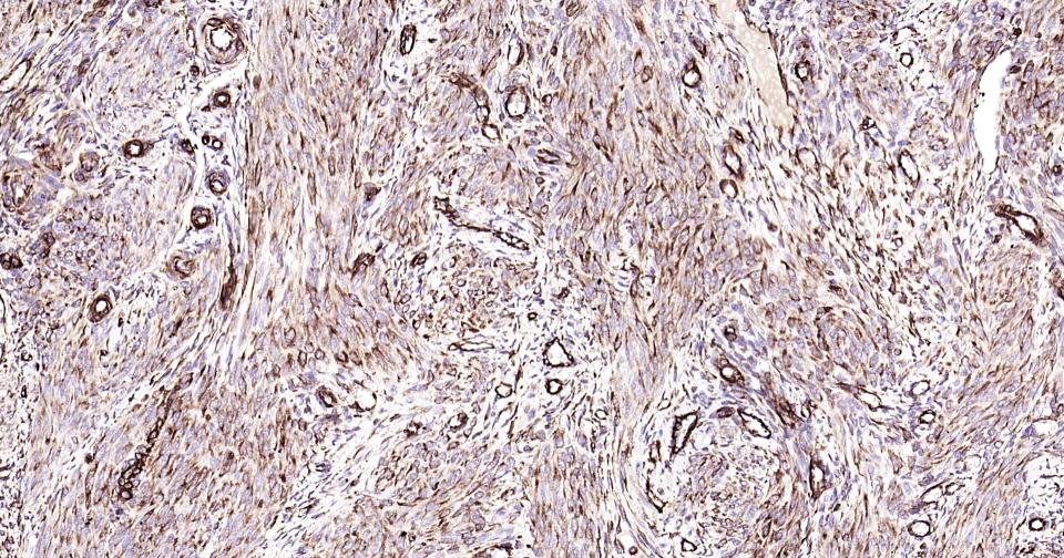
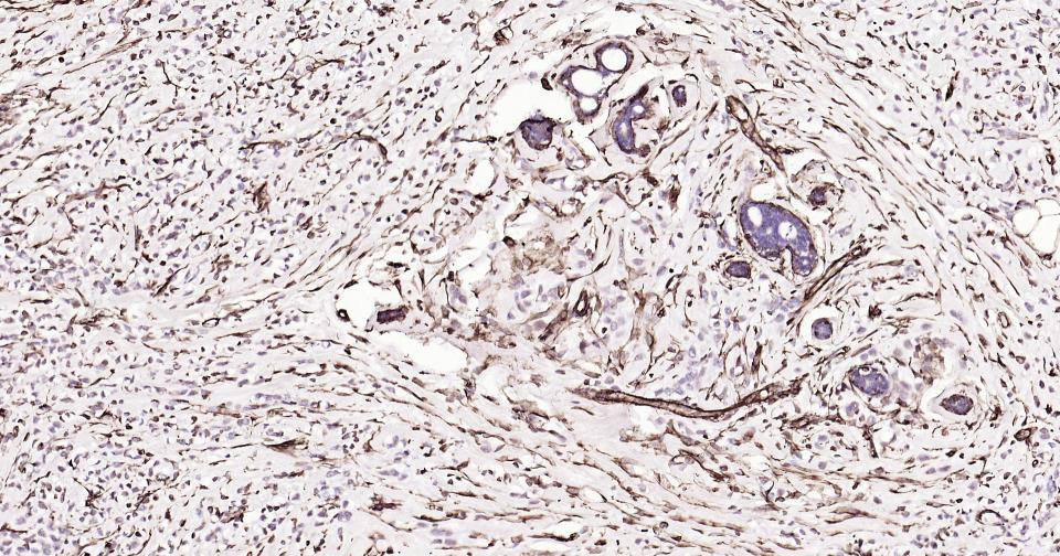
Paraformaldehyde-fixed, paraffin embedded Human Uterus; Antigen retrieval by boiling in sodium citrate buffer (pH6.0) for 15 min; Antibody incubation with Vimentin Polyclonal Antibody, Unconjugated (bs-8533R) at 1:200 overnight at 4°C, followed by conjugation to the SP Kit (Rabbit, SP-0023) and DAB (C-0010) staining.
Paraformaldehyde-fixed, paraffin embedded Human Breast Cancer; Antigen retrieval by boiling in sodium citrate buffer (pH6.0) for 15 min; Antibody incubation with Vimentin Polyclonal Antibody, Unconjugated (bs-8533R) at 1:200 overnight at 4°C, followed by conjugation to the SP Kit (Rabbit, SP-0023) and DAB (C-0010) staining.
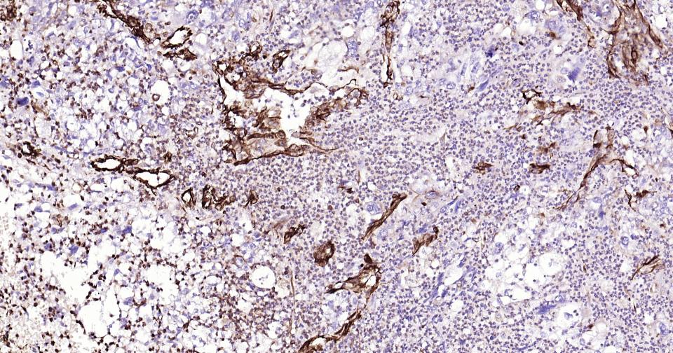
Paraformaldehyde-fixed, paraffin embedded Human Endometrium Cancer; Antigen retrieval by boiling in sodium citrate buffer (pH6.0) for 15 min; Antibody incubation with Vimentin Polyclonal Antibody, Unconjugated (bs-8533R) at 1:200 overnight at 4°C, followed by conjugation to the SP Kit (Rabbit, SP-0023) and DAB (C-0010) staining.
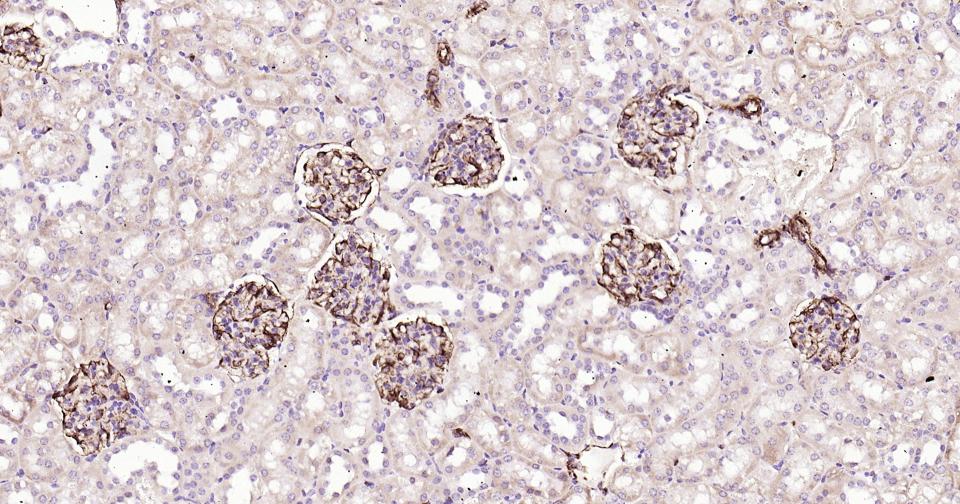
Paraformaldehyde-fixed, paraffin embedded Mouse Kidney; Antigen retrieval by boiling in sodium citrate buffer (pH6.0) for 15 min; Antibody incubation with Vimentin Polyclonal Antibody, Unconjugated (bs-8533R) at 1:200 overnight at 4°C, followed by conjugation to the SP Kit (Rabbit, SP-0023) and DAB (C-0010) staining.
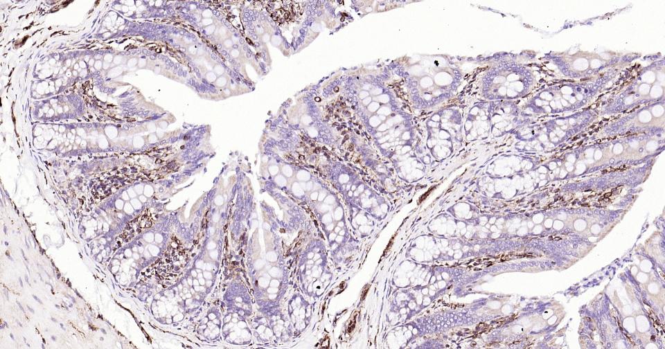
Paraformaldehyde-fixed, paraffin embedded Rat Colon; Antigen retrieval by boiling in sodium citrate buffer (pH6.0) for 15 min; Antibody incubation with Vimentin Polyclonal Antibody, Unconjugated (bs-8533R) at 1:200 overnight at 4°C, followed by conjugation to the SP Kit (Rabbit, SP-0023) and DAB (C-0010) staining.
Paraformaldehyde-fixed, paraffin embedded Mouse Colon; Antigen retrieval by boiling in sodium citrate buffer (pH6.0) for 15 min; Antibody incubation with Vimentin Polyclonal Antibody, Unconjugated (bs-8533R) at 1:200 overnight at 4°C, followed by conjugation to the SP Kit (Rabbit, SP-0023) and DAB (C-0010) staining.
Paraformaldehyde-fixed, paraffin embedded Rat Kidney; Antigen retrieval by boiling in sodium citrate buffer (pH6.0) for 15 min; Antibody incubation with Vimentin Polyclonal Antibody, Unconjugated (bs-8533R) at 1:200 overnight at 4°C, followed by conjugation to the SP Kit (Rabbit, SP-0023) and DAB (C-0010) staining.
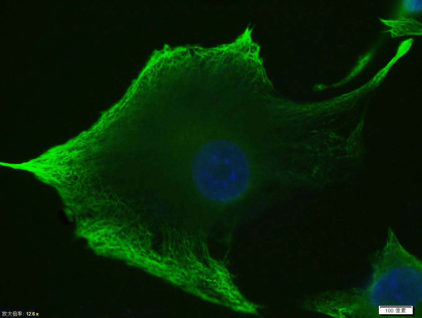
Tissue/cell: U-87MG cell; 4% Paraformaldehyde-fixed; Triton X-100 at room temperature for 20 min; Blocking buffer (normal goat serum, C-0005) at 37°C for 20 min; Antibody incubation with (Vimentin) Polyclonal Antibody, Unconjugated (bs-8533R)antibody (bs-0295G-FITC) at 37°C for 90 minutes, DAPI (blue, C02-04002) was used to stain the cell nuclei.
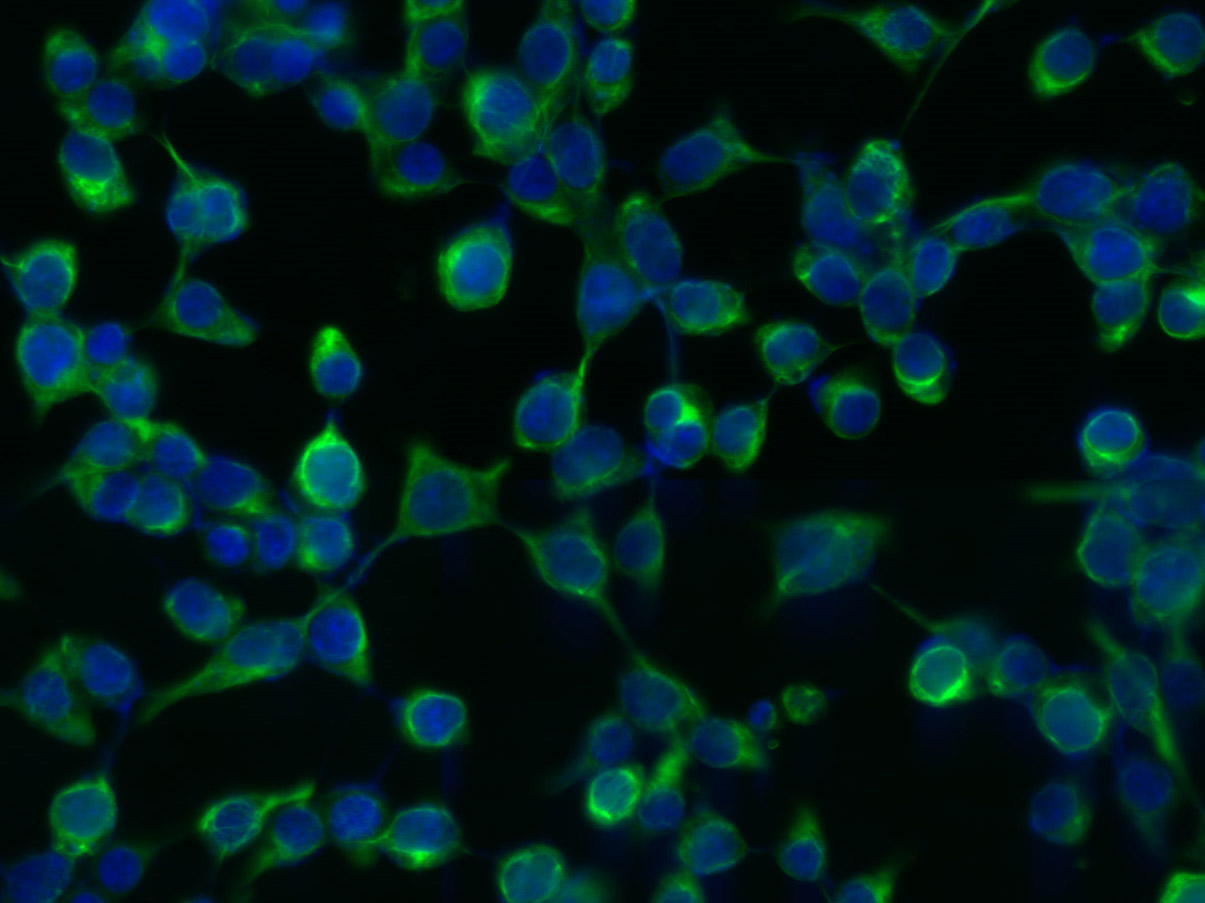
Tissue/cell: 293T cell; 4% Paraformaldehyde-fixed; Triton X-100 at room temperature for 20 min; Blocking buffer (normal goat serum, C-0005) at 37°C for 20 min; Antibody incubation with (Vimentin) Polyclonal Antibody, Unconjugated (bs-8533R) 1:200, 2 hours at 37°C; followed by a conjugated Goat Anti-Rabbit IgG antibody (bs-0295G-FITC) at 37°C for 90 minutes, DAPI (5ug/ml, blue, C-0033) was used to stain the cell nuclei.
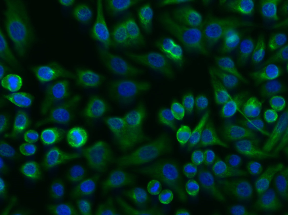
Tissue/cell: FHC cell; 4% Paraformaldehyde-fixed; Triton X-100 at room temperature for 20 min; Blocking buffer (normal goat serum, C-0005) at 37°C for 20 min; Antibody incubation with (Vimentin) Polyclonal Antibody, Unconjugated (bs-8533R) 1:200, 2 hours at 37°C; followed by a conjugated Goat Anti-Rabbit IgG antibody (bs-0295G-FITC) at 37°C for 90 minutes, DAPI (5ug/ml, blue, C-0033) was used to stain the cell nuclei.
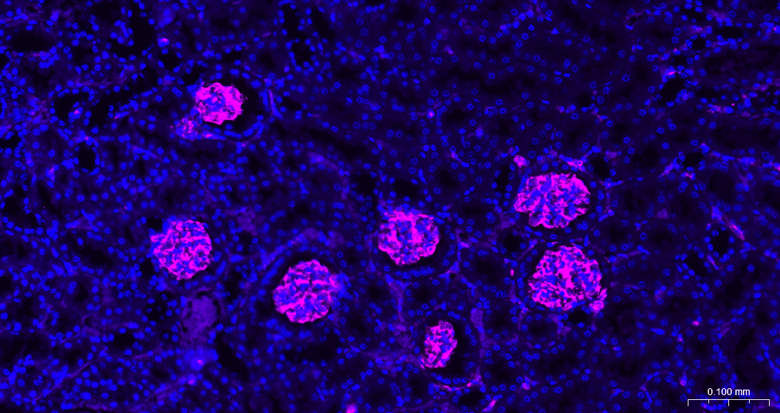
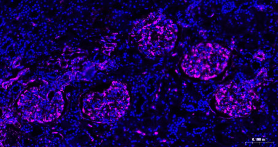
Paraformaldehyde-fixed, paraffin embedded Mouse Kidney; Antigen retrieval by boiling in sodium citrate buffer (pH6.0) for 15 min; The section was incubated with Vimentin Polyclonal Antibody, Unconjugated (bs-8533R) at 1:200 overnight at 4°C. Followed by conjugated Goat Anti-Rabbit IgG antibody (Rose Red, bs-40295G-BF647), DAPI (blue, C02-04002) was used to stain the cell nuclei.
Paraformaldehyde-fixed, paraffin embedded Rat Kidney; Antigen retrieval by boiling in sodium citrate buffer (pH6.0) for 15 min; The section was incubated with Vimentin Polyclonal Antibody, Unconjugated (bs-8533R) at 1:200 overnight at 4°C. Followed by conjugated Goat Anti-Rabbit IgG antibody (Rose Red, bs-40295G-BF647), DAPI (blue, C02-04002) was used to stain the cell nuclei.
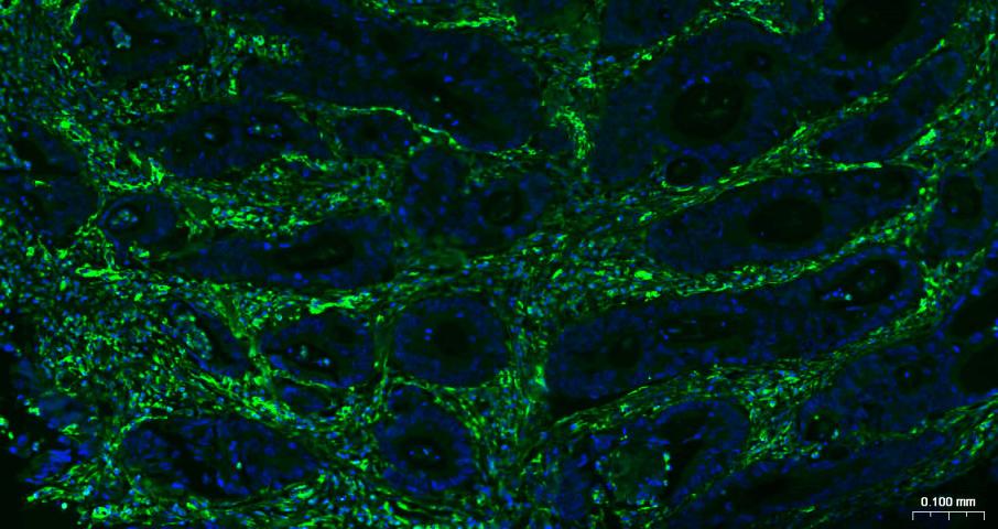
Paraformaldehyde-fixed, paraffin embedded Human Colon Cancer; Antigen retrieval by boiling in sodium citrate buffer (pH6.0) for 15 min; Antibody incubation with Vimentin Polyclonal Antibody, Unconjugated (bs-8533R) at 1:200 overnight at 4°C. Followed by conjugated Goat Anti-Rabbit IgG antibody (green, bs-0295G-BF488), DAPI (blue, C02-04002) was used to stain the cell nuclei.
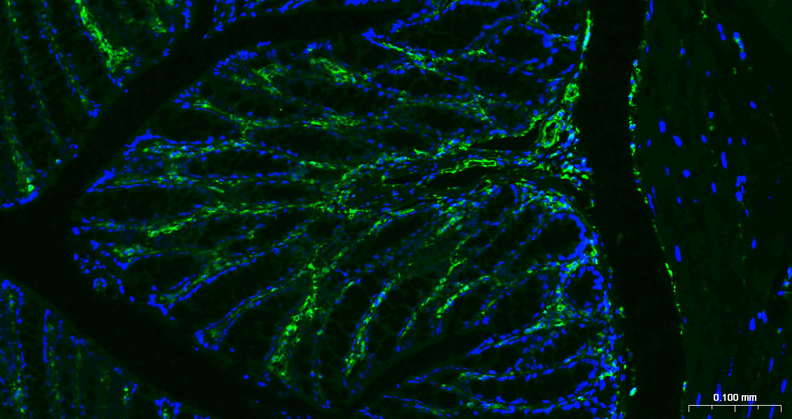
Paraformaldehyde-fixed, paraffin embedded Mouse Colon; Antigen retrieval by boiling in sodium citrate buffer (pH6.0) for 15 min; Antibody incubation with Vimentin Polyclonal Antibody, Unconjugated (bs-8533R) at 1:200 overnight at 4°C. Followed by conjugated Goat Anti-Rabbit IgG antibody (green, bs-0295G-BF488), DAPI (blue, C02-04002) was used to stain the cell nuclei.
Paraformaldehyde-fixed, paraffin embedded Rat Colon; Antigen retrieval by boiling in sodium citrate buffer (pH6.0) for 15 min; Antibody incubation with Vimentin Polyclonal Antibody, Unconjugated (bs-8533R) at 1:200 overnight at 4°C. Followed by conjugated Goat Anti-Rabbit IgG antibody (green, bs-0295G-BF488), DAPI (blue, C02-04002) was used to stain the cell nuclei.
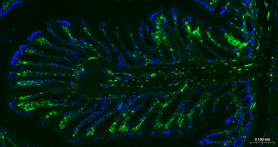
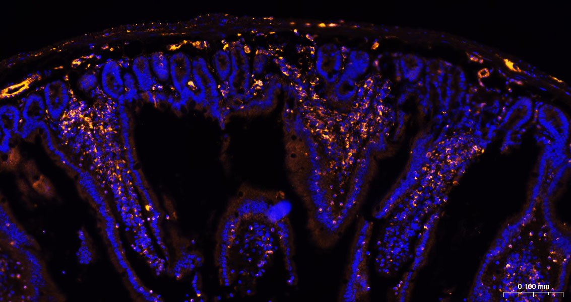
Paraformaldehyde-fixed, paraffin embedded Mouse Small Intestine; Antigen retrieval by boiling in sodium citrate buffer (pH6.0) for 15 min; The section was incubated with Vimentin Polyclonal Antibody, Unconjugated (bs-8533R) at 1:200 overnight at 4°C. Followed by conjugated Goat Anti-Rabbit IgG antibody (Rose Red, bs-40295G-BF647), DAPI (blue, C02-04002) was used to stain the cell nuclei.
Paraformaldehyde-fixed, paraffin embedded Rat Small Intestine; Antigen retrieval by boiling in sodium citrate buffer (pH6.0) for 15 min; The section was incubated with Vimentin Polyclonal Antibody, Unconjugated (bs-8533R) at 1:200 overnight at 4°C. Followed by conjugated Goat Anti-Rabbit IgG antibody (Rose Red, bs-40295G-BF647), DAPI (blue, C02-04002) was used to stain the cell nuclei.
Paraformaldehyde-fixed, paraffin embedded Human Kidney; Antigen retrieval by boiling in sodium citrate buffer (pH6.0) for 15 min; The section was incubated with Vimentin Polyclonal Antibody, Unconjugated (bs-8533R) at 1:200 overnight at 4°C. Followed by conjugated Goat Anti-Rabbit IgG antibody (Rose Red, bs-40295G-BF647), DAPI (blue, C02-04002) was used to stain the cell nuclei.
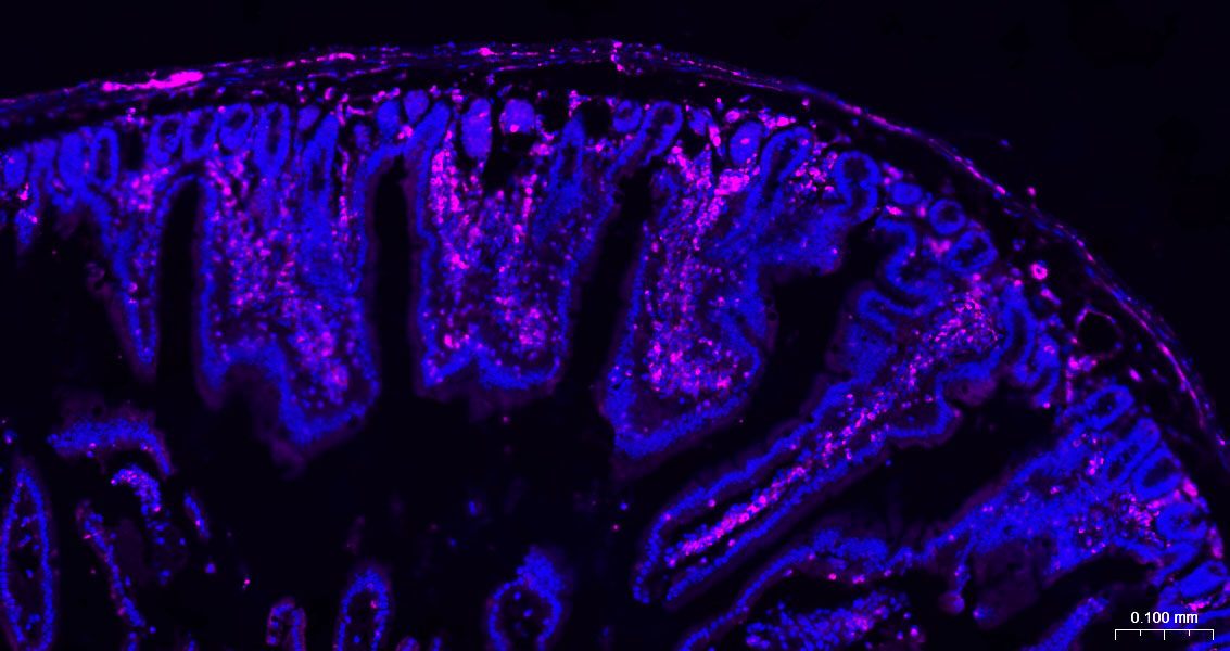
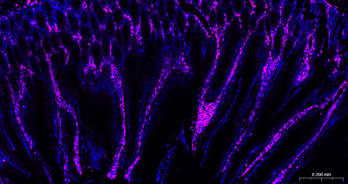
Paraformaldehyde-fixed, paraffin embedded Mouse Small Intestine ; Antigen retrieval by boiling in sodium citrate buffer (pH6.0) for 15min; The section was incubated with Vimentin Polyclonal Antibody, Unconjugated (bs-8533R) at 1:200 overnight at 4°C, followed by a conjugated Donkey Anti-Rabbit IgG antibody (bs-0295D-BF555) for 90 minutes, and DAPI for nuclei staining.
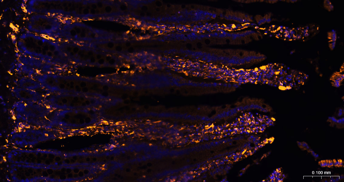
Paraformaldehyde-fixed, paraffin embedded Rat Small Intestine ; Antigen retrieval by boiling in sodium citrate buffer (pH6.0) for 15min; The section was incubated with Vimentin Polyclonal Antibody, Unconjugated (bs-8533R) at 1:200 overnight at 4°C, followed by a conjugated Donkey Anti-Rabbit IgG antibody (bs-0295D-BF555) for 90 minutes, and DAPI for nuclei staining.
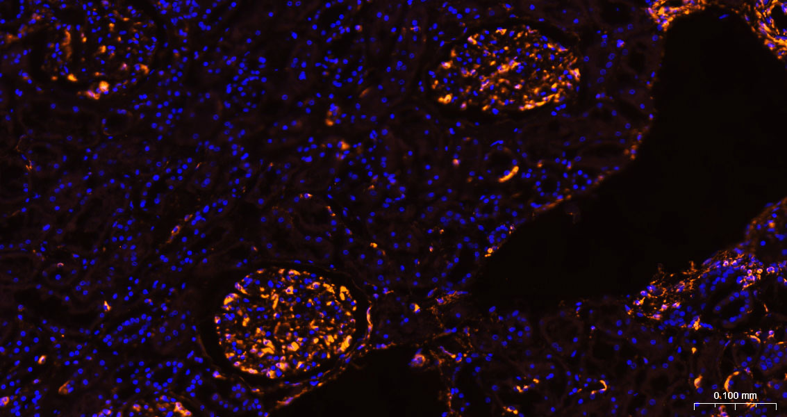
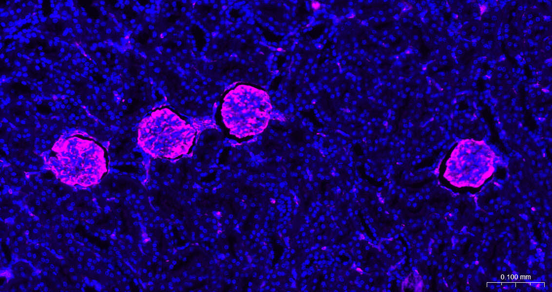
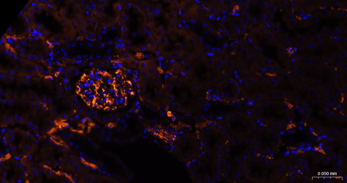
Paraformaldehyde-fixed, paraffin embedded Human Kidney ; Antigen retrieval by boiling in sodium citrate buffer (pH6.0) for 15min; The section was incubated with Vimentin Polyclonal Antibody, Unconjugated (bs-8533R) at 1:200 overnight at 4°C, followed by a conjugated Donkey Anti-Rabbit IgG antibody (bs-0295D-BF555) for 90 minutes, and DAPI for nuclei staining.
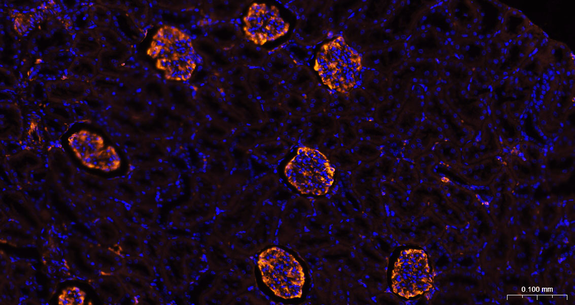
Paraformaldehyde-fixed, paraffin embedded Mouse Kidney ; Antigen retrieval by boiling in sodium citrate buffer (pH6.0) for 15min; The section was incubated with Vimentin Polyclonal Antibody, Unconjugated (bs-8533R) at 1:200 overnight at 4°C, followed by a conjugated Donkey Anti-Rabbit IgG antibody (bs-0295D-BF555) for 90 minutes, and DAPI for nuclei staining.
Paraformaldehyde-fixed, paraffin embedded Rat Kidney ; Antigen retrieval by boiling in sodium citrate buffer (pH6.0) for 15min; The section was incubated with Vimentin Polyclonal Antibody, Unconjugated (bs-8533R) at 1:200 overnight at 4°C, followed by a conjugated Donkey Anti-Rabbit IgG antibody (bs-0295D-BF555) for 90 minutes, and DAPI for nuclei staining.

Blank control:A549.
Primary Antibody (green line): Rabbit Anti-Vimentin antibody (bs-8533R)
Dilution: 1μg /10^6 cells;
Isotype Control Antibody (orange line): Rabbit IgG .
Secondary Antibody : Goat anti-rabbit IgG-AF488
Dilution: 1μg /test.
Protocol
The cells were fixed with 4% PFA (10min at room temperature)and then permeabilized with 90% ice-cold methanol for 20 min at -20℃. The cells were then incubated in 5%BSA to block non-specific protein-protein interactions for 30 min at room temperature .Cells stained with Primary Antibody for 30 min at room temperature. The secondary antibody used for 40 min at room temperature. Acquisition of 20,000 events was performed.
|
| 1、抗体溶解方法 | |
| 2、抗体修复方式 | |
| 3、常用试剂的配制 | |
| 4、免疫组化操作步骤 | |
| 5、免疫组化问题解答 | |
| 6、Western Blotting 操作步骤 | |
| 7、Western Blotting 问题解答 | |
| 8、关于肽链的设计 | |
| 9、多肽的溶解与保存 | |
| 10、酶标抗体效价测定程序 | |


