

| 产品编号 | bs-15455R |
| 英文名称 | HBcAg Rabbit pAb |
| 中文名称 | 乙肝病毒衣壳蛋白抗体 |
| 别 名 | HBSAG_HBVE1; S; L glycoprotein; L-HBsAg (LHB); Large S protein; Large surface protein; Major surface antigen; |
| 研究领域 | 微生物学 细菌及病毒 |
| 抗体来源 | Rabbit |
| 克隆类型 | Polyclonal |
| 克 隆 号 | |
| 交叉反应 | Hepatitis B virus |
| 产品应用 | ICC/IF=1:50-200
not yet tested in other applications. optimal dilutions/concentrations should be determined by the end user. |
| 理论分子量 | 21 kDa |
| 性 状 | Liquid |
| 浓 度 | 1mg/ml |
| 免 疫 原 | KLH conjugated synthetic peptide derived from Hepatitis B Virus Core Antigen: 1-100/185 |
| 亚 型 | IgG |
| 纯化方法 | affinity purified by Protein A |
| 缓 冲 液 | 0.01M TBS (pH7.4) with 1% BSA, 0.02% Proclin300 and 50% Glycerol. |
| 保存条件 | Shipped at 4℃. Store at -20℃ for one year. Avoid repeated freeze/thaw cycles. |
| 注意事项 | This product as supplied is intended for research use only, not for use in human, therapeutic or diagnostic applications. |
| PubMed | PubMed |
| 产品介绍 |
Hepatitis B Virus Core Antigen (HBcAg) is part of the infectious virion containing an inner "core particle" enclosing the viral genome. The icosahedral core particle contains 180 or 240 copies of the core protein. HBcAg is one of the three major clinical antigens of hepatitis B virus but disappears early in the course of infection.
The hepatitis B virus core antigen (HBcAg) is a highly immunogenic subviral particle and functions as both a T-cell-dependent and a T-cell-independent antigen. Therefore, HBcAg may be a promising candidate target for therapeutic vaccine control of chronic HBV infection. Function: Self assembles to form an icosahedral capsid. Mostcapsid appear to be large particles with a icosahedral symmetry ofT=4 and consist of 240 copies of capsid protein, though a fractionforms smaller T=3 particles consisting of 180 capsid proteins.Entering capsid are transported along microtubules to the nucleus.Phosphorylation of the capsid is thought to induce exposure ofnuclear localization signal in the C-terminal portion of the capsidprotein that allows binding to the nuclear pore complex via theimportin (karyopherin-) alpha and beta. Capsids are imported inintact form through the nuclear pore into the nuclear basket, whereit probably binds NUP153. Only capsids that contain the matureviral genome can release the viral DNA and capsid protein into thenucleoplasm. Immature capsids get stucked in the basket. Capsidsencapsulate the pre-genomic RNA and the P protein. Pre-genomic RNAis reverse transcribed into DNA while the capsid is still in thecytoplasm. The capsid can then either be directed to the nucleus,providing more genome for transcription, or bud through theendoplasmic reticulum to provide new virions (By similarity). Encapsidates hepatitis delta genome. Subunit: Homodimerizes, then multimerizes. Interacts with cytosolexposed regions of viral L glycoprotein present in thereticulum-to-Golgi compartment. Interacts with human FLNB. Subcellular Location: Capsid protein: Virion. Host cytoplasm. Post-translational modifications: Phosphorylated by host SRPK1, SRPK2, and maybe protein kinaseC or GAPDH. Phosphorylation is critical for pregenomic RNApackaging. Protein kinase C phosphorylation is stimulated by HBxprotein and may play a role in transport of the viral genome to thenucleus at the late step during viral replication cycle. Similarity: Belongs to the orthohepadnavirus core antigen family. Database links:
|
| 产品图片 |
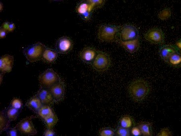
HepG2-NTCP HBV infected; 4% Paraformaldehyde-fixed; Triton X-100 at room temperature for 20 min; Blocking buffer (normal goat serum) at 37°C for 20 min; Antibody incubation with (HBcAg) Polyclonal Antibody, Unconjugated (bs-15455R) 1:100, 90 minutes at 37°C; followed by a conjugated Goat Anti-Rabbit IgG antibody (AF488,1:200) at 37°C for 90 minutes, DAPI (blue)was used to stain the cell nuclei. Maria Francesca Cortese,PhD researcher in Vall d'Hebron Research Institute (VHIR) of Barcelona (Spain).
|
| 1、抗体溶解方法 | |
| 2、抗体修复方式 | |
| 3、常用试剂的配制 | |
| 4、免疫组化操作步骤 | |
| 5、免疫组化问题解答 | |
| 6、Western Blotting 操作步骤 | |
| 7、Western Blotting 问题解答 | |
| 8、关于肽链的设计 | |
| 9、多肽的溶解与保存 | |
| 10、酶标抗体效价测定程序 | |


