

| 产品编号 | bs-12335R |
| 英文名称 | HFE Rabbit pAb |
| 中文名称 | 遗传性血色病蛋白相关蛋白1抗体 |
| 别 名 | HFE_HUMAN; HFE; HLA-H; HLAH; homeostatic iron regulator; hemochromatosis; high Fe |
 |
Specific References (1) | bs-12335R has been referenced in 1 publications.
[IF=5.008] Rychtarcikova, Zuzana, et al. "Tumor-initiating cells of breast and prostate origin show alterations in the expression of genes related to iron metabolism." Oncotarget (2016). WB ; Human.
|
| 研究领域 | 肿瘤 心血管 细胞生物 神经生物学 信号转导 糖尿病 新陈代谢 |
| 抗体来源 | Rabbit |
| 克隆类型 | Polyclonal |
| 交叉反应 | Human (predicted: Mouse,Rat,Sheep,Cow,Dog,Horse) |
| 产品应用 | Flow-Cyt=1μg/Test
not yet tested in other applications. optimal dilutions/concentrations should be determined by the end user. |
| 理论分子量 | 38kDa |
| 细胞定位 | 细胞膜 |
| 性 状 | Liquid |
| 浓 度 | 1mg/ml |
| 免 疫 原 | KLH conjugated synthetic peptide derived from Human HFE/Hemochromatosis: 262-348/348 <Extracellular> |
| 亚 型 | IgG |
| 纯化方法 | affinity purified by Protein A |
| 缓 冲 液 | 0.01M TBS (pH7.4) with 1% BSA, 0.02% Proclin300 and 50% Glycerol. |
| 保存条件 | Shipped at 4℃. Store at -20℃ for one year. Avoid repeated freeze/thaw cycles. |
| 注意事项 | This product as supplied is intended for research use only, not for use in human, therapeutic or diagnostic applications. |
| PubMed | PubMed |
| 产品介绍 |
The features of hemochromatosis include cirrhosis of the liver, diabetes, hypermelanotic pigmentation of the skin, and heart failure. Since hemochromatosis is a relatively easily treated disorder if diagnosed, this is a form of preventable cancer. The HFE protein, which is defective in hereditary hemo-chromatosis, normally is expressed in crypt enterocytes of the duodenum where it has a unique, predominantly intracellular localization. In placenta, the HFE protein co-localizes with and forms a stable association with the transferrin receptor (TfR), providing a link between the HFE protein and iron transport. Immunocytochemistry shows that the HFE protein and TfR both are expressed in the crypt enterocytes. Western blots show that, as is the case in human placenta, the HFE protein in crypt enterocytes is physically associated with the TfR and with β2-microglobulin. It is proposed that HFE has two mutually exclusive activities in cells: inhibition of uptake or inhibition of release of iron and that the balance between serum transferrin saturation and serum transferrin-receptor concentrations determines which of these functions predominates. The gene which encodes HFE maps to human chromosome 6p21.3. Function: Binds to transferrin receptor (TFR) and reduces its affinity for iron-loaded transferrin. Subunit: Binds TFR through the extracellular domain in a pH-dependent manner. Subcellular Location: Membrane; Single-pass type I membrane protein. Tissue Specificity: Expressed in all tissues tested except brain. DISEASE: Defects in HFE are a cause of hemochromatosis (HFE) [MIM:235200]. A disorder of iron metabolism characterized by iron overload. Excess iron is deposited in a variety of organs leading to their failure, and resulting in serious illnesses including cirrhosis, hepatomas, diabetes, cardiomyopathy, arthritis, and hypogonadotropic hypogonadism. Severe effects of the disease usually do not appear until after decades of progressive iron loading. Defects in HFE are associated with variegate porphyria (VP) [MIM:176200]. Porphyrias are inherited defects in the biosynthesis of heme, resulting in the accumulation and increased excretion of porphyrins or porphyrin precursors. They are classified as erythropoietic or hepatic, depending on whether the enzyme deficiency occurs in red blood cells or in the liver. VP is the most common form of porphyria in South Africa. It is characterized by skin hyperpigmentation and hypertrichosis, abdominal pain, tachycardia, hypertension and neuromuscular disturbances. High fecal levels of protoporphyrin and coproporphyrin, increased urine uroporphyrins and iron overload are typical markers of the disease. Note=Iron overload due to HFE mutations is a precipitating or exacerbating factor in variegate porphyria. Defects in HFE are associated with susceptibility to microvascular complications of diabetes type 7 (MVCD7) [MIM:612635]. These are pathological conditions that develop in numerous tissues and organs as a consequence of diabetes mellitus. They include diabetic retinopathy, diabetic nephropathy leading to end-stage renal disease, and diabetic neuropathy. Diabetic retinopathy remains the major cause of new-onset blindness among diabetic adults. It is characterized by vascular permeability and increased tissue ischemia and angiogenesis. Similarity: Belongs to the MHC class I family. Contains 1 Ig-like C1-type (immunoglobulin-like) domain. SWISS: Q30201 Gene ID: 3077 Database links: Entrez Gene: 3077 Human Omim: 613609 Human SwissProt: Q30201 Human Unigene: 233325 Human |
| 产品图片 |
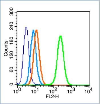
Blank control (blue line): HL60(fixed with 70% ethanol Overnight at 4℃).
Primary Antibody (green line): Rabbit Anti-iHFE antibody (bs-12335R),Dilution: 0.2μg /10^6 cells;
Isotype Control Antibody (orange line): Rabbit IgG .
Secondary Antibody (white blue line): Goat anti-rabbit IgG-PE,Dilution: 1μg /test.
|
| 1、抗体溶解方法 | |
| 2、抗体修复方式 | |
| 3、常用试剂的配制 | |
| 4、免疫组化操作步骤 | |
| 5、免疫组化问题解答 | |
| 6、Western Blotting 操作步骤 | |
| 7、Western Blotting 问题解答 | |
| 8、关于肽链的设计 | |
| 9、多肽的溶解与保存 | |
| 10、酶标抗体效价测定程序 | |


