

| 产品编号 | bs-4917R |
| 英文名称 | Osteocalcin Rabbit pAb |
| 中文名称 | 骨钙蛋白/骨钙素抗体 |
| 别 名 | BGP; OC; OCN; Bglap2; Bgpr; Bgpra; OSTCN_BOVIN; BGLAP; Bone Gla protein (BGP); Gamma-carboxyglutamic acid-containing protein; OSTCN_HUMAN; OSTCN_PIG; OSTCN_RAT; |
 |
Specific References (43) | bs-4917R has been referenced in 43 publications.
|
| 研究领域 | 细胞生物 信号转导 干细胞 细胞外基质 |
| 抗体来源 | Rabbit |
| 克隆类型 | Polyclonal |
| 克 隆 号 | |
| 交叉反应 | Human,Mouse,Rat |
| 产品应用 | WB=1:500-2000,IHC-P=1:100-500,IHC-F=1:100-500,IF=1:100-500,ELISA=1:5000-10000
not yet tested in other applications. optimal dilutions/concentrations should be determined by the end user. |
| 理论分子量 | 11 kDa |
| 检测分子量 | 11 |
| 细胞定位 | 分泌型蛋白 |
| 性 状 | Liquid |
| 浓 度 | 1mg/ml |
| 免 疫 原 | KLH conjugated synthetic peptide derived from human Osteocalcin: 21-100/100 |
| 亚 型 | IgG |
| 纯化方法 | affinity purified by Protein A |
| 缓 冲 液 | 0.01M TBS (pH7.4) with 1% BSA, 0.02% Proclin300 and 50% Glycerol. |
| 保存条件 | Shipped at 4℃. Store at -20℃ for one year. Avoid repeated freeze/thaw cycles. |
| 注意事项 | This product as supplied is intended for research use only, not for use in human, therapeutic or diagnostic applications. |
| PubMed | PubMed |
| 产品介绍 |
Osteocalcin belongs to the osteocalcin/matrix Gla protein family and constitutes 1 to 2% of the total bone protein. It is a 49 amino acid single chain vitamin K dependent protein, made by osteoblasts, and is a major component of the noncollagenous bone matrix. Post translational modification by a vitamin K dependent carboxylase produces three gamma carboxyglutamic acid residues at positions 17, 21 and 24, giving it a high affinity for calcium. It also binds strongly to apatite. Function: Constitutes 1-2% of the total bone protein. It binds strongly to apatite and calcium. Subcellular Location: Secreted. Post-translational modifications: Gamma-carboxyglutamate residues are formed by vitamin K dependent carboxylation. These residues are essential for the binding of calcium. Similarity: Belongs to the osteocalcin/matrix Gla protein family. Contains 1 Gla (gamma-carboxy-glutamate) domain. SWISS: P02818 Gene ID: 632 Database links: Entrez Gene: 632 Human Omim: 112260 Human SwissProt: P02818 Human Unigene: 654541 Human Unigene: 9722 Rat 骨钙素又称骨γ-羧谷氨酸包含蛋白 |
| 产品图片 |
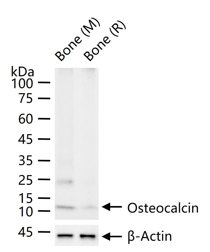
25 ug total protein per lane of various lysates (see on figure) probed with Osteocalcin polyclonal antibody, unconjugated (bs-4917R) at 1:1000 dilution and 4°C overnight incubation. Followed by conjugated secondary antibody incubation at r.t. for 60 min.
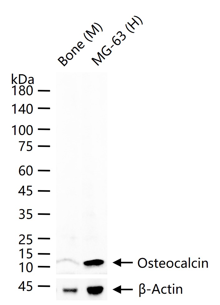
25 ug total protein per lane of various lysates (see on figure) probed with Osteocalcin polyclonal antibody, unconjugated (bs-4917R) at 1:1000 dilution and 4°C overnight incubation. Followed by conjugated secondary antibody incubation at r.t. for 60 min.
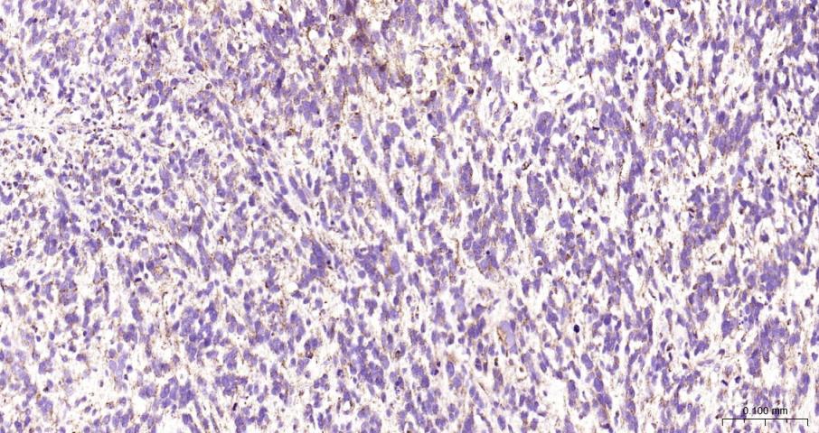
Paraformaldehyde-fixed, paraffin embedded Human Ewing’s sarcoma; Antigen retrieval by boiling in sodium citrate buffer (pH6.0) for 15 min; The section was incubated with Osteocalcin Polyclonal Antibody, Unconjugated (bs-4917R) at 1:200 overnight at 4°C, followed by conjugation to the bs-0295G-HRP and DAB (C-0010) staining.
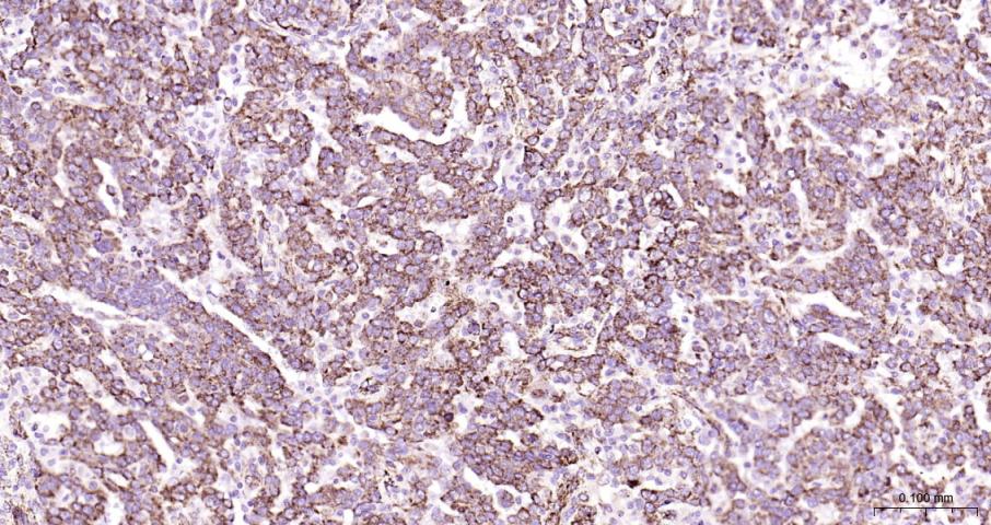
Paraformaldehyde-fixed, paraffin embedded Human Ovarian Cancer; Antigen retrieval by boiling in sodium citrate buffer (pH6.0) for 15 min; The section was incubated with Osteocalcin Polyclonal Antibody, Unconjugated (bs-4917R) at 1:200 overnight at 4°C, followed by conjugation to the bs-0295G-HRP and DAB (C-0010) staining.
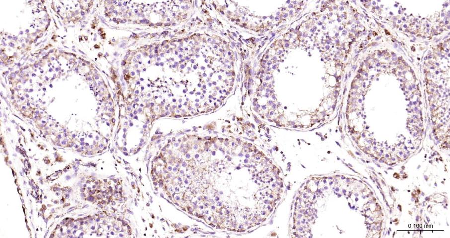
Paraformaldehyde-fixed, paraffin embedded Human Testicles; Antigen retrieval by boiling in sodium citrate buffer (pH6.0) for 15 min; The section was incubated with Osteocalcin Polyclonal Antibody, Unconjugated (bs-4917R) at 1:200 overnight at 4°C, followed by conjugation to the bs-0295G-HRP and DAB (C-0010) staining.
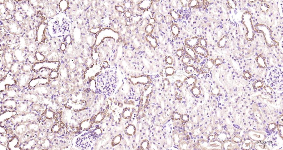
Paraformaldehyde-fixed, paraffin embedded Mouse Kidney; Antigen retrieval by boiling in sodium citrate buffer (pH6.0) for 15 min; The section was incubated with Osteocalcin Polyclonal Antibody, Unconjugated (bs-4917R) at 1:200 overnight at 4°C, followed by conjugation to the bs-0295G-HRP and DAB (C-0010) staining.
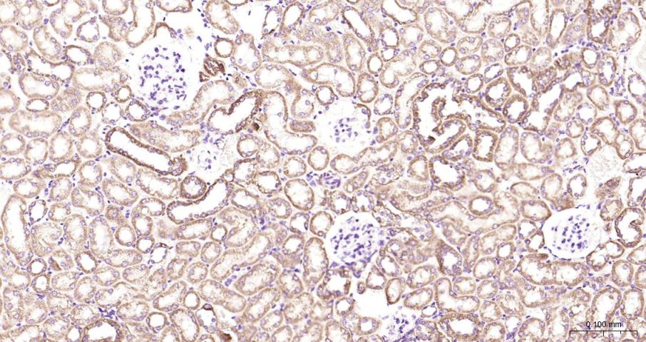
Paraformaldehyde-fixed, paraffin embedded Human Kidney; Antigen retrieval by boiling in sodium citrate buffer (pH6.0) for 15 min; The section was incubated with Osteocalcin Polyclonal Antibody, Unconjugated (bs-4917R) at 1:200 overnight at 4°C, followed by conjugation to the bs-0295G-HRP and DAB (C-0010) staining.
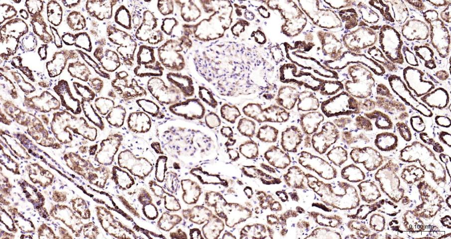
Paraformaldehyde-fixed, paraffin embedded Rat Kidney; Antigen retrieval by boiling in sodium citrate buffer (pH6.0) for 15 min; The section was incubated with Osteocalcin Polyclonal Antibody, Unconjugated (bs-4917R) at 1:200 overnight at 4°C, followed by conjugation to the bs-0295G-HRP and DAB (C-0010) staining.
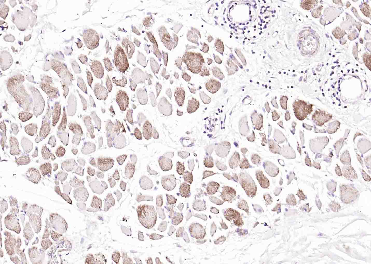
Paraformaldehyde-fixed, paraffin embedded (human skeletal muscle); Antigen retrieval by boiling in sodium citrate buffer (pH6.0) for 15min; Block endogenous peroxidase by 3% hydrogen peroxide for 20 minutes; Blocking buffer (normal goat serum) at 37°C for 30min; Incubation with (Osteocalcin) Polyclonal Antibody, Unconjugated (bs-4917R) at 1:100 overnight at 4°C, followed by operating according to SP Kit(Rabbit) (sp-0023) instructionsand DAB staining.
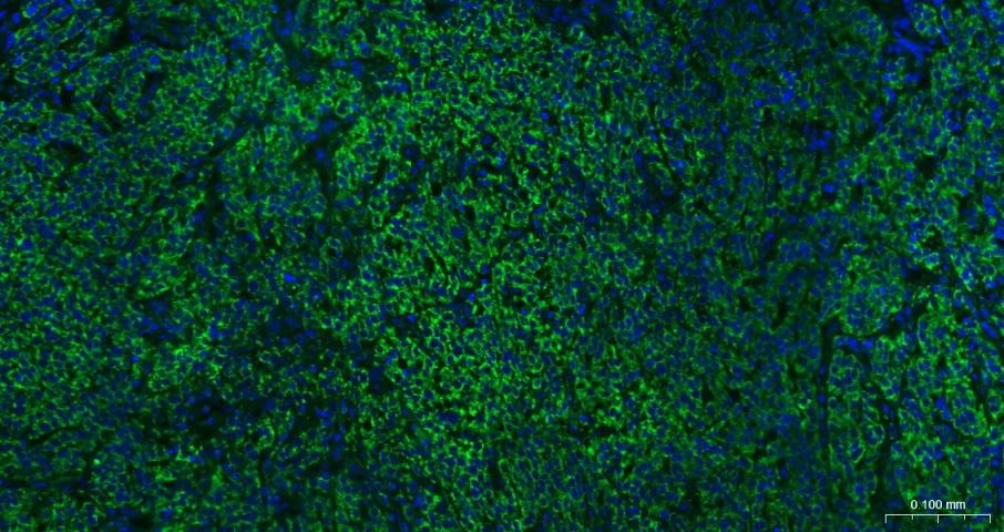
Paraformaldehyde-fixed, paraffin embedded Human Ovarian Cancer; Antigen retrieval by boiling in sodium citrate buffer (pH6.0) for 15 min; The section was incubated with Osteocalcin Polyclonal Antibody, Unconjugated (bs-4917R) at 1:200 overnight at 4°C. Followed by conjugated Goat Anti-Rabbit IgG antibody (green, bs-0295G-BF488), DAPI (blue, C02-04002) was used to stain the cell nuclei.
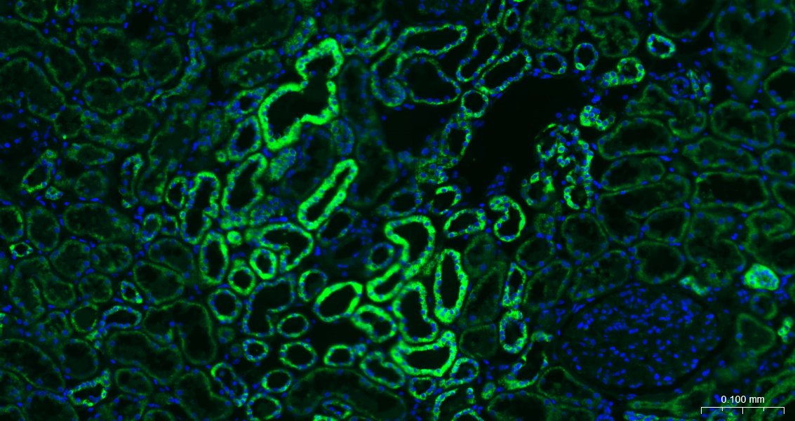
Paraformaldehyde-fixed, paraffin embedded Human Kidney; Antigen retrieval by boiling in sodium citrate buffer (pH6.0) for 15 min; The section was incubated with Osteocalcin Polyclonal Antibody, Unconjugated (bs-4917R) at 1:200 overnight at 4°C. Followed by conjugated Goat Anti-Rabbit IgG antibody (green, bs-0295G-BF488), DAPI (blue, C02-04002) was used to stain the cell nuclei.
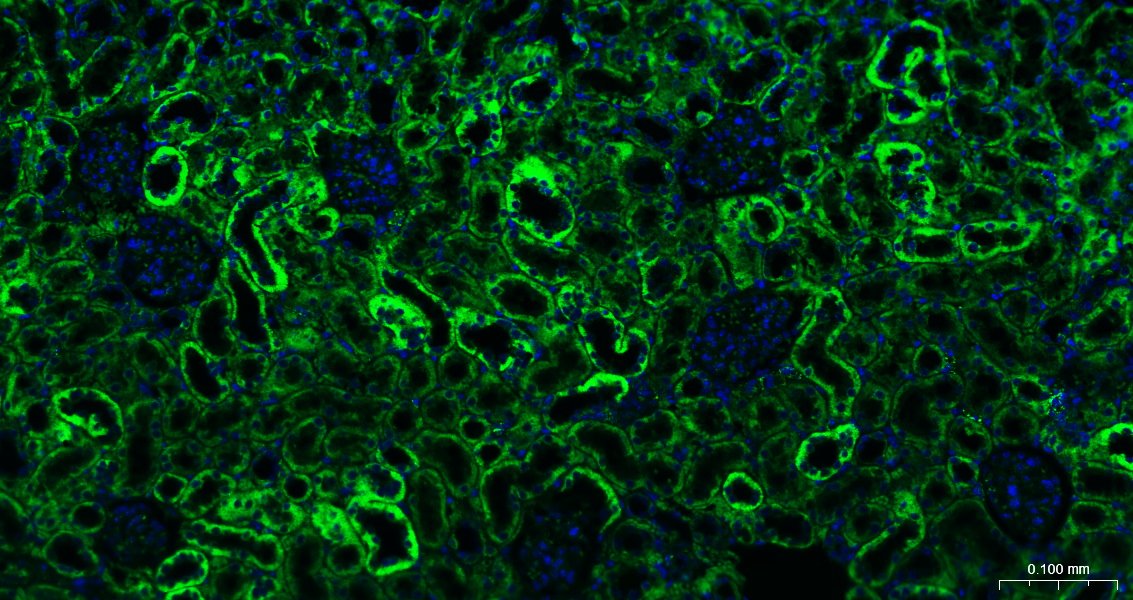
Paraformaldehyde-fixed, paraffin embedded Mouse Kidney; Antigen retrieval by boiling in sodium citrate buffer (pH6.0) for 15 min; The section was incubated with Osteocalcin Polyclonal Antibody, Unconjugated (bs-4917R) at 1:200 overnight at 4°C. Followed by conjugated Goat Anti-Rabbit IgG antibody (green, bs-0295G-BF488), DAPI (blue, C02-04002) was used to stain the cell nuclei.
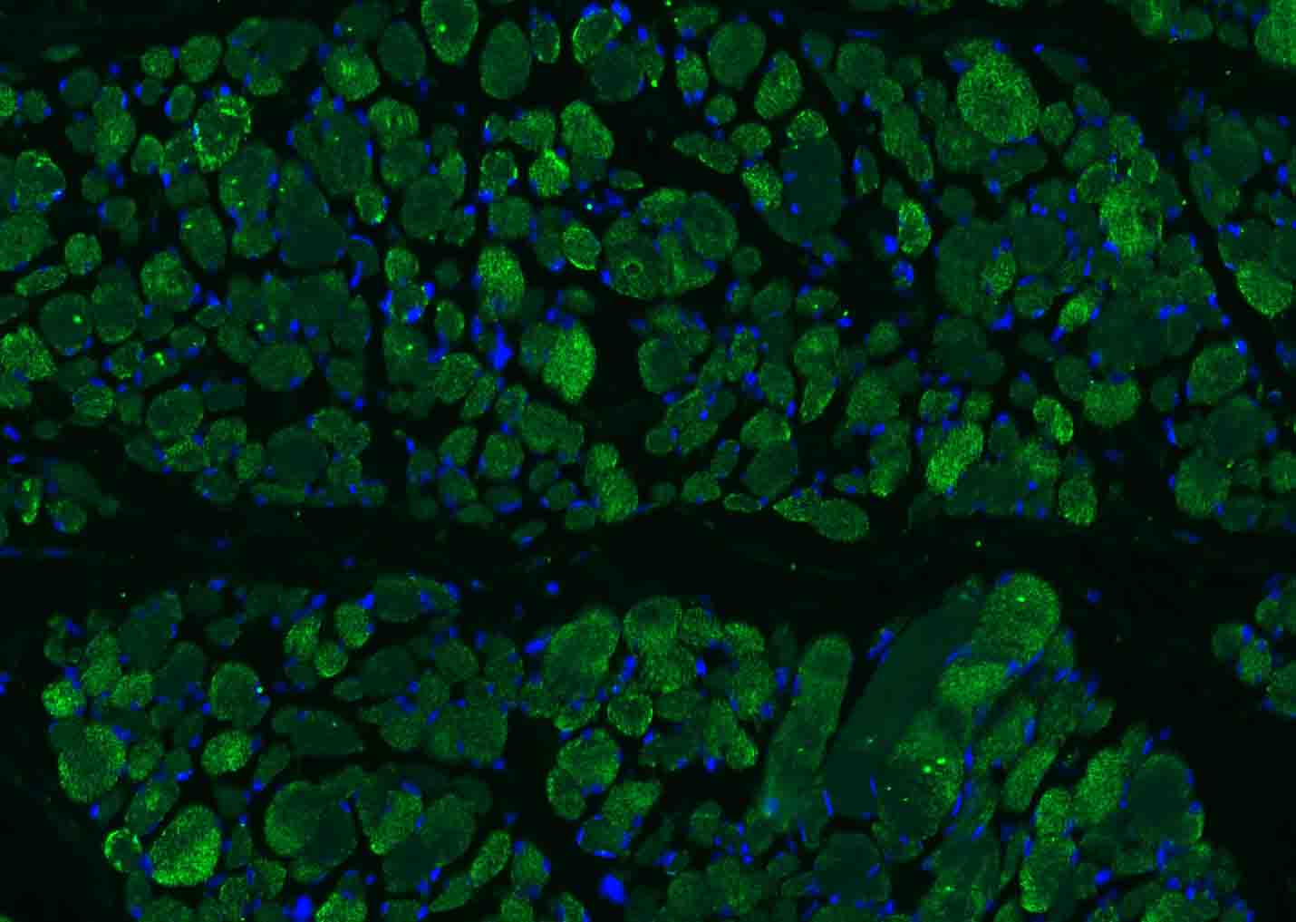
Paraformaldehyde-fixed, paraffin embedded (human skeletal muscle); Antigen retrieval by boiling in sodium citrate buffer (pH6.0) for 15min; Blocking buffer (normal goat serum) at 37°C for 30min; Incubation with (Osteocalcin) Polyclonal Antibody, Unconjugated (bs-4917R) at 1:100 overnight at 4°C, followed by a conjugated Goat Anti-Rabbit IgG antibody (bs-0295G-AF488) for 90 minutes, and DAPI for nuclei stainin
|
| 1、抗体溶解方法 | |
| 2、抗体修复方式 | |
| 3、常用试剂的配制 | |
| 4、免疫组化操作步骤 | |
| 5、免疫组化问题解答 | |
| 6、Western Blotting 操作步骤 | |
| 7、Western Blotting 问题解答 | |
| 8、关于肽链的设计 | |
| 9、多肽的溶解与保存 | |
| 10、酶标抗体效价测定程序 | |


