

| 产品编号 | bs-4770R |
| 英文名称 | CD133 Rabbit pAb |
| 中文名称 | 造血干细胞抗原CD133抗体 |
| 别 名 | AC133; CD133; CORD12; MCDR2; MSTP061; PROML1; RP41; STGD4; 4932416E19Rik; Prom; Prom-1; PROM1_HUMAN; PROM1; Antigen AC133; Prominin-like protein 1; PROM1_MOUSE; Antigen AC133 homolog; |
 |
Specific References (6) | bs-4770R has been referenced in 6 publications.
[IF=5.587] Qi et al. Targeting the Wnt-Regulatory Protein CTNNBIP1 by microRNA-214 Enhances the Stemness and Self-Renewal of Cancer Stem-Like Cells in Lung Adenocarcinomas. (2015) Stem.Cells. 33(12):3423-36 IHC,IF ; Human.
[IF=4.486] Wang L et al. Lung CSC‐derived exosomal miR‐210‐3p contributes to a pro‐metastatic phenotype in lung cancer by targeting FGFRL1. J Cell Mol Med. 2020 Jun;24(11):6324-6339. WB/IF ; Human.
[IF=4.432] Yang Yang. et al. PT109, a novel multi-kinase inhibitor suppresses glioblastoma multiforme through cell reprogramming: Involvement of PTBP1/PKM1/2 pathway. Eur J Pharmacol. 2022 Apr;920:174837 WB ; Human.
[IF=3.811] Sun Z et al. Glioblastoma Stem Cell-Derived Exosomes Enhance Stemness and Tumorigenicity of Glioma Cells by Transferring Notch1 Protein. Cell Mol Neurobiol. 2019 Dec 18. WB ; Human&Mouse.
[IF=3.25] Qin, Jianhua, et al. "Probing impaired neurogenesis in human brain organoids exposed to alcohol." Integrative Biology(2017). IHC-F ; Human.
[IF=2.26] Yu, Xian-huan, et al. "Clinicopathological characteristics of 20 cases of hepatocellular carcinoma with bile duct tumor thrombi." Digestive diseases and sciences 56.1 (2011): 252-259. IHC-P ; Human.
|
| 研究领域 | 肿瘤 细胞生物 免疫学 干细胞 细胞类型标志物 |
| 抗体来源 | Rabbit |
| 克隆类型 | Polyclonal |
| 克 隆 号 | |
| 交叉反应 | Human,Mouse (predicted: Rat) |
| 产品应用 | WB=1:500-2000,Flow-Cyt=1μg/Test
not yet tested in other applications. optimal dilutions/concentrations should be determined by the end user. |
| 理论分子量 | 95 kDa |
| 检测分子量 | 120 |
| 细胞定位 | 细胞膜 |
| 性 状 | Liquid |
| 浓 度 | 1mg/ml |
| 免 疫 原 | KLH conjugated synthetic peptide derived from human CD133: 508-552/865 <Extracellular> |
| 亚 型 | IgG |
| 纯化方法 | affinity purified by Protein A |
| 缓 冲 液 | 0.01M TBS (pH7.4) with 1% BSA, 0.02% Proclin300 and 50% Glycerol. |
| 保存条件 | Shipped at 4℃. Store at -20℃ for one year. Avoid repeated freeze/thaw cycles. |
| 注意事项 | This product as supplied is intended for research use only, not for use in human, therapeutic or diagnostic applications. |
| PubMed | PubMed |
| 产品介绍 |
This gene encodes a pentaspan transmembrane glycoprotein. The protein localizes to membrane protrusions and is often expressed on adult stem cells, where it is thought to function in maintaining stem cell properties by suppressing differentiation. Mutations in this gene have been shown to result in retinitis pigmentosa and Stargardt disease. Expression of this gene is also associated with several types of cancer. This gene is expressed from at least five alternative promoters that are expressed in a tissue-dependent manner. Multiple transcript variants encoding different isoforms have been found for this gene. [provided by RefSeq, Mar 2009] Function: Binds cholesterol in cholesterol-containing plasma membrane microdomains. Proposed to play a role in apical plasma membrane organization of epithelial cells. During early retinal development acts as a key regulator of disk morphogenesis. Involved in regulation of MAPK and Akt signaling pathways. In neuroblastoma cells suppresses cell differentiation such as neurite outgrowth in a RET-dependent manner. Subunit: Interacts with CDHR1 and with actin filaments. Subcellular Location: Cell projection, cilium, photoreceptor outer segment. Isoform 1: Apical cell membrane; Multi-pass membrane protein. Cell projection, microvillus membrane; Multi-pass membrane protein. Note=Found in extracellular membrane particles in various body fluids such as cerebrospinal fluid, saliva, seminal fluid and urine. Tissue Specificity: Isoform 1 is selectively expressed on CD34 hematopoietic stem and progenitor cells in adult and fetal bone marrow, fetal liver, cord blood and adult peripheral blood. Isoform 1 is not detected on other blood cells. Isoform 1 is also expressed in a number of non-lymphoid tissues including retina, pancreas, placenta, kidney, liver, lung, brain and heart. Found in saliva within small membrane particles. Isoform 2 is predominantly expressed in fetal liver, skeletal muscle, kidney, and heart as well as adult pancreas, kidney, liver, lung, and placenta. Isoform 2 is highly expressed in fetal liver, low in bone marrow, and barely detectable in peripheral blood. Isoform 2 is expressed on hematopoietic stem cells and in epidermal basal cells (at protein level). Expressed in adult retina by rod and cone photoreceptor cells (at protein level). Post-translational modifications: Isoform 1 and isoform 2 are glycosylated. DISEASE: Defects in PROM1 are the cause of retinitis pigmentosa type 41 (RP41) [MIM:612095]; also known as retinal degeneration autosomal recessive prominin-related. RP is a retinal dystrophy belonging to the group of pigmentary retinopathies. RP is characterized by retinal pigment deposits visible on fundus examination and primary loss of rod photoreceptor cells followed by secondary loss of cone photoreceptors. Patients typically have night vision blindness and loss of midperipheral visual field. As their condition progresses, they lose their far peripheral visual field and eventually central vision as well. Defects in PROM1 are the cause of cone-rod dystrophy type 12 (CORD12) [MIM:612657]. CORD12 is an inherited retinal dystrophy characterized by retinal pigment deposits visible on fundus examination, predominantly in the macular region, and initial loss of cone photoreceptors followed by rod degeneration. This leads to decreased visual acuity and sensitivity in the central visual field, followed by loss of peripheral vision. Severe loss of vision occurs earlier than in retinitis pigmentosa. Defects in PROM1 are the cause of Stargardt disease type 4 (STGD4) [MIM:603786]. Stargardt disease is the most common hereditary macular degeneration. It is characterized by decreased central vision, atrophy of the macula and underlying retinal pigment epithelium, and frequent presence of prominent flecks in the posterior pole of the retina. Defects in PROM1 are the cause of retinal macular dystrophy type 2 (MCDR2) [MIM:608051]. MCDR2 is a bull's-eye macular dystrophy characterized by bilateral annular atrophy of retinal pigment epithelium at the macula. Similarity: Belongs to the prominin family. SWISS: O43490 Gene ID: 8842 Database links: Entrez Gene: 8842 Human Entrez Gene: 19126 Mouse Omim: 604365 Human SwissProt: O43490 Human SwissProt: O54990 Mouse Unigene: 614734 Human Unigene: 6250 Mouse Unigene: 144589 Rat 一般认为,VEGFR2(血管内皮生长因子受体2)是HSCs(造血干细胞)的特异性的表面标志。近来经研究发现CD133分子是HSCs(造血干细胞)特异性标志。CD133即AC133,是一个新发现的HSCs(造血干细胞)表面标志,在HSCs(造血干细胞)分化成熟过程中,CD133的含量迅速降低。EPCs(血管内皮前体细胞)区别于成熟内皮细胞的主要标志是CD133。 经研究发现内皮细胞不能结合CD133的抗体。证实分化成熟的内皮细胞不具有CD133。这些说明CD133可以作为EPCs(血管内皮前体细胞)区别于成熟内皮细胞的一个表面标志. |
| 产品图片 |
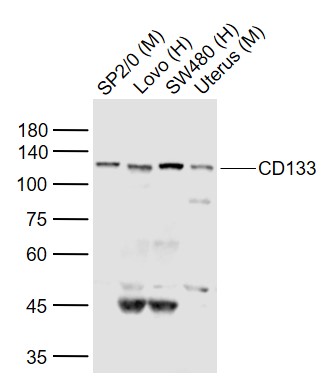
Sample: Lane 1: SP2/0 (Mouse) Cell Lysate at 30 ug Lane 2: Lovo (Human) Cell Lysate at 30 ug Lane 3: SW480 (Human) Cell Lysate at 30 ug Lane 4: Uterus (Mouse) Lysate at 40 ug Primary: Anti-CD133 (bs-4770R) at 1/300 dilution Secondary: IRDye800CW Goat Anti-Rabbit IgG at 1/20000 dilution Predicted band size: 95 kD Observed band size: 120 kD
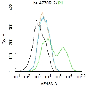
Blank control:Mouse kidney.
Primary Antibody (green line): Rabbit Anti-CD133 antibody (bs-4770R)
Dilution: 2μg /10^6 cells;
Isotype Control Antibody (orange line): Rabbit IgG .
Secondary Antibody : Goat anti-rabbit IgG-AF488
Dilution: 1μg /test.
Protocol
The cells were incubated in 5%BSA to block non-specific protein-protein interactions for 30 min at room temperature .Cells stained with Primary Antibody for 30 min at room temperature. The secondary antibody used for 40 min at room temperature. Acquisition of 20,000 events was performed.
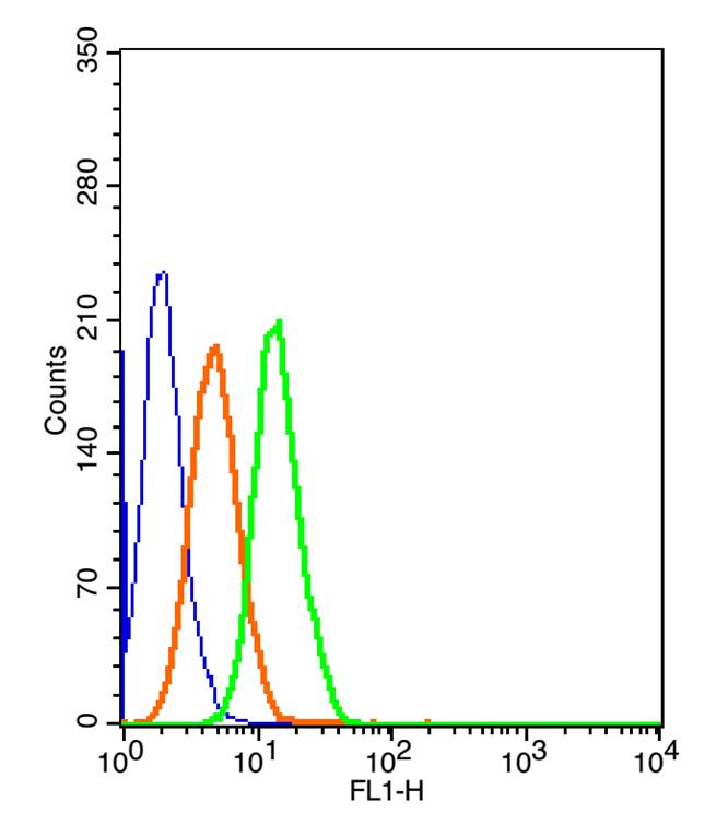
The blue histogram is unstained cells(HepG 2).
The Orange histogram is cells stained with Rabbit IgG/FITC (bs-0295P-FITC)
The green histogram is cells stained with Rabbit Anti-CD133/FITC Conjugated antibody (bs-4770R-FITC).
Isotype control: Cell lines treated with Rabbit IgG/FITC (bs-0295P-FITC) instead of the primary antibody to confirm that primary antibody binding is 2μg/5μg/1μg in 100μL 1 X PBS containing 0.5% BSA.
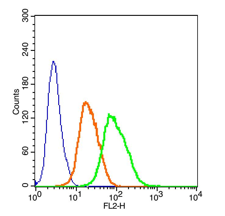
The blue histogram is unstained cells(HepG 2).
The Orange histogram is cells stained with Rabbit IgG/PE (bs-0295P-PE)
The green histogram is cells stained with Rabbit Anti-CD133/PE Conjugated antibody (bs-4770R-PE).
Isotype control: Cell lines treated with Rabbit IgG/PE (bs-0295P-PE) instead of the primary antibody to confirm that primary antibody binding is specific. 2μg/5μg/10μg in 100μL 1 X PBS containing 0.5% BSA.
|
| 1、抗体溶解方法 | |
| 2、抗体修复方式 | |
| 3、常用试剂的配制 | |
| 4、免疫组化操作步骤 | |
| 5、免疫组化问题解答 | |
| 6、Western Blotting 操作步骤 | |
| 7、Western Blotting 问题解答 | |
| 8、关于肽链的设计 | |
| 9、多肽的溶解与保存 | |
| 10、酶标抗体效价测定程序 | |


