

| 产品编号 | bs-6983R |
| 英文名称 | WT1 Rabbit pAb |
| 中文名称 | 肾母细胞瘤蛋白抗体 |
| 别 名 | AWT1; GUD; NPHS4; WAGR; WIT-2; WT-1; WT33; D630046I19Rik; WT1_HUMAN; WT1; WT1_MOUSE; WT1_RAT; |
 |
Specific References (5) | bs-6983R has been referenced in 5 publications.
[IF=2.74] Rui Feng. et al. The ameliorative effect of melatonin on LPS-induced Sertoli cells inflammatory and tight junctions damage via suppression of the TLR4/MyD88/NF-κB signaling pathway in newborn calf. Theriogenology. 2022 Feb;179:103 IF ; Bovine.
[IF=2.33] Xiao, Tangli, et al. "Rapamycin promotes podocyte autophagy and ameliorates renal injury in diabetic mice." Molecular and Cellular Biochemistry (2014): 1-10. IHC-P ; Mouse.
[IF=2.299] Wang X et al. 3, 3′, 5-Triiodo-L-thyronine affects polarity proteins of bovine Sertoli cells via WT1/non-canonical Wnt signaling pathway. Theriogenology. 2020 Feb 21;148:8-17. WB ; Bovine.
[IF=2.299] Wang X et al. Influence of Wilms' tumor suppressor gene WT1 on bovine Sertoli cells polarity and tight junctions via non-canonical WNT signaling pathway. Theriogenology. 2019 Jul 8;138:84-93. ICF ; Cow.
[IF=1.723] Wang X et al. Wilms' tumour 1 (WT1) negatively regulates the expression of connexin 43 via a non-canonical Wnt signalling pathway in cultured bovine Sertoli cells. Reprod Fertil Dev. 2020 Feb 6. WB&ICF ; Bovine.
|
| 研究领域 | 肿瘤 细胞生物 免疫学 发育生物学 肿瘤细胞生物标志物 表观遗传学 |
| 抗体来源 | Rabbit |
| 克隆类型 | Polyclonal |
| 克 隆 号 | |
| 交叉反应 | Human,Mouse (predicted: Rat,Pig,Sheep,Cow,Chicken,Dog) |
| 产品应用 | WB=1:500-2000,IHC-P=1:100-500,IHC-F=1:100-500,IF=1:100-500,Flow-Cyt=1ug/test
not yet tested in other applications. optimal dilutions/concentrations should be determined by the end user. |
| 理论分子量 | 55 kDa |
| 检测分子量 | 48 |
| 细胞定位 | 细胞核 细胞浆 |
| 性 状 | Liquid |
| 浓 度 | 1mg/ml |
| 免 疫 原 | KLH conjugated synthetic peptide derived from human WT1: 301-400/449 |
| 亚 型 | IgG |
| 纯化方法 | affinity purified by Protein A |
| 缓 冲 液 | 0.01M TBS (pH7.4) with 1% BSA, 0.02% Proclin300 and 50% Glycerol. |
| 保存条件 | Shipped at 4℃. Store at -20℃ for one year. Avoid repeated freeze/thaw cycles. |
| 注意事项 | This product as supplied is intended for research use only, not for use in human, therapeutic or diagnostic applications. |
| PubMed | PubMed |
| 产品介绍 |
Transcription factor that plays an important role in cellular development and cell survival. Regulates the expression of numerous target genes, including EPO. Plays an essential role for development of the urogenital system. Recognizes and binds to the DNA sequence 5'-CGCCCCCGC-3'. It has a tumor suppressor as well as an oncogenic role in tumor formation. Function may be isoform-specific: isoforms lacking the KTS motif may act as transcription factors. Isoforms containing the KTS motif may bind mRNA and play a role in mRNA metabolism or splicing. Isoform 1 has lower affinity for DNA, and can bind RNA. Function: Transcription factor that plays an important role in cellular development and cell survival. Regulates the expression of numerous target genes, including EPO. Plays an essential role for development of the urogenital system. Recognizes and binds to the DNA sequence 5'-CGCCCCCGC-3'. It has a tumor suppressor as well as an oncogenic role in tumor formation. Function may be isoform-specific: isoforms lacking the KTS motif may act as transcription factors. Isoforms containing the KTS motif may bind mRNA and play a role in mRNA metabolism or splicing. Isoform 1 has lower affinity for DNA, and can bind RNA. Subunit: Homodimer. Interacts with WTIP. Interacts with actively translating polysomes. Detected in nuclear ribonucleoprotein (mRNP) particles. Interacts with HNRNPU via the zinc-finger region. Interacts with U2AF2. Interacts with CITED2. Interacts with ZNF224 via the zinc-finger region. Interacts with WTAP and SRY. Interacts with FAM123B/WTX. Interacts with RBM4. Subcellular Location: Nucleus. Nucleus, nucleolus. Cytoplasm. Note=Shuttles between nucleus and cytoplasm. Isoform 1: Nucleus speckle. Isoform 4: Nucleus, nucleoplasm. Tissue Specificity: Expressed in the kidney and a subset of hematopoietic cells. DISEASE: Defects in WT1 are the cause of Frasier syndrome (FS) [MIM:136680]. FS is characterized by a slowly progressing nephropathy leading to renal failure in adolescence or early adulthood, male pseudohermaphroditism, and no Wilms tumor. As for histological findings of the kidneys, focal glomerular sclerosis is often observed. There is phenotypic overlap with Denys-Drash syndrome. Inheritance is autosomal dominant. Defects in WT1 are the cause of Wilms tumor 1 (WT1) [MIM:194070]. WT is an embryonal malignancy of the kidney that affects approximately 1 in 10'000 infants and young children. It occurs both in sporadic and hereditary forms. Defects in WT1 are the cause of Denys-Drash syndrome (DDS) [MIM:194080]. DDS is a typical nephropathy characterized by diffuse mesangial sclerosis, genital abnormalities, and/or Wilms tumor. There is phenotypic overlap with WAGR syndrome and Frasier syndrome. Inheritance is autosomal dominant, but most cases are sporadic. Similarity: Belongs to the EGR C2H2-type zinc-finger protein family. Contains 4 C2H2-type zinc fingers. SWISS: P19544 Gene ID: 7490 Database links: Entrez Gene: 7490 Human Entrez Gene: 22431 Mouse Omim: 607102 Human SwissProt: P19544 Human SwissProt: P22561 Mouse Unigene: 591980 Human Unigene: 389339 Mouse Unigene: 92531 Rat
|
| 产品图片 |
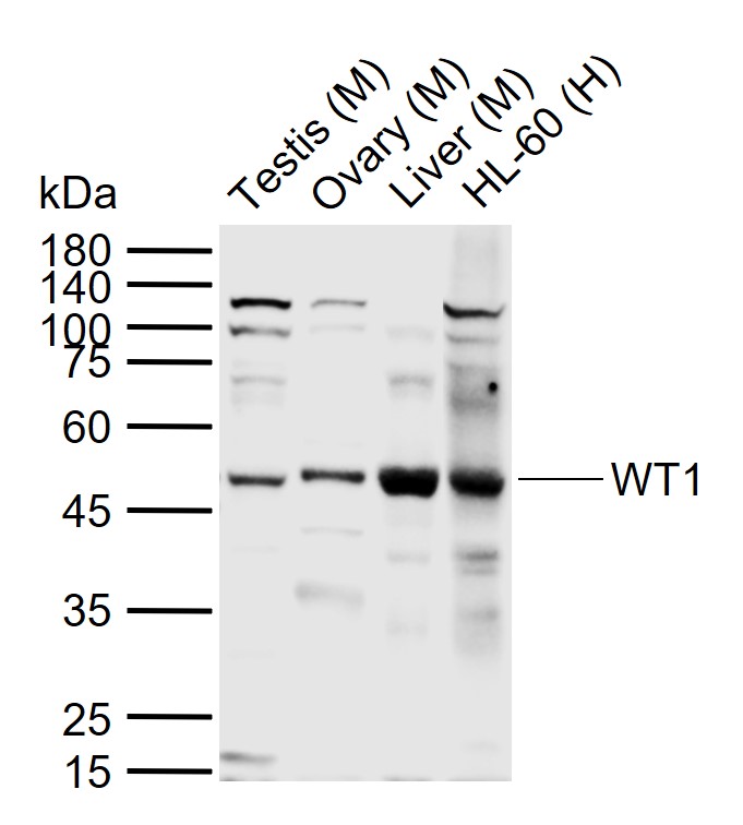
Sample:
Lane 1: Mouse Testis tissue lysates
Lane 2: Mouse Ovary tissue lysates
Lane 3: Mouse Liver tissue lysates
Lane 4: Human HL-60 cell lysates
Primary: Anti-WT1 (bs-6983R) at 1/1000 dilution
Secondary: IRDye800CW Goat Anti-Rabbit IgG at 1/20000 dilution
Predicted band size: 55 kDa
Observed band size: 48 kDa
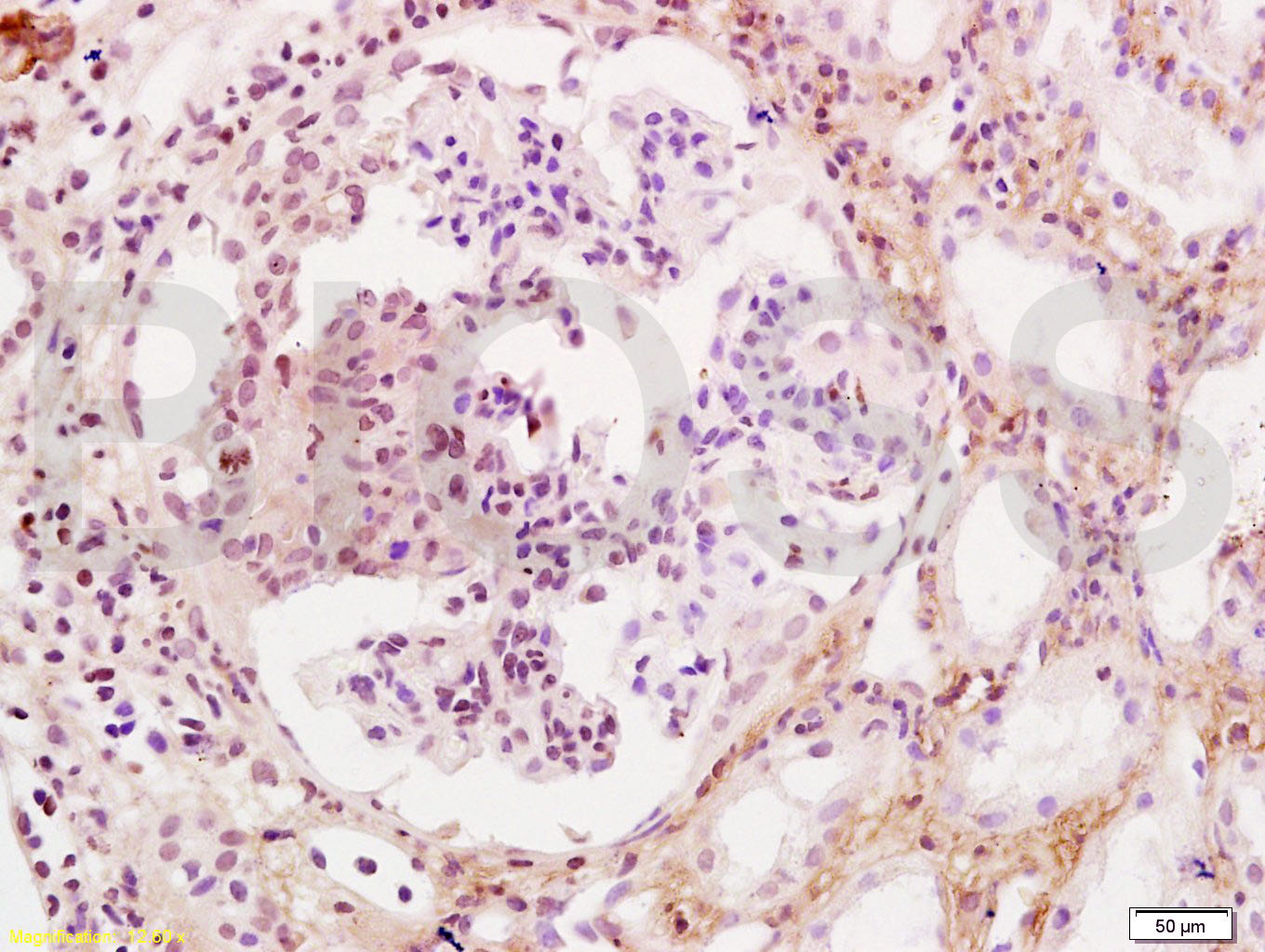
Tissue/cell: human kidney tissue; 4% Paraformaldehyde-fixed and paraffin-embedded;
Antigen retrieval: citrate buffer ( 0.01M, pH 6.0 ), Boiling bathing for 15min; Block endogenous peroxidase by 3% Hydrogen peroxide for 30min; Blocking buffer (normal goat serum,C-0005) at 37℃ for 20 min;
Incubation: Anti-WT-1/Wilms Tumor Protein Polyclonal Antibody, Unconjugated(bs-6983R) 1:200, overnight at 4°C, followed by conjugation to the secondary antibody(SP-0023) and DAB(C-0010) staining
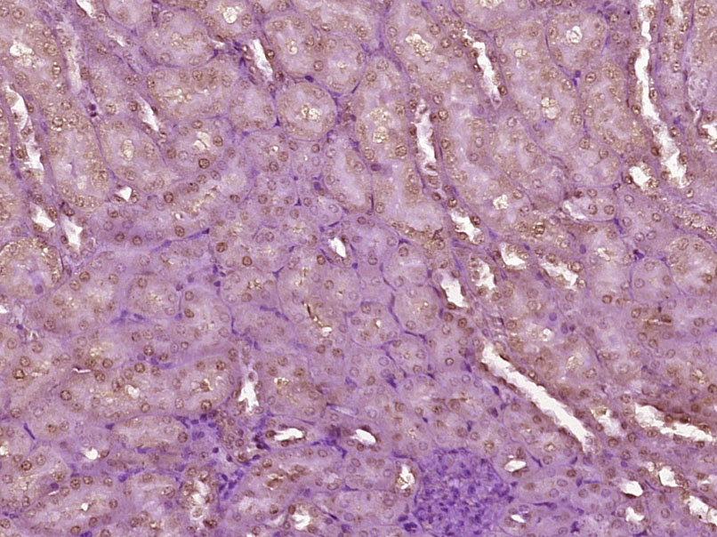
Paraformaldehyde-fixed, paraffin embedded (Mouse kidney); Antigen retrieval by boiling in sodium citrate buffer (pH6.0) for 15min; Block endogenous peroxidase by 3% hydrogen peroxide for 20 minutes; Blocking buffer (normal goat serum) at 37°C for 30min; Antibody incubation with (Wilms Tumor Protein) Polyclonal Antibody, Unconjugated (bs-6983R) at 1:400 overnight at 4°C, followed by operating according to SP Kit(Rabbit) (sp-0023) instructionsand DAB staining.

Paraformaldehyde-fixed, paraffin embedded (Mouse testis); Antigen retrieval by boiling in sodium citrate buffer (pH6.0) for 15min; Block endogenous peroxidase by 3% hydrogen peroxide for 20 minutes; Blocking buffer (normal goat serum) at 37°C for 30min; Antibody incubation with (Wilms Tumor Protein) Polyclonal Antibody, Unconjugated (bs-6983R) at 1:400 overnight at 4°C, followed by a conjugated Goat Anti-Rabbit IgG antibody (bs-0295G-FITC) for 90 minutes, and DAPI for nuclei staining.
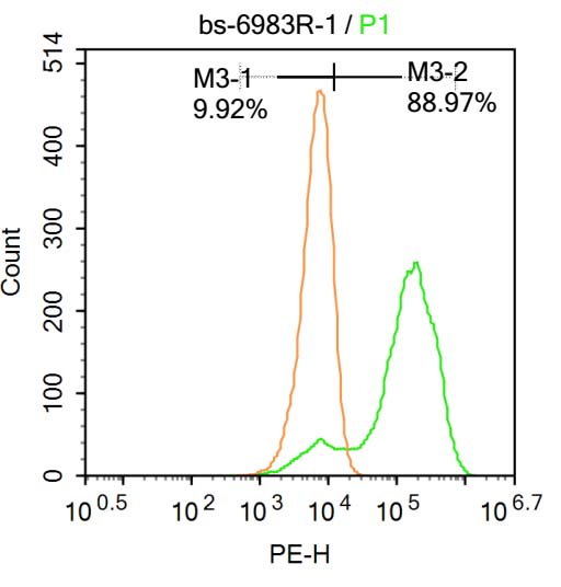
Blank control:Molt-4.
Primary Antibody (green line): Rabbit Anti-Wilms Tumor Protein antibody (bs-6983R)
Dilution: 1μg /10^6 cells;
Isotype Control Antibody (orange line): Rabbit IgG .
Secondary Antibody : Goat anti-rabbit IgG-AF647
Dilution: 1μg /test.
Protocol
The cells were fixed with 4% PFA (10min at room temperature)and then permeabilized with 90% ice-cold methanol for 20 min at-20℃. The cells were then incubated in 5%BSA to block non-specific protein-protein interactions for 30 min at at room temperature .Cells stained with Primary Antibody for 30 min at room temperature. The secondary antibody used for 40 min at room temperature. Acquisition of 20,000 events was performed.
|
| 1、抗体溶解方法 | |
| 2、抗体修复方式 | |
| 3、常用试剂的配制 | |
| 4、免疫组化操作步骤 | |
| 5、免疫组化问题解答 | |
| 6、Western Blotting 操作步骤 | |
| 7、Western Blotting 问题解答 | |
| 8、关于肽链的设计 | |
| 9、多肽的溶解与保存 | |
| 10、酶标抗体效价测定程序 | |


