

| 产品编号 | bs-6316R |
| 英文名称 | PTGER1 Rabbit pAb |
| 中文名称 | 前列腺素EP1受体抗体 |
| 别 名 | EP1; Ptgerep1; PE2R1_HUMAN; PTGER1; PGE receptor EP1 subtype; PGE2 receptor EP1 subtype; Prostanoid EP1 receptor; PE2R1_MOUSE; PE2R1_RAT; |
 |
Specific References (3) | bs-6316R has been referenced in 3 publications.
[IF=4.872] Wu F et al. Manganese exposure caused reproductive toxicity of male mice involving activation of GnRH secretion in the hypothalamus by prostaglandin E2 receptors EP1 and EP2. Ecotoxicol Environ Saf
. 2020 Sep 15;201:110712. WB ; Mouse.
[IF=3.23] Takemiya et al. Microsomal Prostaglandin E Synthase-1 Facilitates an Intercellular Interaction between CD4⁺ T Cells through IL-1β Autocrine Function in Experimental Autoimmune Encephalomyelitis. (2017) Int.J.Mol.Sci. 18 IHC-F ; Mouse.
[IF=2.253] Chen Z et al. Seasonal expressions of prostaglandin E synthases and receptors in the prostate of the wild ground squirrel (Spermophilus dauricus). Prostaglandins and Other Lipid Mediators 148 (2020) 106412. IHC-P ; squirrel.
|
| 研究领域 | 细胞生物 信号转导 转录调节因子 G蛋白偶联受体 G蛋白信号 |
| 抗体来源 | Rabbit |
| 克隆类型 | Polyclonal |
| 克 隆 号 | |
| 交叉反应 | Mouse,Rat (predicted: Human,Dog) |
| 产品应用 | WB=1:500-2000,IHC-P=1:100-500,IHC-F=1:100-500,IF=1:100-500,Flow-Cyt=1ug/Test
not yet tested in other applications. optimal dilutions/concentrations should be determined by the end user. |
| 理论分子量 | 42 kDa |
| 检测分子量 | 42-45 |
| 细胞定位 | 细胞膜 |
| 性 状 | Liquid |
| 浓 度 | 1mg/ml |
| 免 疫 原 | KLH conjugated synthetic peptide derived from human Prostaglandin E Receptor EP1: 61-160/402 <Extracellular> |
| 亚 型 | IgG |
| 纯化方法 | affinity purified by Protein A |
| 缓 冲 液 | 0.01M TBS (pH7.4) with 1% BSA, 0.02% Proclin300 and 50% Glycerol. |
| 保存条件 | Shipped at 4℃. Store at -20℃ for one year. Avoid repeated freeze/thaw cycles. |
| 注意事项 | This product as supplied is intended for research use only, not for use in human, therapeutic or diagnostic applications. |
| PubMed | PubMed |
| 产品介绍 |
PTGER1 is a subtype 1 receptor for prostaglandin E2 (PGE2). This receptor is coupled to the phosphatidylinositol-calcium second messenger system by G(q) proteins. PTGER1 may be an important modulator of renal function and is implicated in the smooth muscle contractile response to PGE2 in various tissues. Subcellular Location: Cell membrane; Multi-pass membrane protein. Tissue Specificity: Abundant in kidney. Lower level expression in lung, skeletal muscle and spleen, lowest expression in testis and not detected in liver brain and heart. Similarity: Belongs to the G-protein coupled receptor 1 family. SWISS: P34995 Gene ID: 5731 Database links: Entrez Gene: 5731 Human Entrez Gene: 19216 Mouse Omim: 176802 Human SwissProt: P34995 Human SwissProt: P35375 Mouse Unigene: 159360 Human Unigene: 474430 Mouse Unigene: 11423 Rat |
| 产品图片 |
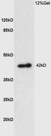
Sample: Brain (Rat) Lysate at 40 ug
Primary: Anti-Prostaglandin E Receptor EP1 (bs-6316R) at 1/300 dilution
Secondary: HRP conjugated Goat-Anti-rabbit IgG (bs-0295G-HRP) at 1/5000 dilution
Predicted band size: 42 kD
Observed band size: 42 kD
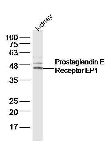
Sample: Kidney (Mouse) Lysate at 40 ug
Primary: Anti-Prostaglandin E Receptor EP1 (bs-6316R) at 1/300 dilution
Secondary: IRDye800CW Goat Anti-Rabbit IgG at 1/20000 dilution
Predicted band size: 42 kD
Observed band size: 45 kD

Tissue/cell: rat brain tissue;4% Paraformaldehyde-fixed and paraffin-embedded;
Antigen retrieval: citrate buffer ( 0.01M, pH 6.0 ), Boiling bathing for 15min; Blocking buffer (normal goat serum,C-0005) at 37℃ for 20 min;
Incubation: Anti-PTGER1 Polyclonal Antibody, Unconjugated(bs-6316R) 1:200, overnight at 4°C; The secondary antibody was Goat Anti-Rabbit IgG, Cy3 conjugated(bs-0295G-Cy3)used at 1:200 dilution for 40 minutes at 37°C. DAPI(5ug/ml,blue,C-0033) was used to stain the cell nuclei
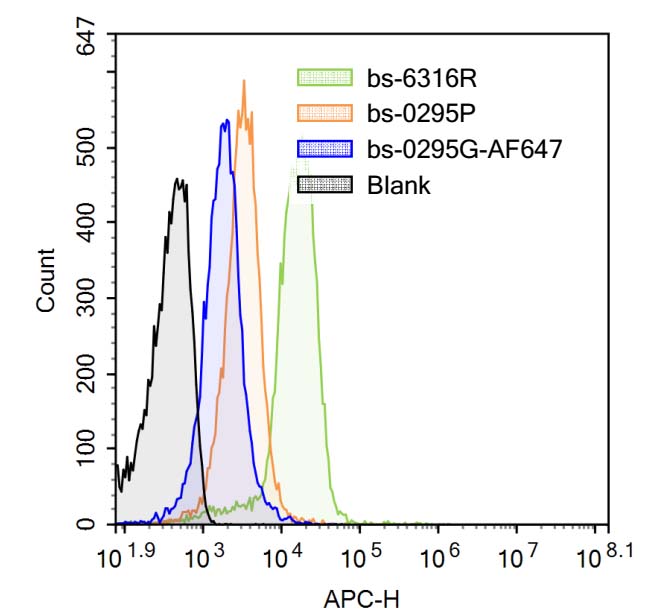
Blank control (Black line): Mouse blood (Black).
Primary Antibody (green line): Rabbit Anti-Prostaglandin E Receptor EP1 antibody (bs-6316R)
Dilution: 3μg /10^6 cells;
Isotype Control Antibody (orange line): Rabbit IgG .
Secondary Antibody (white blue line): Goat anti-rabbit IgG-AF647
Dilution: 1μg /test.
Protocol
The cells were fixed with 4% PFA (10min at room temperature)and then were incubated in 5%BSA to block non-specific protein-protein interactions for 30 min at room temperature .Cells stained with Primary Antibody for 30 min at room temperature. The secondary antibody used for 40 min at room temperature. Acquisition of 20,000 events was performed.
|
| 1、抗体溶解方法 | |
| 2、抗体修复方式 | |
| 3、常用试剂的配制 | |
| 4、免疫组化操作步骤 | |
| 5、免疫组化问题解答 | |
| 6、Western Blotting 操作步骤 | |
| 7、Western Blotting 问题解答 | |
| 8、关于肽链的设计 | |
| 9、多肽的溶解与保存 | |
| 10、酶标抗体效价测定程序 | |


