

| 产品编号 | bs-4690R |
| 英文名称 | Tissue factor Rabbit pAb |
| 中文名称 | 组织因子(CD142)抗体 |
| 别 名 | TF_HUMAN; F3; TF; Coagulation factor III; Thromboplastin; TF_MOUSE; Cf-3; Cf3; TF_RAT; |
 |
Specific References (6) | bs-4690R has been referenced in 6 publications.
[IF=23.394] Fletcher, Russell B., et al. "Deconstructing Olfactory Stem Cell Trajectories at Single-Cell Resolution." Cell Stem Cell (2017). FCM ; Mouse.
[IF=7.13] Gleeson, Birgitta M., et al. "Bone marrow‐derived mesenchymal stem cells have innate procoagulant activity and cause microvascular obstruction following intracoronary delivery: Amelioration by anti‐thrombin therapy." STEM CELLS (2015). Pig.
[IF=6.048] Takabayashi T et al. Increased expression of L‐plastin in nasal polyp of patients with nonsteroidal anti‐inflammatory drug exacerbated respiratory disease.(2018) Allergy IHC ; Human.
[IF=4.249] Zha C et al. Anti-β2GPI/β2GPI induces neutrophil extracellular traps formation to promote thrombogenesis via the TLR4/MyD88/MAPKs axis activation.Neuropharmacology. 2018 Aug;138:140-150. ICF ; Human.
[IF=2.98] Milano, M., et al. "Particulate matter phagocytosis induces tissue factor in differentiating macrophages." Journal of Applied Toxicology 36.1 (2016): 151-160 WB ; Rat.
[IF=2.65] Wang Shijun. et al. Investigations on the Changes of Serum Proteins in Rabbits after Trimeresurus stejnegeri Venom Injection via Mass Spectrometry-Based Proteomics. EVID-BASED COMPL ALT. 2022;2022:9239662 WB ; Rabbit.
|
| 研究领域 | 细胞生物 生长因子和激素 细胞表面分子 |
| 抗体来源 | Rabbit |
| 克隆类型 | Polyclonal |
| 克 隆 号 | |
| 交叉反应 | Human,Mouse,Rat (predicted: Rabbit,Pig,Cow,Dog,GuineaPig,Horse) |
| 产品应用 | WB=1:500-2000,IHC-P=1:100-500,IHC-F=1:100-500,IF=1:100-500
not yet tested in other applications. optimal dilutions/concentrations should be determined by the end user. |
| 理论分子量 | 29 kDa |
| 检测分子量 | 50 |
| 细胞定位 | 细胞膜 分泌型蛋白 |
| 性 状 | Liquid |
| 浓 度 | 1mg/ml |
| 免 疫 原 | KLH conjugated synthetic peptide derived from human Tissue factor: 32-100/295 <Extracellular> |
| 亚 型 | IgG |
| 纯化方法 | affinity purified by Protein A |
| 缓 冲 液 | 0.01M TBS (pH7.4) with 1% BSA, 0.02% Proclin300 and 50% Glycerol. |
| 保存条件 | Shipped at 4℃. Store at -20℃ for one year. Avoid repeated freeze/thaw cycles. |
| 注意事项 | This product as supplied is intended for research use only, not for use in human, therapeutic or diagnostic applications. |
| PubMed | PubMed |
| 产品介绍 |
This gene encodes coagulation factor III which is a cell surface glycoprotein. This factor enables cells to initiate the blood coagulation cascades, and it functions as the high-affinity receptor for the coagulation factor VII. The resulting complex provides a catalytic event that is responsible for initiation of the coagulation protease cascades by specific limited proteolysis. Unlike the other cofactors of these protease cascades, which circulate as nonfunctional precursors, this factor is a potent initiator that is fully functional when expressed on cell surfaces. There are 3 distinct domains of this factor: extracellular, transmembrane, and cytoplasmic. This protein is the only one in the coagulation pathway for which a congenital deficiency has not been described. Alternate splicing results in multiple transcript variants.[provided by RefSeq, May 2010] Function: Initiates blood coagulation by forming a complex with circulating factor VII or VIIa. The [TF:VIIa] complex activates factors IX or X by specific limited protolysis. TF plays a role in normal hemostasis by initiating the cell-surface assembly and propagation of the coagulation protease cascade. Subunit: Interacts with HSPE; the interaction, inhibited by heparin, promotes the generation of activated factor X and activates coagulation in the presence of activated factor VII. Subcellular Location: Isoform 1: Membrane; Single-pass type I membrane protein. Isoform 2: Secreted. Tissue Specificity: Lung, placenta and pancreas. Similarity: Belongs to the tissue factor family. SWISS: P13726 Gene ID: 2152 Database links: Entrez Gene: 2152 Human Omim: 134390 Human SwissProt: P13726 Human Unigene: 62192 Human |
| 产品图片 |
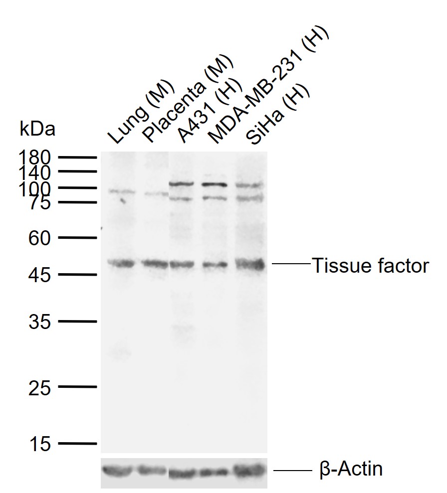
Sample:
Lane 1: Mouse Lung tissue lysates
Lane 2: Mouse Placenta tissue lysates
Lane 3: Human A431 cell lysates
Lane 4: Human MDA-MB-231 cell lysates
Lane 5: Human SiHa cell lysates
Primary: Anti-Tissue factor (bs-4690R) at 1/1000 dilution
Secondary: IRDye800CW Goat Anti-Rabbit IgG at 1/20000 dilution
Predicted band size: 29 kDa
Observed band size: 50 kDa
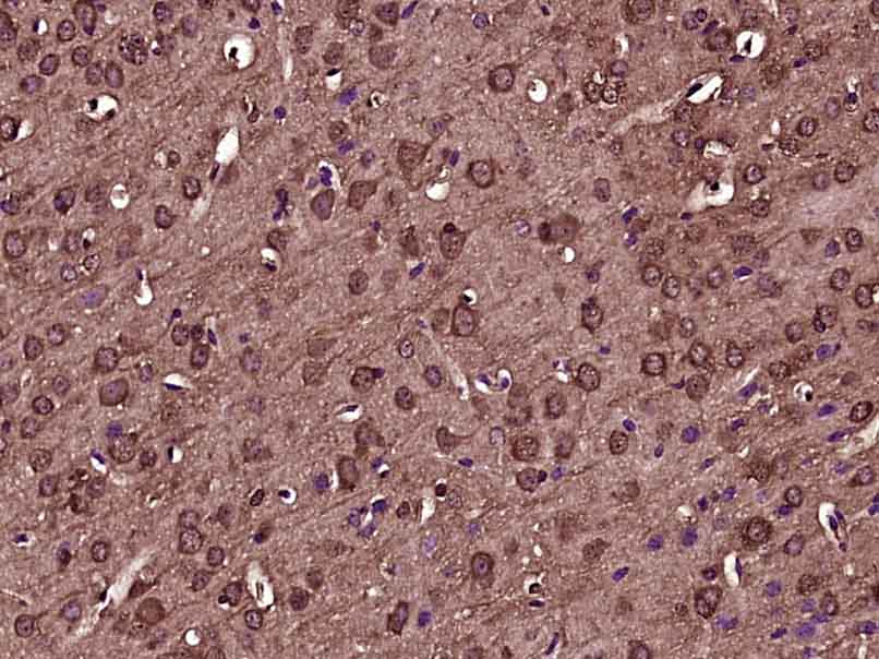
Paraformaldehyde-fixed, paraffin embedded (Mouse brain); Antigen retrieval by boiling in sodium citrate buffer (pH6.0) for 15min; Block endogenous peroxidase by 3% hydrogen peroxide for 20 minutes; Blocking buffer (normal goat serum) at 37°C for 30min; Antibody incubation with (Tissue factor) Polyclonal Antibody, Unconjugated (bs-4690R) at 1:400 overnight at 4°C, followed by operating according to SP Kit(Rabbit) (sp-0023) instructionsand DAB staining.
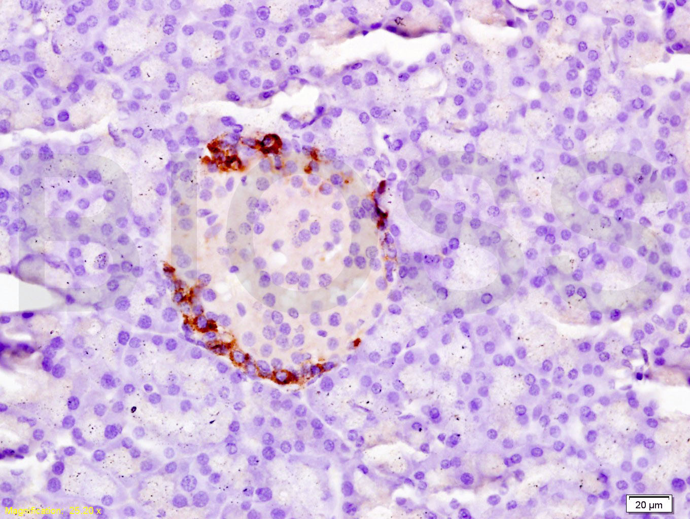
Tissue/cell: rat pancreas tissue; 4% Paraformaldehyde-fixed and paraffin-embedded;
Antigen retrieval: citrate buffer ( 0.01M, pH 6.0 ), Boiling bathing for 15min; Block endogenous peroxidase by 3% Hydrogen peroxide for 30min; Blocking buffer (normal goat serum,C-0005) at 37℃ for 20 min;
Incubation: Anti-Tissue factor/CD142/F3 Polyclonal Antibody, Unconjugated(bs-4690R) 1:200, overnight at 4°C, followed by conjugation to the secondary antibody(SP-0023) and DAB(C-0010) staining
|
| 1、抗体溶解方法 | |
| 2、抗体修复方式 | |
| 3、常用试剂的配制 | |
| 4、免疫组化操作步骤 | |
| 5、免疫组化问题解答 | |
| 6、Western Blotting 操作步骤 | |
| 7、Western Blotting 问题解答 | |
| 8、关于肽链的设计 | |
| 9、多肽的溶解与保存 | |
| 10、酶标抗体效价测定程序 | |


