

| 产品编号 | bs-5435R |
| 英文名称 | phospho-TAK1 (Ser192) Rabbit pAb |
| 中文名称 | 磷酸化转化生长因子β活化激酶1 |
| 别 名 | CSCF; FMD2; MEKK7; TAK1; TGF1a; B430101B05; M3K7_HUMAN; MAP3K7; Transforming growth factor-beta-activated kinase 1 (TGF-beta-activated kinase 1); 2.7.11.25; M3K7_MOUSE; M3K7_RAT; mitogen-activated protein kinase kinase kinase 7; TGF-beta activated kinase 1; MAP3K7 | TAK1 (phospho-S192); p-TAK1; phospho-TAK1; p-MAP3K7; MAP3K7 | TAK1 (phospho-Ser192) |
 |
Specific References (2) | bs-5435R has been referenced in 2 publications.
[IF=4.225] Jianwei Fan. et al. Baicalin Ameliorates Pancreatic Fibrosis by Inhibiting the Activation of Pancreatic Stellate Cells in Mice with Chronic Pancreatitis. Front Pharmacol. 2020; 11: 607133
[IF=2.21] Su, Xuesong, et al. "The PPARβ/δ Agonist GW501516 Attenuates Peritonitis in Peritoneal Fibrosis via Inhibition of TAK1–NFκB Pathway in Rats."Inflammation 37.3 (2014): 729-737. WB ; Rat.
|
| 产品类型 | 磷酸化抗体 |
| 研究领域 | 肿瘤 心血管 免疫学 信号转导 激酶和磷酸酶 |
| 抗体来源 | Rabbit |
| 克隆类型 | Polyclonal |
| 交叉反应 | Human (predicted: Mouse,Rat,Rabbit,Pig,Cow,Chicken,Horse) |
| 产品应用 | IHC-P=1:100-500,IHC-F=1:100-500,IF=1:100-500,ICC/IF=1:100-500
not yet tested in other applications. optimal dilutions/concentrations should be determined by the end user. |
| 理论分子量 | 67kDa |
| 细胞定位 | 细胞浆 细胞膜 |
| 性 状 | Liquid |
| 浓 度 | 1mg/ml |
| 免 疫 原 | KLH conjugated Synthesised phosphopeptide derived from human TAK1 around the phosphorylation site of Ser192: KG(p-S)AA |
| 亚 型 | IgG |
| 纯化方法 | affinity purified by Protein A |
| 缓 冲 液 | 0.01M TBS (pH7.4) with 1% BSA, 0.02% Proclin300 and 50% Glycerol. |
| 保存条件 | Shipped at 4℃. Store at -20℃ for one year. Avoid repeated freeze/thaw cycles. |
| 注意事项 | This product as supplied is intended for research use only, not for use in human, therapeutic or diagnostic applications. |
| PubMed | PubMed |
| 产品介绍 |
TAK1 (or MAP3K7) was shown to participate in regulation of transcription by transforming growth factor beta (TGF beta). TAK1 is stimulated in response to TGF beta and bone morphogenetic protein. These results suggest that TAK1 functions as a mediator in the signaling pathway of TGF beta superfamily members. TAB1 and TAB2 are TAK1 binding proteins that may function as activators of the TAK1 (TGF b activated kinase 1) MAPKKK in TGF b signal transduction. TAB1 induced TAK1 activation promoted the dissociation of active forms of IKKa and IKK b from active TAK1, whereas the IKK mutants remained to interact with active TAK1. TNF a activated endogenous TAK1, and the kinase negative TAK1 acted as a dominant negative inhibitor against TNF a induced NFkB activation. TAK1 was suggested to act as a regulatory kinase of IKKs. Function: Serine/threonine kinase which acts as an essential component of the MAP kinase signal transduction pathway. Plays an important role in the cascades of cellular responses evoked by changes in the environment. Mediates signal transduction of TRAF6, various cytokines including interleukin-1 (IL-1), transforming growth factor-beta (TGFB), TGFB-related factors like BMP2 and BMP4, toll-like receptors (TLR), tumor necrosis factor receptor CD40 and B-cell receptor (BCR). Ceramides are also able to activate MAP3K7/TAK1. Once activated, acts as an upstream activator of the MKK/JNK signal transduction cascade and the p38 MAPK signal transduction cascade through the phosphorylation and activation of several MAP kinase kinases like MAP2K1/MEK1, MAP2K3/MKK3, MAP2K6/MKK6 and MAP2K7/MKK7. These MAP2Ks in turn activate p38 MAPKs, c-jun N-terminal kinases (JNKs) and I-kappa-B kinase complex (IKK). Both p38 MAPK and JNK pathways control the transcription factors activator protein-1 (AP-1), while nuclear factor-kappa B is activated by IKK. MAP3K7 activates also IKBKB and MAPK8/JNK1 in response to TRAF6 signaling and mediates BMP2-induced apoptosis. In osmotic stress signaling, plays a major role in the activation of MAPK8/JNK1, but not that of NF-kappa-B. Promotes TRIM5 capsid-specific restriction activity. Subunit: Binds both upstream activators and downstream substrates in multimolecular complexes. Interacts with TAB1/MAP3K7IP1, TAB2/MAP3K7IP2 and TAB3/MAP3K7IP3. Identified in the TRIKA2 complex composed of MAP3K7/TAK1, TAB1/MAP3K7IP1 and TAB2/MAP3K7IP2. Interacts with PPM1L and PPM1B/PP2CB. Interaction with PP2A and PPP6C leads to its repressed activity. Interacts with TRAF6 and TAB1/MAP3K7IP1; during IL-1 signaling. Interacts with TAOK1 and TAOK2; interaction with TAOK2 interferes with MAP3K7 interaction with IKKA, thus preventing NF-kappa-B activation. Interacts with WDR34 (via WD domains). Interacts with CYLD and RBCK1. Interacts with TGFBR1; induces MAP3K7/TAK1 activation by TRAF6. Interacts with MAPK8IP1 and SMAD6 (By similarity). Interacts with isoform 1 of VRK2. Interacts with DAB2; the interaction is induced by TGF-beta stimulation and may mediate TGF-beta stimulated JNK activation. Interacts with TRIM5. Subcellular Location: Cytoplasm. Cell membrane; Peripheral membrane protein; Cytoplasmic side. Note=Although the majority of MAP3K7/TAK1 is found in the cytosol, when complexed with TAB1/MAP3K7IP1 and TAB2/MAP3K7IP2, it is also localized at the cell membrane. Tissue Specificity: Isoform 1A is the most abundant in ovary, skeletal muscle, spleen and blood mononuclear cells. Isoform 1B is highly expressed in brain, kidney and small intestine. Isoform 1C is the major form in prostate. Isoform 1D is the less abundant form. Post-translational modifications: Association with TAB1/MAP3K7IP1 promotes autophosphorylation at Ser-192 and subsequent activation. Association with TAB2/MAP3K7IP2, itself associated with free unanchored Lys-63 polyubiquitin chain, promotes autophosphorylation and subsequent activation of MAP3K7. Dephosphorylation at Ser-192 by PPM1B/PP2CB and at Thr-187 by PP2A and PPP6C leads to inactivation. Ubiquitinated, leading to proteasomal degradation (By similarity). Requires 'Lys-63'-linked polyubiquitination for autophosphorylation and subsequent activation. 'Lys-63'-linked ubiquitination does not lead to proteasomal degradation. Deubiquitinated by CYLD, a protease that selectively cleaves 'Lys-63'-linked ubiquitin chains. Deubiquitinated by Y.enterocolitica YopP. Similarity: Belongs to the protein kinase superfamily. STE Ser/Thr protein kinase family. MAP kinase kinase kinase subfamily. Contains 1 protein kinase domain. SWISS: O43318 Gene ID: 6885 Database links: Entrez Gene: 6885 Human Entrez Gene: 26409 Mouse Omim: 602614 Human SwissProt: O43318 Human SwissProt: Q62073 Mouse Unigene: 722892 Human Unigene: 258589 Mouse Unigene: 24019 Rat |
| 产品图片 |
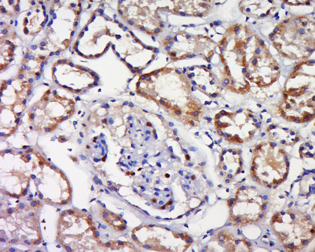
Tissue/cell: human kidney tissue; 4% Paraformaldehyde-fixed and paraffin-embedded;
Antigen retrieval: citrate buffer ( 0.01M, pH 6.0 ), Boiling bathing for 15min; Block endogenous peroxidase by 3% Hydrogen peroxide for 30min; Blocking buffer (normal goat serum,C-0005) at 37℃ for 20 min;
Incubation: Anti-Phospho-TAK1(Ser192)Polyclonal Antibody, Unconjugated(bs-5435R) 1:500, overnight at 4°C, followed by conjugation to the secondary antibody(SP-0023) and DAB(C-0010) staining
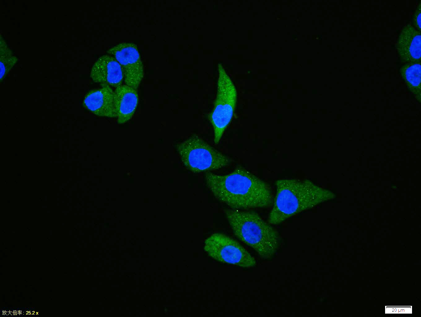
Hela cell; 4% Paraformaldehyde-fixed; Triton X-100 at room temperature for 20 min; Blocking buffer (normal goat serum, C-0005) at 37°C for 20 min; Antibody incubation with (Phospho-TAK1 (Ser192)) polyclonal Antibody, Unconjugated (bs-5435R) 1:100, 90 minutes at 37°C; followed by a conjugated Goat Anti-Rabbit IgG antibody at 37°C for 90 minutes, DAPI (blue, C02-04002) was used to stain the cell nuclei.
|
| 1、抗体溶解方法 | |
| 2、抗体修复方式 | |
| 3、常用试剂的配制 | |
| 4、免疫组化操作步骤 | |
| 5、免疫组化问题解答 | |
| 6、Western Blotting 操作步骤 | |
| 7、Western Blotting 问题解答 | |
| 8、关于肽链的设计 | |
| 9、多肽的溶解与保存 | |
| 10、酶标抗体效价测定程序 | |


