

| 产品编号 | bs-3345R |
| 英文名称 | phospho-PLK1 (Ser137) Rabbit pAb |
| 中文名称 | 磷酸化丝/苏氨酸蛋白激酶Plk1抗体 |
| 别 名 | PLK1 (p-S137); p-PLK1; phospho-PLK1; PLK; STPK13; PLK1_HUMAN; PLK1; Polo-like kinase 1 (PLK-1); Serine/threonine-protein kinase 13 (STPK13); 2.7.11.21; PLK1_MOUSE; PLK1_RAT; |
| 产品类型 | 磷酸化抗体 |
| 研究领域 | 肿瘤 细胞生物 信号转导 转录调节因子 激酶和磷酸酶 |
| 抗体来源 | Rabbit |
| 克隆类型 | Polyclonal |
| 克 隆 号 | |
| 交叉反应 | Human,Rat (predicted: Mouse,Rabbit,Pig,Cow,Chicken,Dog) |
| 产品应用 | WB=1:500-1000,IHC-P=1:100-500,IHC-F=1:100-500,IF=1:100-500,Flow-Cyt=2ug/Test
not yet tested in other applications. optimal dilutions/concentrations should be determined by the end user. |
| 理论分子量 | 68 kDa |
| 细胞定位 | 细胞核 细胞浆 |
| 性 状 | Liquid |
| 浓 度 | 1mg/ml |
| 免 疫 原 | KLH conjugated Synthesised phosphopeptide derived from human PLK1 around the phosphorylation site of Ser137: R(p-S)LL |
| 亚 型 | IgG |
| 纯化方法 | affinity purified by Protein A |
| 缓 冲 液 | 0.01M TBS (pH7.4) with 1% BSA, 0.02% Proclin300 and 50% Glycerol. |
| 保存条件 | Shipped at 4℃. Store at -20℃ for one year. Avoid repeated freeze/thaw cycles. |
| 注意事项 | This product as supplied is intended for research use only, not for use in human, therapeutic or diagnostic applications. |
| PubMed | PubMed |
| 产品介绍 |
PLK1 (polo-like kinase 1) is a member of the serine/threonine protein kinase family, cdc5/polo subfamily. PLK1 contains two polo box domains with a predicted molecular weight of 68 kDa. PLK1 has been shown to regulate cdc2/cyclin B through phosphorylation and activation of cdc25c phosphatase. PLK1 is modified by phosphorylation at Threonine 210. PLK1 may also be required for cell division. Depletion of PLK1 results in apoptosis and deregulation of expression of PKL1 is correlated with development of many malignancies. Function: Serine/threonine-protein kinase that performs several important functions throughout M phase of the cell cycle, including the regulation of centrosome maturation and spindle assembly, the removal of cohesins from chromosome arms, the inactivation of APC/C inhibitors, and the regulation of mitotic exit and cytokinesis. Required for recovery after DNA damage checkpoint and entry into mitosis. Required for kinetochore localization of BUB1B. Phosphorylates SGOL1. Required for spindle pole localization of isoform 3 of SGOL1 and plays a role in regulating its centriole cohesion function. Phosphorylates BORA, and thereby promotes the degradation of BORA. Contributes to the regulation of AURKA function. Regulates TP53 stability through phosphorylation of TOPORS. Phosphorylates NEDD1. NEDD1 phosphorylation promotes subsequent targeting of the gamma-tubulin ring complex (gTuRC) to the centrosome, an important step for spindle formation. Phosphorylates both ECT2 and RACGAP1, and thereby stimulates their interaction that is essential for the cleavage furrow formation. Promotes the central spindle recruitment of ECT2. Subunit: Interacts with CEP170 and EVI5. Interacts and phosphorylates ERCC6L. Interacts with FAM29A. Interacts with SLX4/BTBD12 and TTDN1. Interacts with BUB1B. Interacts (via POLO-box domain) with the phosphorylated form of BUB1, MLF1IP and CDC25C. Interacts with isoform 3 of SGOL1. Interacts with BORA, KIF2A and AURKA. Interacts with TOPORS and CYLD. Interacts with ECT2; the interaction is stimulated upon phosphorylation of ECT2 on 'Thr-444'. Interacts with PRC1. Subcellular Location: Nucleus. Chromosome, centromere, kinetochore. Cytoplasm, cytoskeleton, centrosome. Midbody. Note=During early stages of mitosis, the phosphorylated form is detected on centrosomes and kinetochores. Localizes to the outer kinetochore. Presence of SGOL1 and interaction with the phosphorylated form of BUB1 is required for the kinetochore localization. Tissue Specificity: Placenta and colon. Post-translational modifications: Catalytic activity is enhanced by phosphorylation of Thr-210. Phosphorylation at Thr-210 is first detected on centrosomes in the G2 phase of the cell cycle, peaks in prometaphase and gradually disappears from centrosomes during anaphase. Autophosphorylation and phosphorylation of Ser-137 may not be significant for the activation of PLK1 during mitosis, but may enhance catalytic activity during recovery after DNA damage checkpoint. Ubiquitinated by the anaphase promoting complex/cyclosome (APC/C) in anaphase and following DNA damage, leading to its degradation by the proteasome. Ubiquitination is mediated via its interaction with FZR1/CDH1. Ubiquitination and subsequent degradation prevents entry into mitosis and is essential to maintain an efficient G2 DNA damage checkpoint. Similarity: Belongs to the protein kinase superfamily. Ser/Thr protein kinase family. CDC5/Polo subfamily. Contains 2 POLO box domains. Contains 1 protein kinase domain. SWISS: P53350 Gene ID: 5347 Database links: Entrez Gene: 5347 Human Entrez Gene: 18817 Mouse Omim: 602098 Human SwissProt: P53350 Human SwissProt: Q07832 Mouse Unigene: 592049 Human Unigene: 16525 Mouse Unigene: 11034 Rat |
| 产品图片 |
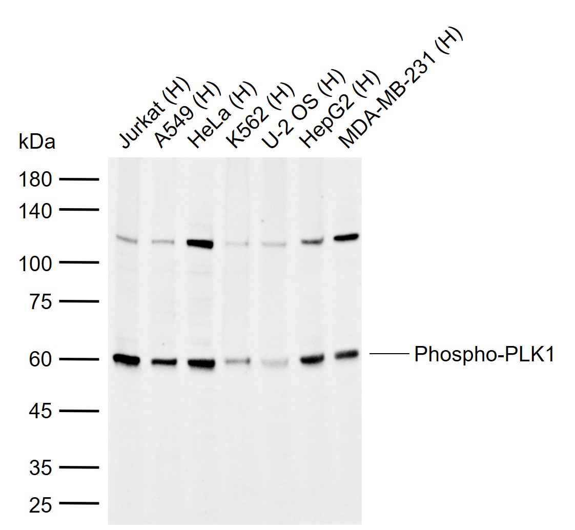
Sample:
Lane 1: Human Jurkat cell lysates
Lane 2: Human A549 cell lysates
Lane 3: Human HeLa cell lysates
Lane 4: Human K562 cell lysates
Lane 5: Human U-2 OS cell lysates
Lane 6: Human HepG2 cell lysates
Lane 7: Human MDA-MB-231 cell lysates
Primary: Anti-Phospho-PLK1 (Ser137) (bs-3345R) at 1/1000 dilution
Secondary: IRDye800CW Goat Anti-Rabbit IgG at 1/20000 dilution
Predicted band size: 68 kDa
Observed band size: 60 kDa
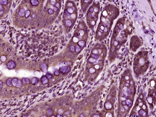
Paraformaldehyde-fixed, paraffin embedded (rat colon tissue); Antigen retrieval by boiling in sodium citrate buffer (pH6.0) for 15min; Block endogenous peroxidase by 3% hydrogen peroxide for 20 minutes; Blocking buffer (normal goat serum) at 37°C for 30min; Antibody incubation with (PLK1 (Ser137)) Polyclonal Antibody, Unconjugated (bs-3345R) at 1:400 overnight at 4°C, followed by operating according to SP Kit(Rabbit) (sp-0023) instructionsand DAB staining.
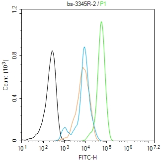
Blank control(black line):Hela.
Primary Antibody (green line): Rabbit Anti-Phospho-PLK1 (Ser137) antibody (bs-3345R)
Dilution:2ug/Test;
Secondary Antibody(white blue line): Goat anti-rabbit IgG-FITC
Dilution: 0.5ug/Test.
Isotype control(orange line): Normal Rabbit IgG
Protocol
The cells were fixed with 4% PFA (10min at room temperature)and then permeabilized with 90% ice-cold methanol for 20 min at -20℃, The cells were then incubated in 5%BSA to block non-specific protein-protein interactions for 30 min at room temperature .Cells stained with Primary Antibody for 30 min at room temperature. The secondary antibody used for 40 min at room temperature. Acquisition of 20,000 events was performed.
|
| 1、抗体溶解方法 | |
| 2、抗体修复方式 | |
| 3、常用试剂的配制 | |
| 4、免疫组化操作步骤 | |
| 5、免疫组化问题解答 | |
| 6、Western Blotting 操作步骤 | |
| 7、Western Blotting 问题解答 | |
| 8、关于肽链的设计 | |
| 9、多肽的溶解与保存 | |
| 10、酶标抗体效价测定程序 | |


