

| 产品编号 | bs-1610R |
| 英文名称 | CK7 Rabbit pAb |
| 中文名称 | 细胞角蛋白7抗体 |
| 别 名 | CK7; K2C7; K7; SCL; K2C7_HUMAN; KRT7; Cytokeratin-7 (CK-7); Keratin-7 (K7); Sarcolectin; Type-II keratin Kb7; |
 |
Specific References (9) | bs-1610R has been referenced in 9 publications.
|
| 研究领域 | 信号转导 |
| 抗体来源 | Rabbit |
| 克隆类型 | Polyclonal |
| 克 隆 号 | |
| 交叉反应 | Human,Mouse,Rat |
| 产品应用 | WB=1:500-2000,IHC-P=1:100-500,IHC-F=1:100-500,IF=1:100-500,Flow-Cyt=0.2µg/Test
not yet tested in other applications. optimal dilutions/concentrations should be determined by the end user. |
| 理论分子量 | 54 kDa |
| 检测分子量 | 54 |
| 细胞定位 | 细胞浆 |
| 性 状 | Liquid |
| 浓 度 | 1mg/ml |
| 免 疫 原 | KLH conjugated synthetic peptide derived from the middle of human CK7: 251-350/469 |
| 亚 型 | IgG |
| 纯化方法 | affinity purified by Protein A |
| 缓 冲 液 | 0.01M TBS (pH7.4) with 1% BSA, 0.02% Proclin300 and 50% Glycerol. |
| 保存条件 | Shipped at 4℃. Store at -20℃ for one year. Avoid repeated freeze/thaw cycles. |
| 注意事项 | This product as supplied is intended for research use only, not for use in human, therapeutic or diagnostic applications. |
| PubMed | PubMed |
| 产品介绍 |
The protein encoded by this gene is a member of the keratin gene family. The type II cytokeratins consist of basic or neutral proteins which are arranged in pairs of heterotypic keratin chains coexpressed during differentiation of simple and stratified epithelial tissues. This type II cytokeratin is specifically expressed in the simple epithelia lining the cavities of the internal organs and in the gland ducts and blood vessels. The genes encoding the type II cytokeratins are clustered in a region of chromosome 12q12-q13. Alternative splicing may result in several transcript variants; however, not all variants have been fully described. [provided by RefSeq, Jul 2008] Function: Blocks interferon-dependent interphase and stimulates DNA synthesis in cells. Involved in the translational regulation of the human papillomavirus type 16 E7 mRNA (HPV16 E7). Subunit: Heterotetramer of two type I and two type II keratins. Interacts with eukaryotic translation initiator factor 3 (eIF3) subunit EIF3S10 and with HPV16 E7. Subcellular Location: Cytoplasm. Tissue Specificity: Expressed in cultured epidermal, bronchial and mesothelial cells but absent in colon, ectocervix and liver. Observed throughout the glandular cells in the junction between stomach and esophagus but is absent in the esophagus. Post-translational modifications: Arg-20 is dimethylated, probably to asymmetric dimethylarginine. Similarity: Belongs to the intermediate filament family. SWISS: P08729 Gene ID: 3855 Database links: Entrez Gene: 3855 Human Omim: 148059 Human SwissProt: P08729 Human Unigene: 411501 Human Unigene: 670221 Human 结构蛋白(Structural Proteins) CK-7是一种 54KDa 的中间丝蛋白,存在于大多数正常组织的腺上皮和移行上皮细胞中。 该抗体与多种良/恶性上皮性肿瘤反应。腺癌中的卵巢、乳腺、肺的腺癌呈阳性反应,而胃肠道的腺癌阴性。移行细胞肿瘤、前列腺癌也呈阳性反应。通常认为 CK7是腺癌和移行上皮细胞癌的比较特异性的标志。 |
| 产品图片 |

Sample:
Skin (Mouse) Lysate at 40 ug
Primary: Anti-CK7 (bs-1610R) at 1/300 dilution
Secondary: IRDye800CW Goat Anti-Rabbit IgG at 1/20000 dilution
Predicted band size: 54 kD
Observed band size: 54 kD
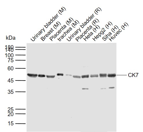
Sample:
Lane 1: Mouse Urinary bladder tissue lysates
Lane 2: Mouse Breast tissue lysates
Lane 3: Mouse Placenta tissue lysates
Lane 4: Mouse trachea tissue lysates
Lane 5: Rat Urinary bladder tissue lysates
Lane 6: Rat Placenta tissue lysates
Lane 7: Human Hela cell lysates
Lane 8: Human Hepg2 cell lysates
Lane 9: Human Siha cell lysates
Lane 10: Human Huvec cell lysates
Primary: Anti- CK7 (bs-1610R) at 1/1000 dilution
Secondary: IRDye800CW Goat Anti-Rabbit IgG at 1/20000 dilution
Predicted band size: 54 kDa
Observed band size: 54 kDa

Sample:
Bronchus (Mouse) Lysate at 40 ug
Primary: Anti-CK7 (bs-1610R) at 1/300 dilution
Secondary: IRDye800CW Goat Anti-Rabbit IgG at 1/20000 dilution
Predicted band size: 54 kD
Observed band size: 54 kD
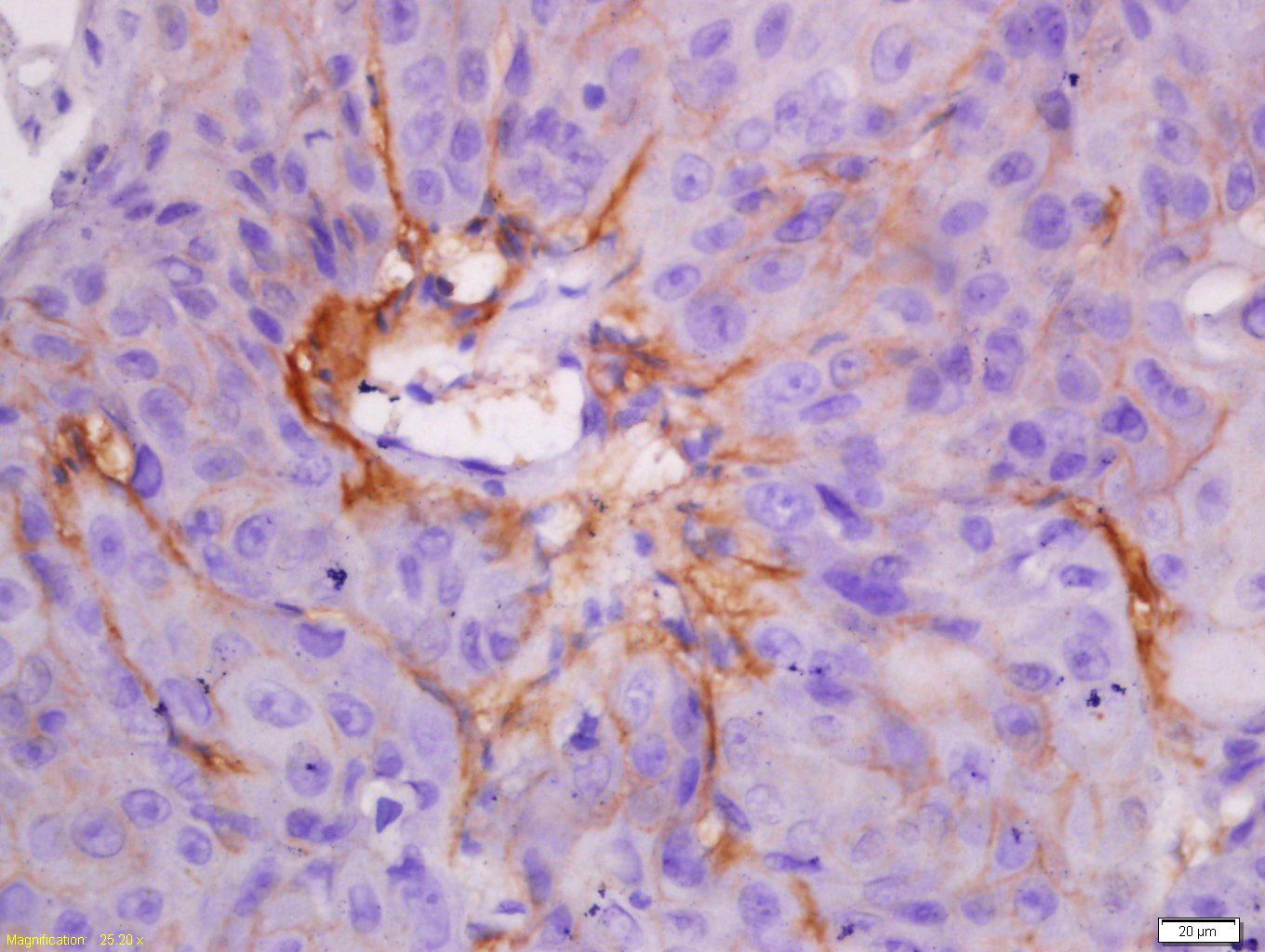
Tissue/cell: human laryngocarcinoma; 4% Paraformaldehyde-fixed and paraffin-embedded;
Antigen retrieval: citrate buffer ( 0.01M, pH 6.0 ), Boiling bathing for 15min; Block endogenous peroxidase by 3% Hydrogen peroxide for 30min; Blocking buffer (normal goat serum,C-0005) at 37℃ for 20 min;
Incubation: Anti-CK7 Polyclonal Antibody, Unconjugated(bs-1610R) 1:200, overnight at 4°C, followed by conjugation to the secondary antibody(SP-0023) and DAB(C-0010) staining
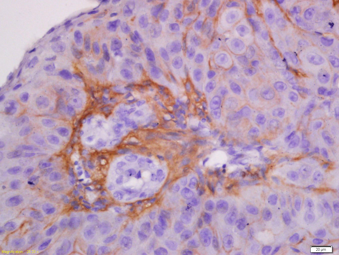
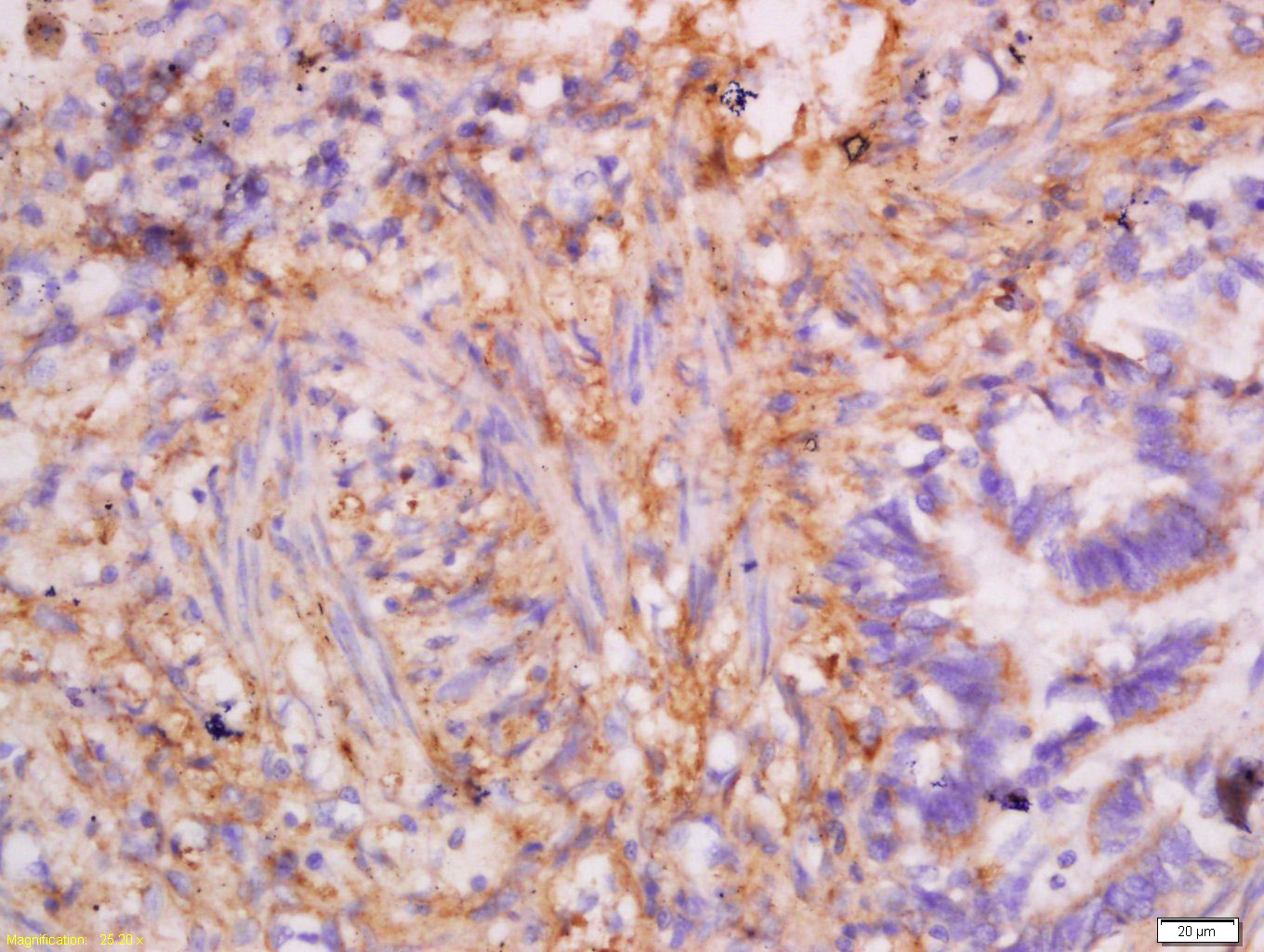
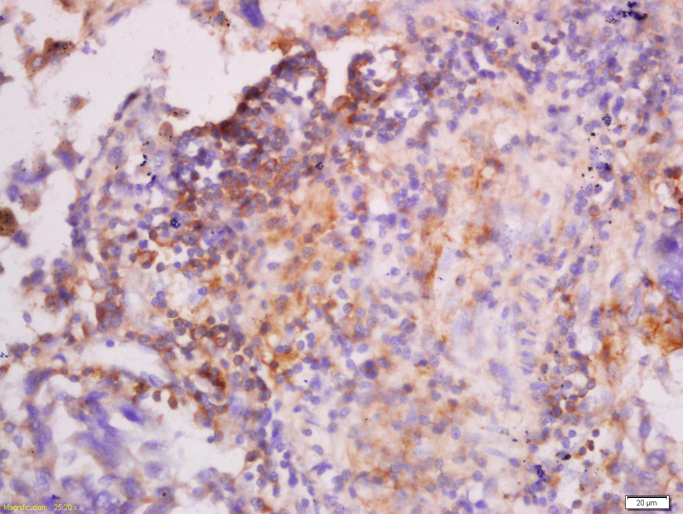
Tissue/cell: human lung carcinoma; 4% Paraformaldehyde-fixed and paraffin-embedded;
Antigen retrieval: citrate buffer ( 0.01M, pH 6.0 ), Boiling bathing for 15min; Block endogenous peroxidase by 3% Hydrogen peroxide for 30min; Blocking buffer (normal goat serum,C-0005) at 37℃ for 20 min;
Incubation: Anti-CK7 Polyclonal Antibody, Unconjugated(bs-1610R) 1:200, overnight at 4°C, followed by conjugation to the secondary antibody(SP-0023) and DAB(C-0010) staining

Tissue/cell: Human esophageal carcinoma; 4% Paraformaldehyde-fixed and paraffin-embedded;
Antigen retrieval: citrate buffer ( 0.01M, pH 6.0 ), Boiling bathing for 15min; Block endogenous peroxidase by 3% Hydrogen peroxide for 30min; Blocking buffer (normal goat serum,C-0005) at 37℃ for 20 min;
Incubation: Anti-CK7 Polyclonal Antibody, Unconjugated(bs-1610R) 1:200, overnight at 4°C, followed by conjugation to the secondary antibody(SP-0023) and DAB(C-0010) staining
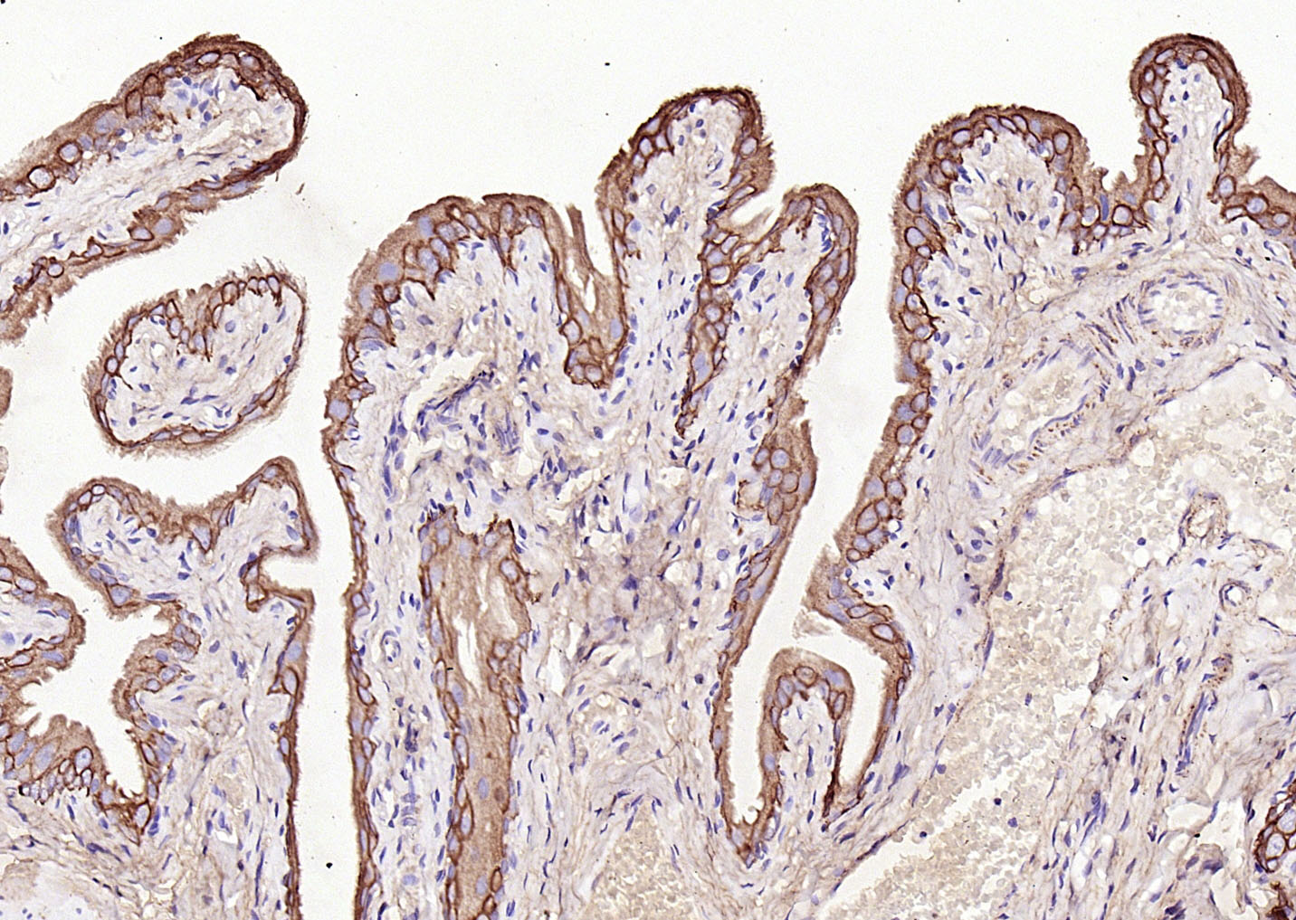
Paraformaldehyde-fixed, paraffin embedded (Rat bladder); Antigen retrieval by boiling in sodium citrate buffer (pH6.0) for 15min; Block endogenous peroxidase by 3% hydrogen peroxide for 20 minutes; Blocking buffer (normal goat serum) at 37°C for 30min; Antibody incubation with (CK7) Polyclonal Antibody, Unconjugated (bs-1610R) at 1:200 overnight at 4°C, followed by operating according to SP Kit(Rabbit) (sp-0023) instructionsand DAB staining.

Paraformaldehyde-fixed, paraffin embedded (Rat bladder); Antigen retrieval by boiling in sodium citrate buffer (pH6.0) for 15min; Block endogenous peroxidase by 3% hydrogen peroxide for 20 minutes; Blocking buffer (normal goat serum) at 37°C for 30min; Antibody incubation with (CK7) Polyclonal Antibody, Unconjugated (bs-1610R) at 1:200 overnight at 4°C, followed by operating according to SP Kit(Rabbit) (sp-0023) instructionsand DAB staining.

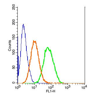
Blank control:Hela.
Primary Antibody (green line): Rabbit Anti-CK7 antibody (bs-1610R)
Dilution: 1ug/Test;
Secondary Antibody : Goat anti-rabbit IgG-FITC
Dilution: 0.5ug/Test.
Protocol
The cells were fixed with 4% PFA (10min at room temperature)and then permeabilized with 90% ice-cold methanol for 20 min at -20℃.The cells were then incubated in 5%BSA to block non-specific protein-protein interactions for 30 min at room temperature .Cells stained with Primary Antibody for 30 min at room temperature. The secondary antibody used for 40 min at room temperature. Acquisition of 20,000 events was performed.
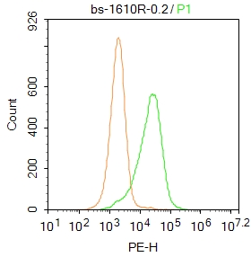
Blank control:A549.
Primary Antibody (green line): Rabbit Anti-CK7 antibody (bs-1610R)
Dilution: 1μg /10^6 cells;
Isotype Control Antibody (orange line): Rabbit IgG .
Secondary Antibody : Goat anti-rabbit IgG-PE
Dilution:0.2μg /test.
Protocol
The cells were fixed with 4% PFA (10min at room temperature)and then permeabilized with 20% PBST for 20 min at room temperature. The cells were then incubated in 5% BSA to block non-specific protein-protein interactions for 30 min at at room temperature .Cells stained with Primary Antibody for 30 min at room temperature. The secondary antibody used for 40 min at room temperature. Acquisition of 20,000 events was performed.
Blank control: Hepg2(blue)
Isotype Control Antibody: Rabbit IgG -FITC(orange);
Primary Antibody Dilution: 1μl in 100 μL1X PBS containing 0.5% BSA(green).
|
| 1、抗体溶解方法 | |
| 2、抗体修复方式 | |
| 3、常用试剂的配制 | |
| 4、免疫组化操作步骤 | |
| 5、免疫组化问题解答 | |
| 6、Western Blotting 操作步骤 | |
| 7、Western Blotting 问题解答 | |
| 8、关于肽链的设计 | |
| 9、多肽的溶解与保存 | |
| 10、酶标抗体效价测定程序 | |


