

| 产品编号 | bs-2189R |
| 英文名称 | UMOD Rabbit pAb |
| 中文名称 | 尿调节蛋白抗体 |
| 别 名 | ADMCKD2; ADTKD1; FJHN; HNFJ; HNFJ1; MCKD2; THGP; THP; UROM_HUMAN; UMOD; Tamm-Horsfall urinary glycoprotein (THP); |
 |
Specific References (1) | bs-2189R has been referenced in 1 publications.
[IF=1.679] Kim et al. Blocking junctional adhesion molecule C promotes the recovery of cisplatin-induced acute kidney injury. (2017) Korean.J.Intern.Me. 32:1053-1061 FCM ; Mouse.
|
| 研究领域 | 细胞生物 免疫学 |
| 抗体来源 | Rabbit |
| 克隆类型 | Polyclonal |
| 克 隆 号 | |
| 交叉反应 | Human,Mouse,Rat |
| 产品应用 | WB=1:500-2000,IHC-P=1:400-800,IHC-F=1:400-800,IF=1:100-500
not yet tested in other applications. optimal dilutions/concentrations should be determined by the end user. |
| 理论分子量 | 61/65 kDa |
| 细胞定位 | 细胞膜 分泌型蛋白 |
| 性 状 | Liquid |
| 浓 度 | 1mg/ml |
| 免 疫 原 | KLH conjugated synthetic peptide derived from mouse UMOD: 351-450/642 |
| 亚 型 | IgG |
| 纯化方法 | affinity purified by Protein A |
| 缓 冲 液 | 0.01M TBS (pH7.4) with 1% BSA, 0.02% Proclin300 and 50% Glycerol. |
| 保存条件 | Shipped at 4℃. Store at -20℃ for one year. Avoid repeated freeze/thaw cycles. |
| 注意事项 | This product as supplied is intended for research use only, not for use in human, therapeutic or diagnostic applications. |
| PubMed | PubMed |
| 产品介绍 |
The protein encoded by this gene is the most abundant protein in mammalian urine under physiological conditions. Its excretion in urine follows proteolytic cleavage of the ectodomain of its glycosyl phosphatidylinosital-anchored counterpart that is situated on the luminal cell surface of the loop of Henle. This protein may act as a constitutive inhibitor of calcium crystallization in renal fluids. Excretion of this protein in urine may provide defense against urinary tract infections caused by uropathogenic bacteria. Defects in this gene are associated with the renal disorders medullary cystic kidney disease-2 (MCKD2), glomerulocystic kidney disease with hyperuricemia and isosthenuria (GCKDHI), and familial juvenile hyperuricemic nephropathy (FJHN). Alternative splicing of this gene results in multiple transcript variants. [provided by RefSeq, Jul 2013]. Function: Uromodulin: Functions in biogenesis and organization of the apical membrane of epithelial cells of the thick ascending limb of Henle's loop (TALH), where it promotes formation of complex filamentous gel-like structure providing the water barrier permeability. May serve as a receptor for binding and endocytosis for cytokines (IL-1, IL-2) and TNF. Facilitates neutrophil migration across renal epithelial (By similarity). Uromodulin, secreted form: Secreted into urine after proteolytically cleaveage. Into the urine, may contribute to colloid osmotic pressure, retards passage of positively charged electrolytes, prevents urinary tract infection and modulates formation of supersaturated salts and their crystals. Subcellular Location: Apical cell membrane; Lipid-anchor, GPI-anchor (By similarity). Basolateral cell membrane; Lipid-anchor, GPI-anchor (By similarity). Cell projection, cilium membrane (By similarity). Note=Only a small fraction is sorts to the basolateral pole of tubular epithelial cells compared to apical localization (By similarity). Uromodulin, secreted form: Secreted (By similarity). Post-translational modifications: N-glycosylated. Similarity: Contains 3 EGF-like domains. Contains 1 ZP domain. Database links: Entrez Gene: 7369 Human Omim: 191845 Human SwissProt: P07911 Human Unigene: 654425 Human |
| 产品图片 |
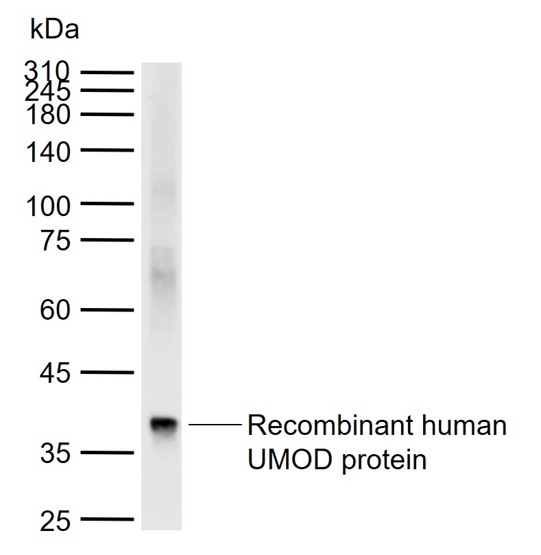
Sample:
Lane 1: Recombinant human UMOD protein, N-His
Primary: Anti-UMOD (bs-2189R) at 1/1000 dilution
Secondary: IRDye800CW Goat Anti-Rabbit IgG at 1/20000 dilution
Predicted band size: 69 kDa
Observed band size: 37 kDa
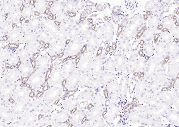
Paraformaldehyde-fixed, paraffin embedded Mouse Kidney; Antigen retrieval by boiling in sodium citrate buffer (pH6.0) for 15 min; Antibody incubation with UMOD Polyclonal Antibody, Unconjugated (bs-2189R) at 1:200 overnight at 4°C, followed by conjugation to the bs-0295G-HRP and DAB (C-0010) staining.
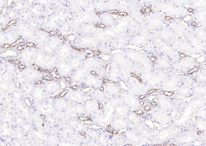
Paraformaldehyde-fixed, paraffin embedded Rat Kidney; Antigen retrieval by boiling in sodium citrate buffer (pH6.0) for 15 min; Antibody incubation with UMOD Polyclonal Antibody, Unconjugated (bs-2189R) at 1:200 overnight at 4°C, followed by conjugation to the bs-0295G-HRP and DAB (C-0010) staining.
|
| 1、抗体溶解方法 | |
| 2、抗体修复方式 | |
| 3、常用试剂的配制 | |
| 4、免疫组化操作步骤 | |
| 5、免疫组化问题解答 | |
| 6、Western Blotting 操作步骤 | |
| 7、Western Blotting 问题解答 | |
| 8、关于肽链的设计 | |
| 9、多肽的溶解与保存 | |
| 10、酶标抗体效价测定程序 | |


