

| 产品编号 | bs-0566R |
| 英文名称 | SMAD7 Rabbit pAb |
| 中文名称 | Smad7抗体 |
| 别 名 | Madh7; SMAD7_HUMAN; SMAD7; MAD homolog 7; Mothers against DPP homolog 7; Mothers against decapentaplegic homolog 8 (MAD homolog 8 | Mothers against DPP homolog 8); SMAD family member 7 (SMAD 7 | Smad7 | hSMAD7); MADH8; SMAD7_MOUSE; SMAD family member 7 (S |
 |
Specific References (18) | bs-0566R has been referenced in 18 publications.
|
| 研究领域 | 肿瘤 细胞生物 信号转导 细胞凋亡 生长因子和激素 激酶和磷酸酶 |
| 抗体来源 | Rabbit |
| 克隆类型 | Polyclonal |
| 克 隆 号 | |
| 交叉反应 | Human,Mouse,Rat (predicted: Pig,Cow) |
| 产品应用 | WB=1:500-2000,IHC-P=1:100-500,IHC-F=1:100-500,IF=1:100-500,Flow-Cyt=1ug/Test,ICC/IF=1:100-500
not yet tested in other applications. optimal dilutions/concentrations should be determined by the end user. |
| 理论分子量 | 46 kDa |
| 检测分子量 | 50 |
| 细胞定位 | 细胞核 细胞浆 |
| 性 状 | Liquid |
| 浓 度 | 1mg/ml |
| 免 疫 原 | KLH conjugated synthetic peptide derived from human Smad7: 1-100/426 |
| 亚 型 | IgG |
| 纯化方法 | affinity purified by Protein A |
| 缓 冲 液 | 0.01M TBS (pH7.4) with 1% BSA, 0.02% Proclin300 and 50% Glycerol. |
| 保存条件 | Shipped at 4℃. Store at -20℃ for one year. Avoid repeated freeze/thaw cycles. |
| 注意事项 | This product as supplied is intended for research use only, not for use in human, therapeutic or diagnostic applications. |
| PubMed | PubMed |
| 产品介绍 |
The protein encoded by this gene is a nuclear protein that binds the E3 ubiquitin ligase SMURF2. Upon binding, this complex translocates to the cytoplasm, where it interacts with TGF-beta receptor type-1 (TGFBR1), leading to the degradation of both the encoded protein and TGFBR1. Expression of this gene is induced by TGFBR1. Variations in this gene are a cause of susceptibility to colorectal cancer type 3 (CRCS3). Several transcript variants encoding different isoforms have been found for this gene. [provided by RefSeq, Jun 2010] Function: Antagonist of signaling by TGF-beta (transforming growth factor) type 1 receptor superfamily members; has been shown to inhibit TGF-beta (Transforming growth factor) and activin signaling by associating with their receptors thus preventing SMAD2 access. Functions as an adapter to recruit SMURF2 to the TGF-beta receptor complex. Also acts by recruiting the PPP1R15A-PP1 complex to TGFBR1, which promotes its dephosphorylation. Positively regulates PDPK1 kinase activity by stimulating its dissociation from the 14-3-3 protein YWHAQ which acts as a negative regulator. Subunit: Interacts with WWP1. Interacts with COPS5. Interacts with NEDD4L. Interacts with STAMBP. Interacts with RNF111, AXIN1 and AXIN2. Interacts with PPP1R15A. Interacts (via MH2 domain) with EP300. Interacts with ACVR1B, SMURF1, SMURF2 and TGFBR1; SMAD7 recruits SMURF1 and SMURF2 to the TGF-beta receptor and regulates its degradation. Interacts with PDPK1 (via PH domain). Subcellular Location: Nucleus. Cytoplasm. Note=Interaction with NEDD4L or RNF111 or induces translocation from the nucleus to the cytoplasm. TGF-beta stimulates its translocation from the nucleus to the cytoplasm. PDPK1 inhibits its translocation from the nucleus to the cytoplasm in response to TGF-beta. Tissue Specificity: Ubiquitous with higher expression in the lung and vascular endothelium. Post-translational modifications: Phosphorylation on Ser-249 does not affect its stability, nuclear localization or inhibitory function in TGFB signaling; however it affects its ability to regulate transcription. Phosphorylated by PDPK1. Ubiquitinated by WWP1 (By similarity). Polyubiquitinated by RNF111, which is enhanced by AXIN1 and promotes proteasomal degradation. In response to TGF-beta, ubiquitinated by SMURF1; which promotes its degradation. Acetylation prevents ubiquitination and degradation mediated by SMURF1. DISEASE: Genetic variations in SMAD7 influence susceptibility to colorectal cancer type 3 (CRCS3) [MIM:612229]. Colorectal cancer consists of tumors or cancer of either the colon or rectum or both. Cancers of the large intestine are the second most common form of cancer found in males and females. Symptoms include rectal bleeding, occult blood in stools, bowel obstruction and weight loss. Treatment is based largely on the extent of cancer penetration into the intestinal wall. Surgical cures are possible if the malignancy is confined to the intestine. Risk can be reduced when following a diet which is low in fat and high in fiber. Similarity: Belongs to the dwarfin/SMAD family. Contains 1 MH1 (MAD homology 1) domain. Contains 1 MH2 (MAD homology 2) domain. SWISS: O15105 Gene ID: 4092 Database links: Entrez Gene: 4092 Human Entrez Gene: 17131 Mouse Omim: 602932 Human SwissProt: O15105 Human SwissProt: O35253 Mouse Unigene: 465087 Human 转录调节因子(Transcriptin Regulators) Smad7是转化生长因子(TGF-β)信号通路的抑制分子,Smad7可干预MAPK信号通路,使ERK和JNK磷酸化活性的平衡失调,导致促增殖作用强于生长抑制作用,从而有助于细胞向恶性方向发展。 |
| 产品图片 |
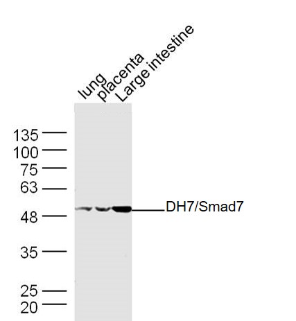
Sample:
Lung(Mouse) Lysate at 30 ug
Placenta(Mouse) Lysate at 30 ug
Large intestine(Mouse) Lysate at 30 ug
Primary: Anti- MADH7/Smad7 (bs-0566R) at 1/300 dilution
Secondary: IRDye800CW Goat Anti-Rabbit IgG at 1/20000 dilution
Predicted band size: 46 kD
Observed band size: 50 kD
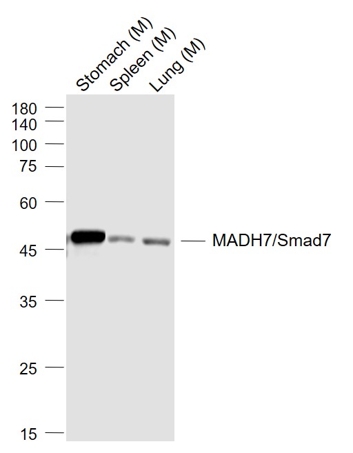
Sample:
Lane 1: Stomach (Mouse) Lysate at 40 ug
Lane 2: Spleen (Mouse) Lysate at 40 ug
Lane 3: Lung (Mouse) Lysate at 40 ug
Primary: Anti-MADH7/Smad7 (bs-0566R) at 1/1000 dilution
Secondary: IRDye800CW Goat Anti-Rabbit IgG at 1/20000 dilution
Predicted band size: 46 kD
Observed band size: 50 kD
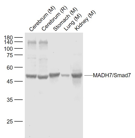
Sample:
Lane 1: Cerebrum (Mouse) Lysate at 40 ug
Lane 2: Cerebrum (Rat) Lysate at 40 ug
Lane 3: Stomach (Mouse) Lysate at 40 ug
Lane 4: Lung (Mouse) Lysate at 40 ug
Lane 5: Kidney (Mouse) Lysate at 40 ug
Primary: Anti-MADH7/Smad7 (bs-0566R) at 1/1000 dilution
Secondary: IRDye800CW Goat Anti-Rabbit IgG at 1/20000 dilution
Predicted band size: 46 kD
Observed band size: 50 kD
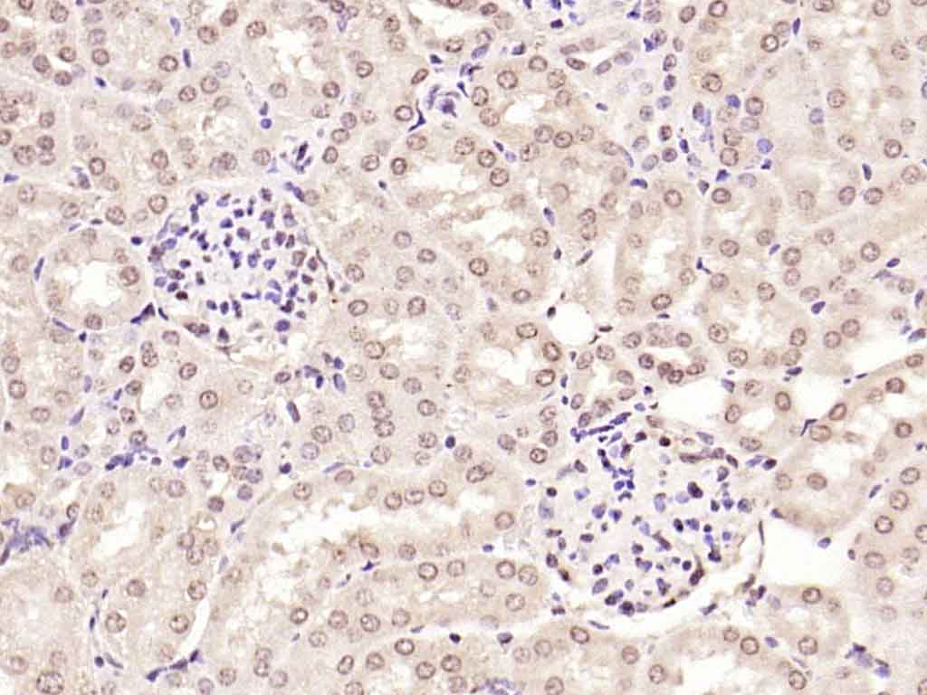
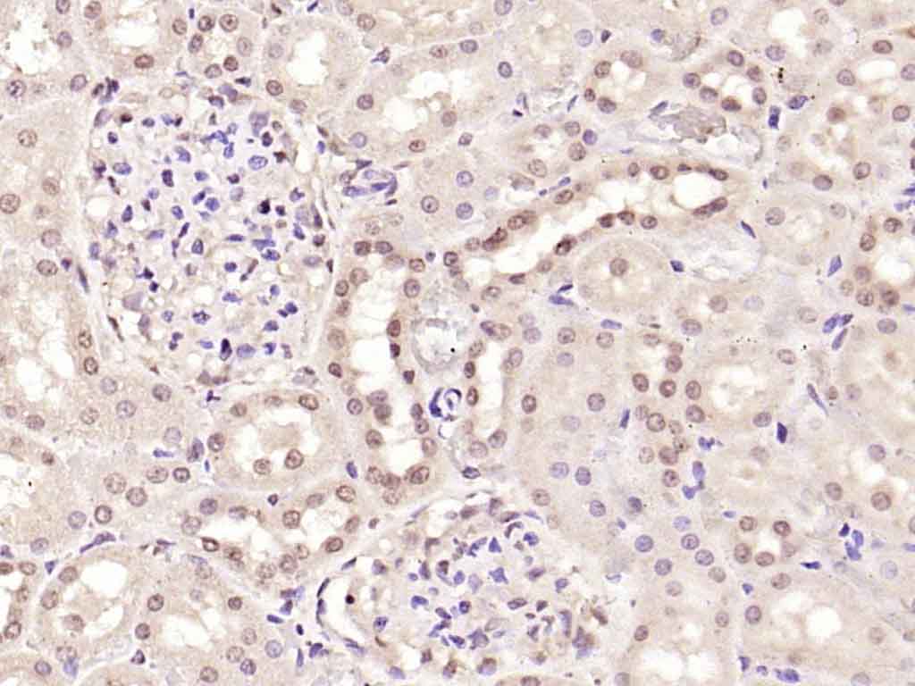
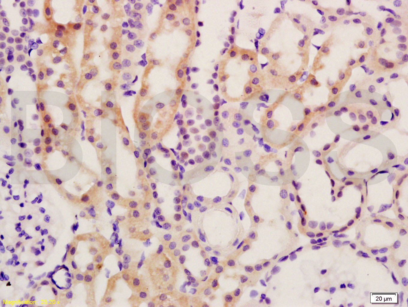
Paraformaldehyde-fixed, paraffin embedded (mouse kidney); Antigen retrieval by boiling in sodium citrate buffer (pH6.0) for 15min; Block endogenous peroxidase by 3% hydrogen peroxide for 20 minutes; Blocking buffer (normal goat serum) at 37°C for 30min; Antibody incubation with (MADH7) Polyclonal Antibody, Unconjugated (bs-0566R) at 1:200 overnight at 4°C, followed by operating according to SP Kit(Rabbit) (sp-0023) instructionsand DAB staining.
Paraformaldehyde-fixed, paraffin embedded (rat kidney); Antigen retrieval by boiling in sodium citrate buffer (pH6.0) for 15min; Block endogenous peroxidase by 3% hydrogen peroxide for 20 minutes; Blocking buffer (normal goat serum) at 37°C for 30min; Antibody incubation with (MADH7) Polyclonal Antibody, Unconjugated (bs-0566R) at 1:200 overnight at 4°C, followed by operating according to SP Kit(Rabbit) (sp-0023) instructionsand DAB staining.
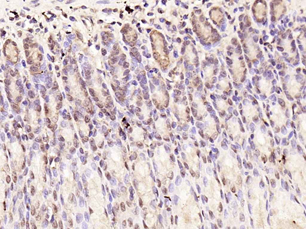
Paraformaldehyde-fixed, paraffin embedded (rat stomach); Antigen retrieval by boiling in sodium citrate buffer (pH6.0) for 15min; Block endogenous peroxidase by 3% hydrogen peroxide for 20 minutes; Blocking buffer (normal goat serum) at 37°C for 30min; Antibody incubation with (MADH7) Polyclonal Antibody, Unconjugated (bs-0566R) at 1:200 overnight at 4°C, followed by operating according to SP Kit(Rabbit) (sp-0023) instructionsand DAB staining.
Paraformaldehyde-fixed, paraffin embedded (rat lung); Antigen retrieval by boiling in sodium citrate buffer (pH6.0) for 15min; Block endogenous peroxidase by 3% hydrogen peroxide for 20 minutes; Blocking buffer (normal goat serum) at 37°C for 30min; Antibody incubation with (Smad7) Polyclonal Antibody, Unconjugated (bs-0566R) at 1:600 overnight at 4°C, followed by a conjugated secondary (sp-0023) for 20 minutes and DAB staining.
Tissue/cell: rat kidney tissue; 4% Paraformaldehyde-fixed and paraffin-embedded;
Antigen retrieval: citrate buffer ( 0.01M, pH 6.0 ), Boiling bathing for 15min; Block endogenous peroxidase by 3% Hydrogen peroxide for 30min; Blocking buffer (normal goat serum,C-0005) at 37℃ for 20 min;
Incubation: Anti-Smad7/Smad6 Polyclonal Antibody, Unconjugated(bs-0071R) 1:200, overnight at 4°C, followed by conjugation to the secondary antibody(SP-0023) and DAB(C-0010) staining
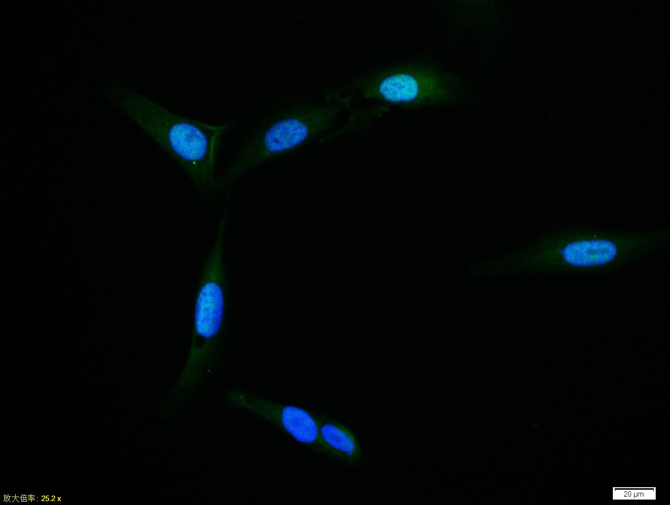
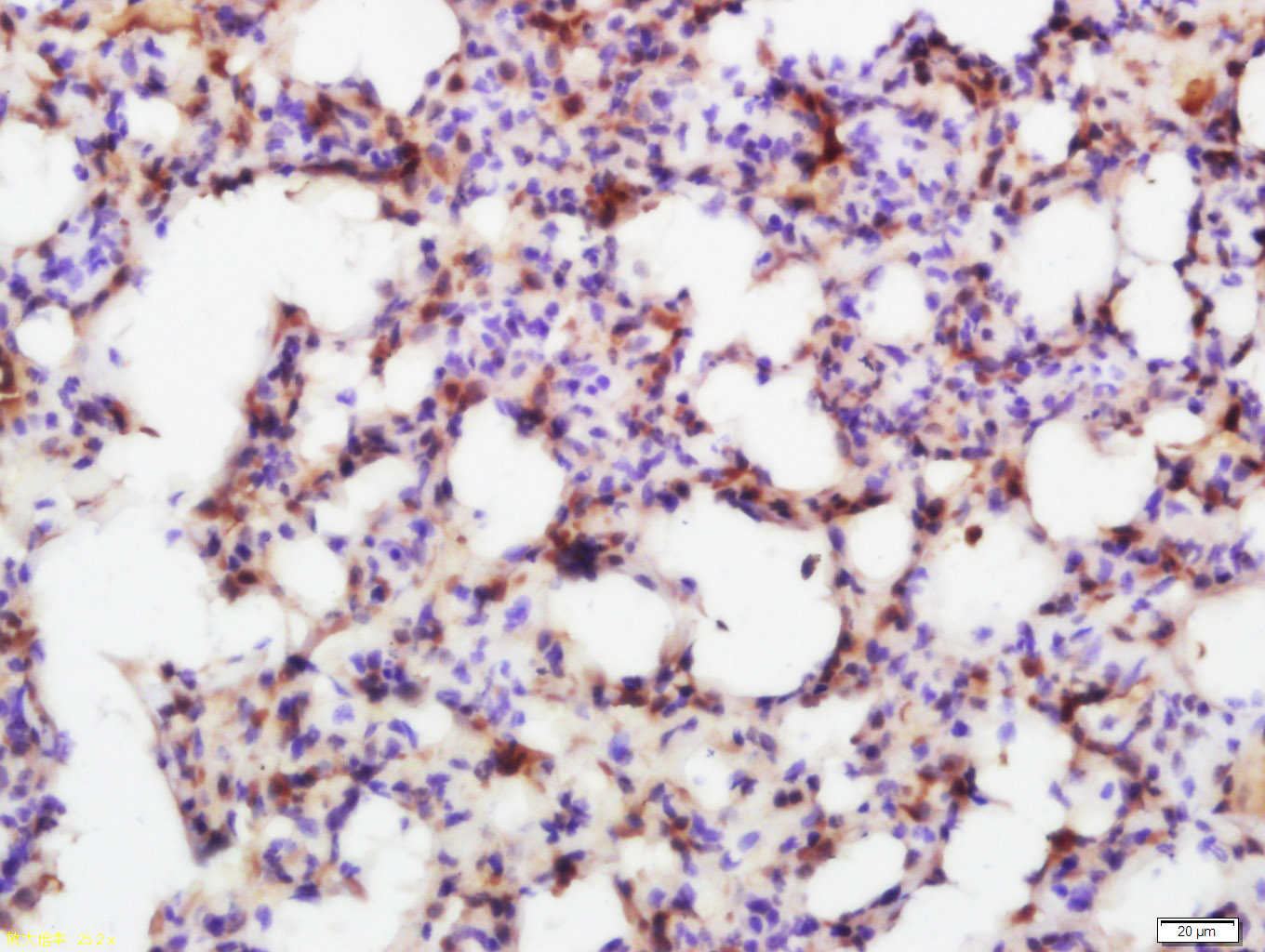
U-2OS cell; 4% Paraformaldehyde-fixed; Triton X-100 at room temperature for 20 min; Blocking buffer (normal goat serum, C-0005) at 37°C for 20 min; Antibody incubation with (MADH7/Smad7) polyclonal Antibody, Unconjugated (bs-0566R) 1:100, 90 minutes at 37°C; followed by a conjugated Goat Anti-Rabbit IgG antibody at 37°C for 90 minutes, DAPI (blue, C02-04002) was used to stain the cell nuclei.
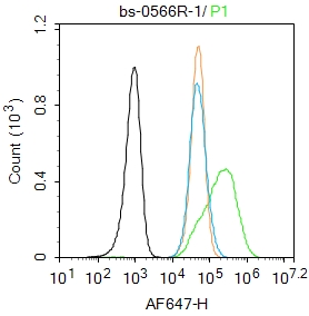
Blank control: SH-SY5Y.
Primary Antibody (green line): Rabbit Anti-MADH7/Smad7 antibody (bs-0566R)
Dilution: 1μg /10^6 cells;
Isotype Control Antibody (orange line): Rabbit IgG .
Secondary Antibody : Goat anti-rabbit IgG-AF647
Dilution: 1μg /test.
Protocol
The cells were fixed with 4% PFA (10min at room temperature)and then permeabilized with 90% ice-cold methanol for 20 min at-20℃.The cells were then incubated in 5%BSA to block non-specific protein-protein interactions for 30 min at room temperature .Cells stained with Primary Antibody for 30 min at room temperature. The secondary antibody used for 40 min at room temperature. Acquisition of 20,000 events was performed.
|
| 1、抗体溶解方法 | |
| 2、抗体修复方式 | |
| 3、常用试剂的配制 | |
| 4、免疫组化操作步骤 | |
| 5、免疫组化问题解答 | |
| 6、Western Blotting 操作步骤 | |
| 7、Western Blotting 问题解答 | |
| 8、关于肽链的设计 | |
| 9、多肽的溶解与保存 | |
| 10、酶标抗体效价测定程序 | |


