

| 产品编号 | bs-0530R |
| 英文名称 | PPAR gamma Rabbit pAb |
| 中文名称 | 过氧化酶活化增生受体γ抗体 |
| 别 名 | CIMT1; FPLD3; GLM1; NR1C3; PPARG1; PPARG2; PPARG5; PPARgamma; PPAR-gamma; PPAR-gamma2; PPARgamma2; PPARG_HUMAN; PPARG; Nuclear receptor subfamily 1 group C member 3; PPARG_MOUSE; PPARG_RAT; |
 |
Specific References (44) | bs-0530R has been referenced in 44 publications.
|
| 研究领域 | 免疫学 信号转导 转录调节因子 激酶和磷酸酶 糖尿病 内分泌病 |
| 抗体来源 | Rabbit |
| 克隆类型 | Polyclonal |
| 克 隆 号 | |
| 交叉反应 | Human,Mouse,Rat (predicted: Rabbit,Pig,Sheep,Cow,Zebrafish,Chicken) |
| 产品应用 | WB=1:500-2000,Flow-Cyt=1μg/Test,ICC/IF=1:100-500
not yet tested in other applications. optimal dilutions/concentrations should be determined by the end user. |
| 理论分子量 | 57 kDa |
| 检测分子量 | 52 |
| 细胞定位 | 细胞核 |
| 性 状 | Liquid |
| 浓 度 | 1mg/ml |
| 免 疫 原 | KLH conjugated synthetic peptide derived from human PPAR Gamma: 101-200/505 |
| 亚 型 | IgG |
| 纯化方法 | affinity purified by Protein A |
| 缓 冲 液 | 0.01M TBS (pH7.4) with 1% BSA, 0.02% Proclin300 and 50% Glycerol. |
| 保存条件 | Shipped at 4℃. Store at -20℃ for one year. Avoid repeated freeze/thaw cycles. |
| 注意事项 | This product as supplied is intended for research use only, not for use in human, therapeutic or diagnostic applications. |
| PubMed | PubMed |
| 产品介绍 |
This gene encodes a member of the peroxisome proliferator-activated receptor (PPAR) subfamily of nuclear receptors. PPARs form heterodimers with retinoid X receptors (RXRs) and these heterodimers regulate transcription of various genes. Three subtypes of PPARs are known: PPAR-alpha, PPAR-delta, and PPAR-gamma. The protein encoded by this gene is PPAR-gamma and is a regulator of adipocyte differentiation. Additionally, PPAR-gamma has been implicated in the pathology of numerous diseases including obesity, diabetes, atherosclerosis and cancer. Alternatively spliced transcript variants that encode different isoforms have been described. [provided by RefSeq, Jul 2008] Function: Receptor that binds peroxisome proliferators such as hypolipidemic drugs and fatty acids. Once activated by a ligand, the receptor binds to a promoter element in the gene for acyl-CoA oxidase and activates its transcription. It therefore controls the peroxisomal beta-oxidation pathway of fatty acids. Key regulator of adipocyte differentiation and glucose homeostasis. Subunit: Forms a heterodimer with the retinoic acid receptor RXRA called adipocyte-specific transcription factor ARF6. Interacts with NCOA6 coactivator, leading to a strong increase in transcription of target genes. Interacts with coactivator PPARBP, leading to a mild increase in transcription of target genes. Interacts with FAM120B. Interacts with PRDM16 (By similarity). Interacts with NOCA7 in a ligand-inducible manner. Interacts with NCOA1 LXXLL motifs. Interacts with TGFB1I1. Interacts with DNTTIP2. Interacts with PRMT2. Subcellular Location: Nucleus. Tissue Specificity: Highest expression in adipose tissue. Lower in skeletal muscle, spleen, heart and liver. Also detectable in placenta, lung and ovary. DISEASE: Note=Defects in PPARG can lead to type 2 insulin-resistant diabetes and hyptertension. PPARG mutations may be associated with colon cancer. Defects in PPARG may be associated with susceptibility to obesity (OBESITY) [MIM:601665]. It is a condition characterized by an increase of body weight beyond the limitation of skeletal and physical requirements, as the result of excessive accumulation of body fat. Defects in PPARG are the cause of familial partial lipodystrophy type 3 (FPLD3) [MIM:604367]. Familial partial lipodystrophies (FPLD) are a heterogeneous group of genetic disorders characterized by marked loss of subcutaneous (sc) fat from the extremities. Affected individuals show an increased preponderance of insulin resistance, diabetes mellitus and dyslipidemia. Genetic variations in PPARG can be associated with susceptibility to glioma type 1 (GLM1) [MIM:137800]. Gliomas are central nervous system neoplasms derived from glial cells and comprise astrocytomas, glioblastoma multiforme, oligodendrogliomas, and ependymomas. Note=Polymorphic PPARG alleles have been found to be significantly over-represented among a cohort of American patients with sporadic glioblastoma multiforme suggesting a possible contribution to disease susceptibility. Similarity: Belongs to the nuclear hormone receptor family. NR1 subfamily. Contains 1 nuclear receptor DNA-binding domain. SWISS: P37231 Gene ID: 5468 Database links: Entrez Gene: 5468 Human Entrez Gene: 19016 Mouse SwissProt: P37231 Human SwissProt: P37238 Mouse Unigene: 162646 Human Unigene: 3020 Mouse Unigene: 23443 Rat 类固醇受体(Steroid Receptors) 过氧化物酶体增殖物激活受体γ(PPARγ)主要存在于白色脂肪组织,PPARγ对于脂肪生成、血糖稳定、炎症反应、动脉粥样硬化和肿瘤等的发生都起到重要的作用。主要在脂肪细胞内表达。PPARγ是噻唑烷二酮类药物(TZDs)作用的药靶,又是脂肪细胞分化的重要调节因子。经研究发现,PPARγ在肥胖及胰岛素抵抗的发病机制中具有十分重要的意义,是治疗糖尿病、肥胖等代谢性疾病的重要药靶。 过氧化物酶体增殖物激活受体γ(PPARγ)属Ⅱ型核受体超家族成员,主要在脂肪细胞内表达。PPARγ是噻唑烷二酮类药物(TZDs)作用的药靶,又是脂肪细胞分化的重要调节因子。现有研究(包括一次于美国加州大学进行的研究)发现PPARγ在肥胖及胰岛素抵抗的发病机制中具有十分重要的意义,是治疗糖尿病、肥胖等代谢性疾病的重要药靶。目前,该受体蛋白质水平的筛选模式已经建立,并正在建立该受体的报告基因的细胞水平筛选评价模式。 |
| 产品图片 |
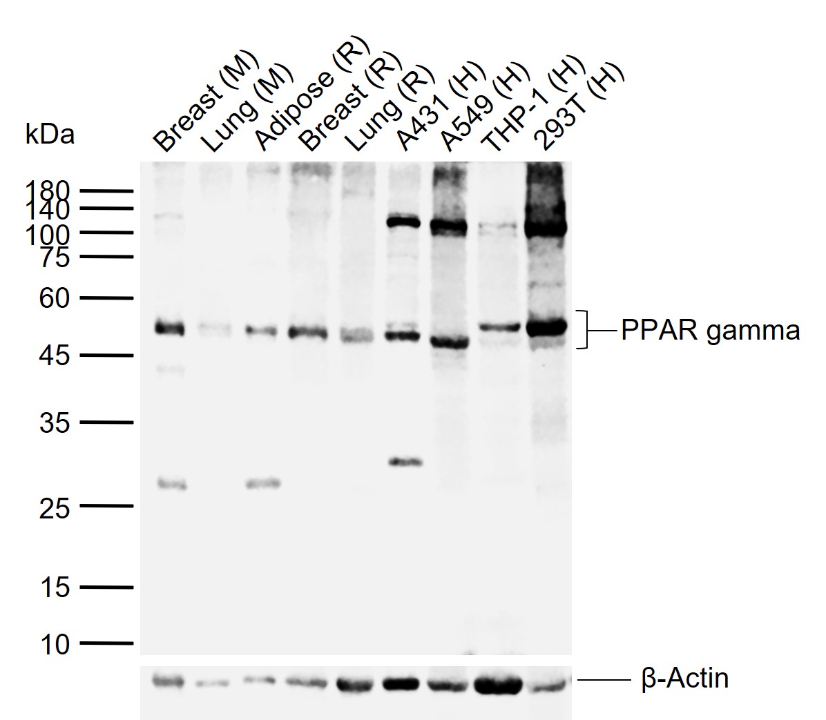
Sample:
Lane 1: Mouse Breast tissue lysates
Lane 2: Mouse Lung tissue lysates
Lane 3: Rat Adipose tissue lysates
Lane 4: Rat Breast tissue lysates
Lane 5: Rat Lung tissue lysates
Lane 6: Human A431 cell lysates
Lane 7: Human A549 cell lysates
Lane 8: Human THP-1 cell lysates
Lane 9: Human 293T cell lysates
Primary: Anti-PPAR gamma (bs-0530R) at 1/500 dilution
Secondary: IRDye800CW Goat Anti-Rabbit IgG at 1/20000 dilution
Predicted band size: 57 kDa
Observed band size: 52 kDa

Tissue/cell: mouse lung tissue; 4% Paraformaldehyde-fixed and paraffin-embedded;
Antigen retrieval: citrate buffer ( 0.01M, pH 6.0 ), Boiling bathing for 15min; Block endogenous peroxidase by 3% Hydrogen peroxide for 30min; Blocking buffer (normal goat serum,C-0005) at 37℃ for 20 min;
Incubation: Anti-PPAR Gamma Polyclonal Antibody, Unconjugated(bs-0530R) 1:200, overnight at 4°C, followed by conjugation to the secondary antibody(SP-0023) and DAB(C-0010) staining
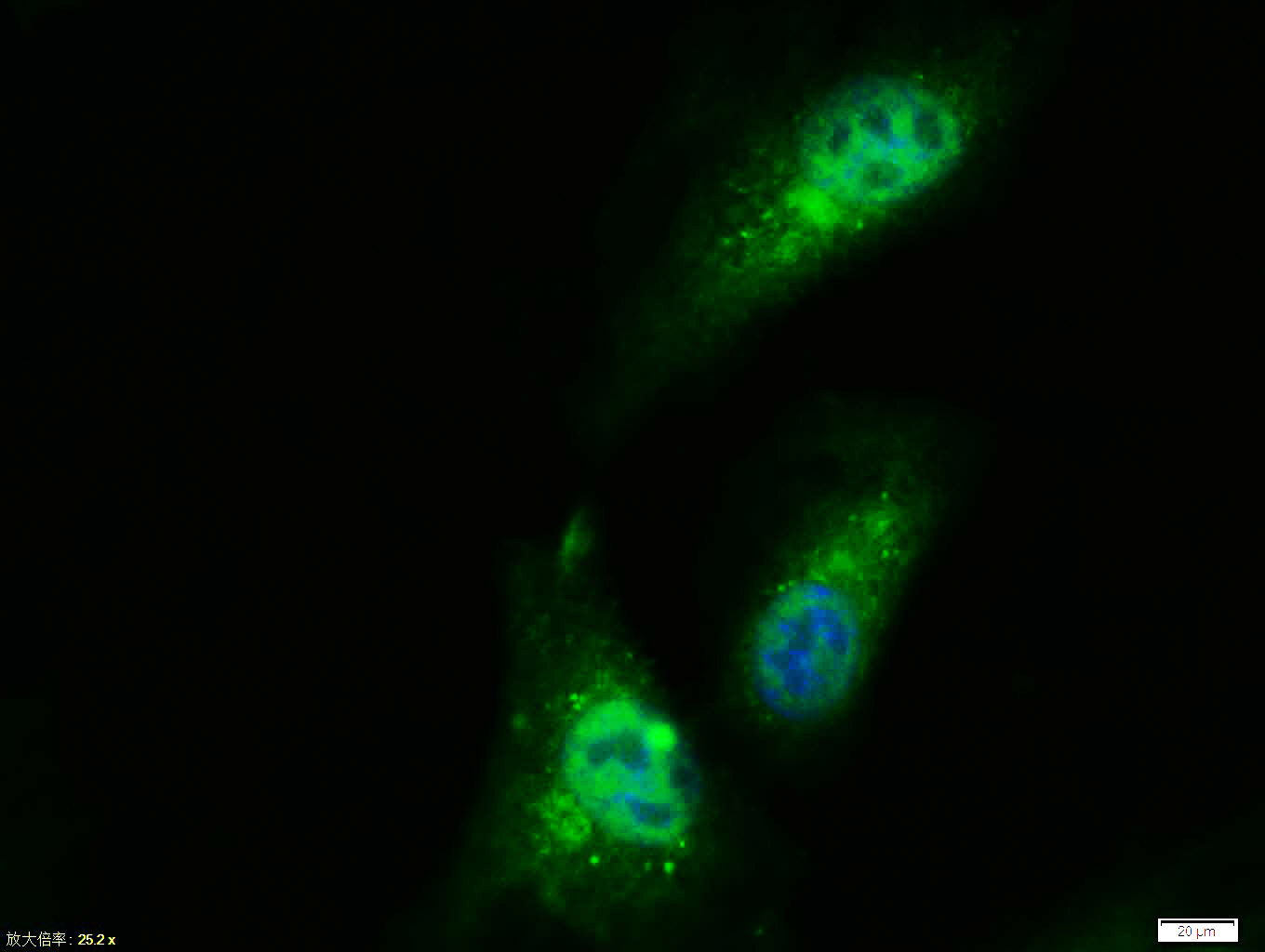
Tissue/cell: A549 cell; 4% Paraformaldehyde-fixed; Triton X-100 at room temperature for 20 min; Blocking buffer (normal goat serum, C-0005) at 37°C for 20 min; Antibody incubation with (PPAR gamma) polyclonal Antibody, Unconjugated (bs-0530R) 1:100, 90 minutes at 37°C; followed by a FITC conjugated Goat Anti-Rabbit IgG antibody at 37°C for 90 minutes, DAPI (blue, C02-04002) was used to stain the cell nuclei.

Blank control (blue line): U251
Primary Antibody (green line): Rabbit Anti-PPARG/PPAR gamma antibody (bs-0530R)
Dilution: 1μg /10^6 cells;
Isotype Control Antibody (orange line): Rabbit IgG .
Secondary Antibody (white blue line): Goat anti-rabbit IgG-FITC
Dilution: 1μg /test.
Protocol
The cells were fixed with 70% ethanol (Overnight at 4℃) and then permeabilized with 90% ice-cold methanol for 30 min on ice. Cells stained with Primary Antibody for 30 min at room temperature. The cells were then incubated in 1 X PBS/2%BSA/10% goat serum to block non-specific protein-protein interactions followed by the antibody for 15 min at room temperature. The secondary antibody used for 40 min at room temperature. Acquisition of 20,000 events was performed.

Blank control: A431.
Primary Antibody (green line): Rabbit Anti-PPAR gamma antibody (bs-0530R)
Dilution: 1μg /10^6 cells;
Isotype Control Antibody (orange line): Rabbit IgG .
Secondary Antibody : Goat anti-rabbit IgG-AF647
Dilution: 1μg /test.
Protocol
The cells were fixed with 4% PFA (10min at room temperature)and then permeabilized with 90% ice-cold methanol for 20 min at-20℃. The cells were then incubated in 5%BSA to block non-specific protein-protein interactions for 30 min at room temperature .Cells stained with Primary Antibody for 30 min at room temperature. The secondary antibody used for 40 min at room temperature. Acquisition of 20,000 events was performed.
|
| 1、抗体溶解方法 | |
| 2、抗体修复方式 | |
| 3、常用试剂的配制 | |
| 4、免疫组化操作步骤 | |
| 5、免疫组化问题解答 | |
| 6、Western Blotting 操作步骤 | |
| 7、Western Blotting 问题解答 | |
| 8、关于肽链的设计 | |
| 9、多肽的溶解与保存 | |
| 10、酶标抗体效价测定程序 | |


