

| 产品编号 | bs-0165R |
| 英文名称 | EGFR Rabbit pAb |
| 中文名称 | 表皮生长因子受体抗体 |
| 别 名 | ERBB; ERBB1; ERRP; HER1; NISBD2; NNCIS; PIG61; mENA; 9030024J15Rik; Errb1; Wa5; wa-2; wa2; ErbB-1; EGFR_HUMAN; EGFR; Proto-oncogene c-ErbB-1; Receptor tyrosine-protein kinase erbB-1; 2.7.10.1; EGFR_MOUSE; |
 |
Specific References (10) | bs-0165R has been referenced in 10 publications.
|
| 研究领域 | 肿瘤 细胞生物 免疫学 生长因子和激素 |
| 抗体来源 | Rabbit |
| 克隆类型 | Polyclonal |
| 克 隆 号 | |
| 交叉反应 | Human,Rat (predicted: Mouse,Pig,Dog) |
| 产品应用 | WB=1:500-2000,IHC-P=1:100-500,IHC-F=1:100-500,IF=1:100-500,Flow-Cyt=1μg/Test
not yet tested in other applications. optimal dilutions/concentrations should be determined by the end user. |
| 理论分子量 | 130 kDa |
| 检测分子量 | 170 |
| 细胞定位 | 细胞核 细胞浆 细胞膜 分泌型蛋白 |
| 性 状 | Liquid |
| 浓 度 | 1mg/ml |
| 免 疫 原 | KLH conjugated synthetic peptide derived from human EGFR: 951-1050/1210 <Cytoplasmic> |
| 亚 型 | IgG |
| 纯化方法 | affinity purified by Protein A |
| 缓 冲 液 | 0.01M TBS (pH7.4) with 1% BSA, 0.02% Proclin300 and 50% Glycerol. |
| 保存条件 | Shipped at 4℃. Store at -20℃ for one year. Avoid repeated freeze/thaw cycles. |
| 注意事项 | This product as supplied is intended for research use only, not for use in human, therapeutic or diagnostic applications. |
| PubMed | PubMed |
| 产品介绍 |
The protein encoded by this gene is a transmembrane glycoprotein that is a member of the protein kinase superfamily. This protein is a receptor for members of the epidermal growth factor family. EGFR is a cell surface protein that binds to epidermal growth factor. Binding of the protein to a ligand induces receptor dimerization and tyrosine autophosphorylation and leads to cell proliferation. Mutations in this gene are associated with lung cancer. Multiple alternatively spliced transcript variants that encode different protein isoforms have been found for this gene. [provided by RefSeq, Jul 2010] Function: Receptor tyrosine kinase binding ligands of the EGF family and activating several signaling cascades to convert extracellular cues into appropriate cellular responses. Known ligands include EGF, TGFA/TGF-alpha, amphiregulin, epigen/EPGN, BTC/betacellulin, epiregulin/EREG and HBEGF/heparin-binding EGF. Ligand binding triggers receptor homo- and/or heterodimerization and autophosphorylation on key cytoplasmic residues. The phosphorylated receptor recruits adapter proteins like GRB2 which in turn activates complex downstream signaling cascades. Activates at least 4 major downstream signaling cascades including the RAS-RAF-MEK-ERK, PI3 kinase-AKT, PLCgamma-PKC and STATs modules. May also activate the NF-kappa-B signaling cascade. Also directly phosphorylates other proteins like RGS16, activating its GTPase activity and probably coupling the EGF receptor signaling to the G protein-coupled receptor signaling. Also phosphorylates MUC1 and increases its interaction with SRC and CTNNB1/beta-catenin. Isoform 2 may act as an antagonist of EGF action. Subunit: Binding of the ligand triggers homo- and/or heterodimerization of the receptor triggering its autophosphorylation. Heterodimer with ERBB2. Interacts with ERRFI1; inhibits dimerization of the kinase domain and autophosphorylation. Part of a complex with ERBB2 and either PIK3C2A or PIK3C2B. Interacts with GRB2; an adapter protein coupling the receptor to downstream signaling pathways. Interacts with GAB2; involved in signaling downstream of EGFR. Interacts with STAT3; mediates EGFR downstream signaling in cell proliferation. Interacts with RIPK1; involved in NF-kappa-B activation. Interacts (autophosphorylated) with CBL; involved in EGFR ubiquitination and regulation. Interacts with SOCS5; regulates EGFR degradation through TCEB1- and TCEB2-mediated ubiquitination and proteasomal degradation. Interacts with PRMT5; methylates EGFR and enhances interaction with PTPN6. Interacts (phosphorylated) with PTPN6; inhibits EGFR-dependent activation of MAPK/ERK. Interacts with COPG; essential for regulation of EGF-dependent nuclear transport of EGFR by retrograde trafficking from the Golgi to the ER. Interacts with TNK2; this interaction is dependent on EGF stimulation and kinase activity of EGFR. Interacts with PCNA; positively regulates PCNA. Interacts with PELP1. Interacts with MUC1. Interacts with AP2M1. Interacts with FER. May interact with EPS8; mediates EPS8 phosphorylation. Interacts (via SH2 domains) with GRB2, NCK1 and NCK2. Subcellular Location: Cell membrane; Single-pass type I membrane protein. Endoplasmic reticulum membrane; Single-pass type I membrane protein. Golgi apparatus membrane; Single-pass type I membrane protein. Nucleus membrane; Single-pass type I membrane protein. Endosome. Endosome membrane. Note=In response to EGF, translocated from the cell membrane to the nucleus via Golgi and ER. Endocytosed upon activation by ligand. Isoform 2: Secreted. Tissue Specificity: Ubiquitously expressed. Isoform 2 is also expressed in ovarian cancers. Post-translational modifications: Phosphorylation at Ser-695 is partial and occurs only if Thr-693 is phosphorylated. Phosphorylation at Thr-678 and Thr-693 by PRKD1 inhibits EGF-induced MAPK8/JNK1 activation. Dephosphorylation by PTPRJ prevents endocytosis and stabilizes the receptor at the plasma membrane. Autophosphorylation at Tyr-1197 is stimulated by methylation at Arg-1199 and enhances interaction with PTPN6. Autophosphorylation at Tyr-1092 and/or Tyr-1110 recruits STAT3. Monoubiquitinated and polyubiquitinated upon EGF stimulation; which does not affect tyrosine kinase activity or signaling capacity but may play a role in lysosomal targeting. Polyubiquitin linkage is mainly through 'Lys-63', but linkage through 'Lys-48', 'Lys-11' and 'Lys-29' also occur. Methylated. Methylation at Arg-1199 by PRMT5 positively stimulates phosphorylation at Tyr-1197. DISEASE: Defects in EGFR are associated with lung cancer (LNCR) [MIM:211980]. LNCR is a common malignancy affecting tissues of the lung. The most common form of lung cancer is non-small cell lung cancer (NSCLC) that can be divided into 3 major histologic subtypes: squamous cell carcinoma, adenocarcinoma, and large cell lung cancer. NSCLC is often diagnosed at an advanced stage and has a poor prognosis. Similarity: Belongs to the protein kinase superfamily. Tyr protein kinase family. EGF receptor subfamily. Contains 1 protein kinase domain. SWISS: P00533 Gene ID: 1956 Database links: Entrez Gene: 1956 Human Entrez Gene: 13649 Mouse Omim: 131550 Human SwissProt: P00533 Human SwissProt: Q01279 Mouse Unigene: 488293 Human Unigene: 420648 Mouse Unigene: 439882 Mouse Unigene: 8534 Mouse Unigene: 37227 Rat 细胞膜受体(Membrane Receptors) EGFR-血管内皮生长因子受体EGFR是一类分子量为170kDa的糖蛋白,在生长的细胞包括中瘤细胞中跨越细胞质膜,表现有蛋白激酶活性。 EGFR是一种细胞膜受体激酶,对血管内皮生长因子有高度的亲和性,主要功能是参与血管内皮细胞生长和血管生成的调控,主要用于各种恶性肿瘤的研究. 与其配体表皮生长因子或尿抑胃素结合后则被激活,从而启动DNA及蛋白质的合成。再不进行有丝分裂的细胞中并不存在,但在胃中例外。 大量研究报道显示,EGFR高表达的肿瘤生存降低、转移风险增高、预后不良。在很多肿瘤中都存在着EGFR表达或过度表达。这些疾病包括:结直肠癌(CRC)、头颈部鳞状细胞癌(SCCHN)、乳腺癌、卵巢癌、宫颈癌、食道癌、胰腺癌、膀胱癌、前列腺癌和非小细胞肺癌等。研究表明,EGFR表达的肿瘤恶性程度增高、侵袭性强,这类肿瘤患者往往生存降低、转移风险增高、预后不良。 |
| 产品图片 |
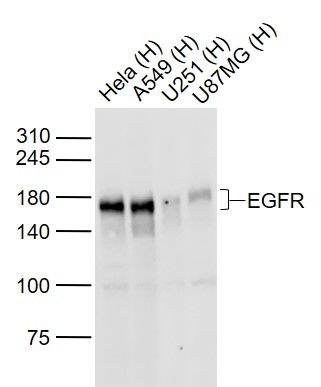
Sample:
Lane 1: Hela (Human) Cell Lysate at 30 ug
Lane 2: A549 (Human) Cell Lysate at 30 ug
Lane 3: U251 (Human) Cell Lysate at 30 ug
Lane 4: U87MG (Human) Cell Lysate at 30 ug
Primary: Anti-EGFR (bs-34018R) at 1/1000 dilution
Secondary: IRDye800CW Goat Anti-Rabbit IgG at 1/20000 dilution
Predicted band size: 170 kD
Observed band size: 170 kD
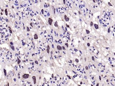
Paraformaldehyde-fixed, paraffin embedded (rat placenta); Antigen retrieval by boiling in sodium citrate buffer (pH6.0) for 15min; Block endogenous peroxidase by 3% hydrogen peroxide for 20 minutes; Blocking buffer (normal goat serum) at 37°C for 30min; Antibody incubation with (EGFR) Polyclonal Antibody, Unconjugated (bs-0165R) at 1:200 overnight at 4°C, followed by operating according to SP Kit(Rabbit) (sp-0023) instructionsand DAB staining.
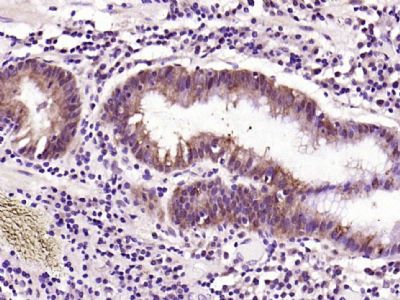
Paraformaldehyde-fixed, paraffin embedded (human gastric carcinoma); Antigen retrieval by boiling in sodium citrate buffer (pH6.0) for 15min; Block endogenous peroxidase by 3% hydrogen peroxide for 20 minutes; Blocking buffer (normal goat serum) at 37°C for 30min; Antibody incubation with (EGFR) Polyclonal Antibody, Unconjugated (bs-0165R) at 1:200 overnight at 4°C, followed by operating according to SP Kit(Rabbit) (sp-0023) instructionsand DAB staining.
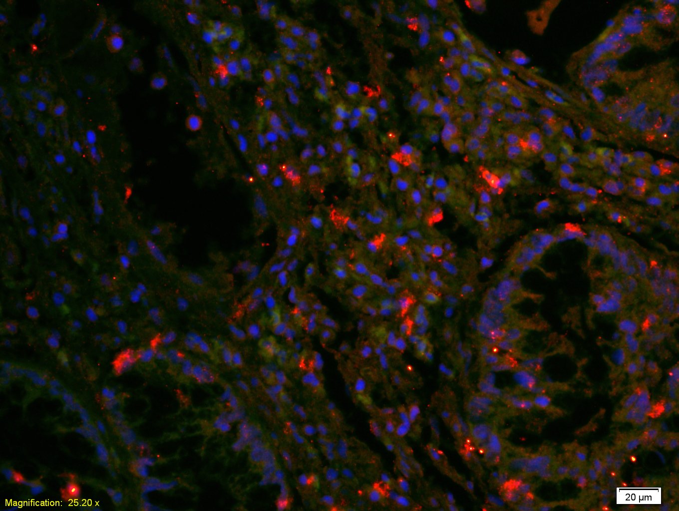
Tissue/cell: human rectal carcinoma;4% Paraformaldehyde-fixed and paraffin-embedded;
Antigen retrieval: citrate buffer ( 0.01M, pH 6.0 ), Boiling bathing for 15min; Blocking buffer (normal goat serum,C-0005) at 37℃ for 20 min;
Incubation: Anti-EGFR Polyclonal Antibody, Unconjugated(bs-0165R) 1:200, overnight at 4°C; The secondary antibody was Goat Anti-Rabbit IgG, Cy3 conjugated(bs-0295G-Cy3)used at 1:200 dilution for 40 minutes at 37°C. DAPI(5ug/ml,blue,C-0033) was used to stain the cell nuclei

Blank control: HUVEC cells(blue). Primary Antibody:Rabbit Anti-EGFR antibody(bs-0165R), Dilution: 1μg in 100 μL 1X PBS containing 0.5% BSA; Isotype Control Antibody: Rabbit IgG(orange) ,used under the same conditions ); Secondary Antibody: Goat anti-rabbit IgG-PE(white blue), Dilution: 1:200 in 1 X PBS containing 0.5% BSA.
Protocol
The cells were fixed with 2% paraformaldehyde (10 min) , then permeabilized with 90% ice-cold methanol for 30 min on ice. Primary antibody (bs-0165R,1μg /1x10^6 cells) were incubated for 30 min on the ice, followed by 1 X PBS containing 0.5% BSA + 10% goat serum (15 min) to block non-specific protein-protein interactions. Then the Goat Anti-rabbit IgG/PE antibody was added into the blocking buffer mentioned above to react with the primary antibody at 1/200 dilution for 30 min on ice. Acquisition of 20,000 events was performed.
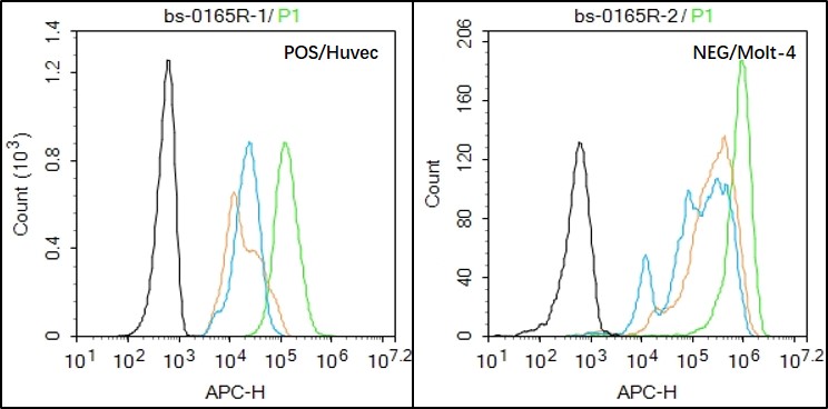
Black line : Positive blank control (HUVEC); Negative blank control (Molt-4)
Green line : Primary Antibody (Rabbit Anti-EGFR antibody (bs-0165R) )
Orange line:Isotype Control Antibody (Rabbit IgG) .
Blue line : Secondary Antibody (Goat anti-rabbit IgG-AF647)
HUVEC(Positive)and Molt-4(Negative control)cells (black) were fixed with 4% PFA for 10min at room temperature,permeabilized with 90% ice-cold methanol for 20 min at -20℃, and incubated in 5% BSA blocking buffer for 30 min at room temperature. Cells were then stained with EGFR Antibody(bs-0165R)at 1:50 dilution in blocking buffer and incubated for 30 min at room temperature, washed twice with 2% BSA in PBS, followed by secondary antibody(blue) incubation for 40 min at room temperature. Acquisitions of 20,000 events were performed. Cells stained with primary antibody (green), and isotype control (orange).
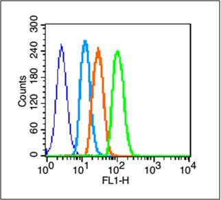
Blank control (blue line): A431 (blue).
Primary Antibody (green line): Rabbit Anti-EGFRantibody (bs-0165R)
Dilution: 3μg /10^6 cells;
Isotype Control Antibody (orange line): Rabbit IgG .
Secondary Antibody (white blue line): Goat anti-rabbit IgG-FITC
Dilution: 1μg /test.
Protocol
The cells were fixed with 2% paraformaldehyde (10 min) , then permeabilized with 90% ice-cold methanol for 30 min on ice. Cells stained with Primary Antibody for 30 min at room temperature. The cells were then incubated in 1 X PBS/2%BSA/10% goat serum to block non-specific protein-protein interactions followed by the antibody for 15 min at room temperature. The secondary antibody used for 40 min at room temperature. Acquisition of 20,000 events was performed.
|
| 1、抗体溶解方法 | |
| 2、抗体修复方式 | |
| 3、常用试剂的配制 | |
| 4、免疫组化操作步骤 | |
| 5、免疫组化问题解答 | |
| 6、Western Blotting 操作步骤 | |
| 7、Western Blotting 问题解答 | |
| 8、关于肽链的设计 | |
| 9、多肽的溶解与保存 | |
| 10、酶标抗体效价测定程序 | |


