

| 产品编号 | bs-0633R |
| 英文名称 | CDK4 Rabbit pAb |
| 中文名称 | 周期素依赖性激酶4抗体 |
| 别 名 | CMM3; PSK-J3; Crk3; CDK4_HUMAN; CDK4; Cell division protein kinase 4; 2.7.11.22; CDK4_MOUSE; CDK4_RAT; |
 |
Specific References (15) | bs-0633R has been referenced in 15 publications.
|
| 研究领域 | 肿瘤 细胞生物 染色质和核信号 信号转导 细胞周期蛋白 激酶和磷酸酶 |
| 抗体来源 | Rabbit |
| 克隆类型 | Polyclonal |
| 克 隆 号 | |
| 交叉反应 | Human,Mouse,Rat (predicted: Pig,Cow) |
| 产品应用 | WB=1:500-2000,IHC-P=1:100-500,IHC-F=1:100-500,IF=1:100-500,Flow-Cyt=1μg/Test,ICC/IF=1:100-500
not yet tested in other applications. optimal dilutions/concentrations should be determined by the end user. |
| 理论分子量 | 34 kDa |
| 检测分子量 | 34 |
| 细胞定位 | 细胞核 细胞浆 细胞膜 |
| 性 状 | Liquid |
| 浓 度 | 1mg/ml |
| 免 疫 原 | KLH conjugated synthetic peptide derived from human CDK4: 241-303/303 |
| 亚 型 | IgG |
| 纯化方法 | affinity purified by Protein A |
| 缓 冲 液 | 0.01M TBS (pH7.4) with 1% BSA, 0.02% Proclin300 and 50% Glycerol. |
| 保存条件 | Shipped at 4℃. Store at -20℃ for one year. Avoid repeated freeze/thaw cycles. |
| 注意事项 | This product as supplied is intended for research use only, not for use in human, therapeutic or diagnostic applications. |
| PubMed | PubMed |
| 产品介绍 |
The protein encoded by this gene is a member of the Ser/Thr protein kinase family. This protein is highly similar to the gene products of S. cerevisiae cdc28 and S. pombe cdc2. It is a catalytic subunit of the protein kinase complex that is important for cell cycle G1 phase progression. The activity of this kinase is restricted to the G1-S phase, which is controlled by the regulatory subunits D-type cyclins and CDK inhibitor p16(INK4a). This kinase was shown to be responsible for the phosphorylation of retinoblastoma gene product (Rb). Mutations in this gene as well as in its related proteins including D-type cyclins, p16(INK4a) and Rb were all found to be associated with tumorigenesis of a variety of cancers. Multiple polyadenylation sites of this gene have been reported. [provided by RefSeq, Jul 2008] Function: Ser/Thr-kinase component of cyclin D-CDK4 (DC) complexes that phosphorylate and inhibit members of the retinoblastoma (RB) protein family including RB1 and regulate the cell-cycle during G(1)/S transition. Phosphorylation of RB1 allows dissociation of the transcription factor E2F from the RB/E2F complexes and the subsequent transcription of E2F target genes which are responsible for the progression through the G(1) phase. Hypophosphorylates RB1 in early G(1) phase. Cyclin D-CDK4 complexes are major integrators of various mitogenenic and antimitogenic signals. Also phosphorylates SMAD3 in a cell-cycle-dependent manner and represses its transcriptional activity. Component of the ternary complex, cyclin D/CDK4/CDKN1B, required for nuclear translocation and activity of the cyclin D-CDK4 complex. Subunit: Component of the D-CDK4 complex, composed of CDK4 and some D-type G1 cyclin (CCND1, CCND2 or CCND3). Interacts directly in the complex with CCND1, CCND2 or CCND3. Interacts with SEI1 and ZNF655. Forms a ternary complex, cyclin D-CDK4-CDKN1B, involved in modulating CDK4 enzymatic activity. Interacts directly with CDKN1B (phosphorylated on 'Tyr-88' and 'Tyr-89'); the interaction allows assembly of the cyclin D-CDK4 complex, Thr-172 phosphorylation, nuclear translocation and enhances the cyclin D-CDK4 complex activity. CDK4 activity is either inhibited or enhanced depending on stoichiometry of complex. The non-tyrosine-phosphorylated form of CDKN1B prevents T-loop phosphorylation of CDK4 producing inactive CDK4. Interacts (unphosphorylated form) with CDK2. Also forms ternary complexes with CDKN1A or CDKN2A. Interacts directly with CDKN1A (via its N-terminal); the interaction promotes the assembly of the cyclin D-CDK4 complex, its nuclear translocation and promotes the cyclin D-dependent enzyme activity of CDK4. Subcellular Location: Cytoplasm. Nucleus. Membrane. Cytoplasmic when non-complexed. Forms a cyclin D-CDK4 complex in the cytoplasm as cells progress through G(1) phase. The complex accumulates on the nuclear membrane and enters the nucleus on transition from G(1) to S phase. Also present in nucleoli and heterochromatin lumps. Colocalizes with RB1 after release into the nucleus. Post-translational modifications: Phosphorylation at Thr-172 is required for enzymatic activity. Phosphorylated, in vitro, at this site by CCNH-CDK7, but, in vivo, appears to be phosphorylated by a proline-directed kinase. In the cyclin D-CDK4-CDKN1B complex, this phosphorylation and consequent CDK4 enzyme activity, is dependent on the tyrosine phosphorylation state of CDKN1B. Thus, in proliferating cells, CDK4 within the complex is phosphorylated on Thr-172 in the T-loop. In resting cells, phosphorylation on Thr-172 is prevented by the non-tyrosine-phosphorylated form of CDKN1B. DISEASE: Defects in CDK4 are a cause of susceptibility to cutaneous malignant melanoma type 3 (CMM3) [MIM:609048]. Malignant melanoma is a malignant neoplasm of melanocytes, arising de novo or from a pre-existing benign nevus, which occurs most often in the skin but also may involve other sites. Similarity: Belongs to the protein kinase superfamily. CMGC Ser/Thr protein kinase family. CDC2/CDKX subfamily. Contains 1 protein kinase domain. SWISS: P11802 Gene ID: 1019 Database links: Entrez Gene: 1019 Human Entrez Gene: 12567 Mouse Omim: 123829 Human SwissProt: P11802 Human SwissProt: P30285 Mouse Unigene: 95577 Human Unigene: 6839 Mouse Unigene: 6115 Rat Cdk4为周期素依赖激酶4,主要参与细胞周期的调控,在细胞分化、有丝分裂中起重要作用。目前主要用于各种肿瘤的研究。 |
| 产品图片 |
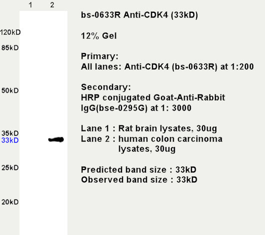
Sample:
Brain (Rat) Lysate at 30 ug
Colon carcinoma(Human) lysate at 30 ug
Primary: Anti- CDK4 (bs-0633R) at 1/200 dilution
Secondary: HRP conjugated Goat-Anti-rabbit IgG (bs-0295G-HRP) at 1/3000 dilution
Predicted band size: 34 kD
Observed band size: 34 kD
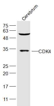
Sample:
Cerebrum (Mouse) Lysate at 40 ug
Primary: Anti-CDK4 (bs-0633R) at 1/300 dilution
Secondary: IRDye800CW Goat Anti-Rabbit IgG at 1/20000 dilution
Predicted band size: 34 kD
Observed band size: 34 kD
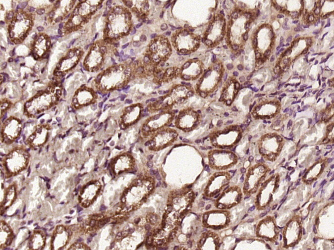
Paraformaldehyde-fixed, paraffin embedded (rat kidney tissue); Antigen retrieval by boiling in sodium citrate buffer (pH6.0) for 15min; Block endogenous peroxidase by 3% hydrogen peroxide for 20 minutes; Blocking buffer (normal goat serum) at 37°C for 30min; Antibody incubation with (CDK4) Polyclonal Antibody, Unconjugated (bs-0633R) at 1:400 overnight at 4°C, followed by operating according to SP Kit(Rabbit) (sp-0023) instructionsand DAB staining.

Paraformaldehyde-fixed, paraffin embedded (rat brain); Antigen retrieval by boiling in sodium citrate buffer (pH6.0) for 15min; Block endogenous peroxidase by 3% hydrogen peroxide for 20 minutes; Blocking buffer (normal goat serum) at 37°C for 30min; Antibody incubation with (CDK4) Polyclonal Antibody, Unconjugated (bs-0633R) at 1:200 overnight at 4°C, followed by operating according to SP Kit(Rabbit) (sp-0023) instructionsand DAB staining.
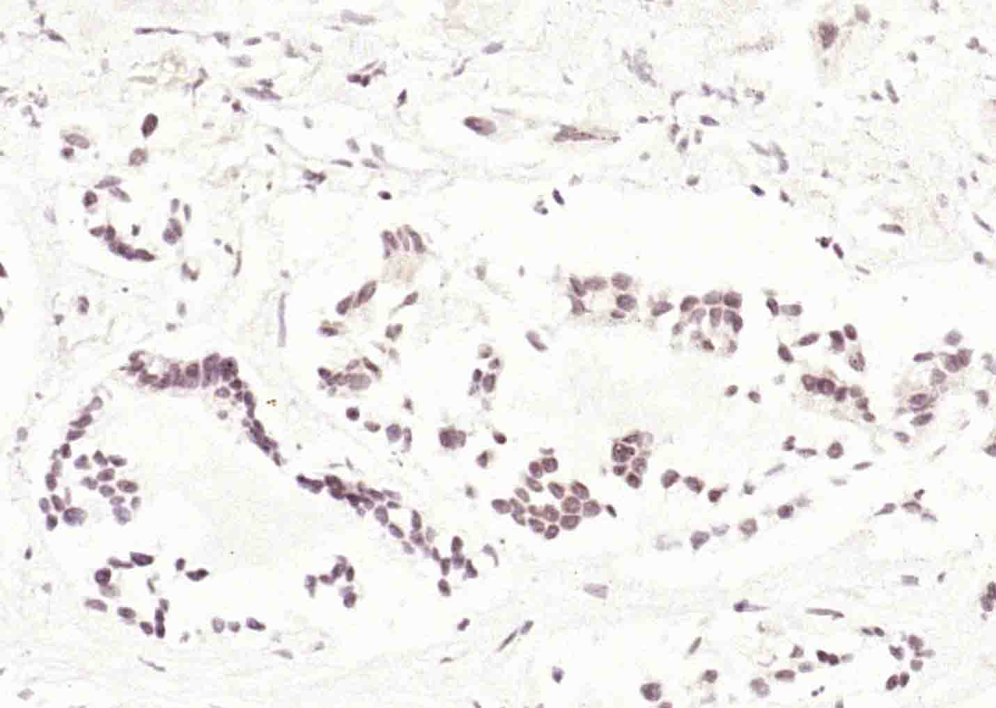
Paraformaldehyde-fixed, paraffin embedded (human gastric carcinoma); Antigen retrieval by boiling in sodium citrate buffer (pH6.0) for 15min; Block endogenous peroxidase by 3% hydrogen peroxide for 20 minutes; Blocking buffer (normal goat serum) at 37°C for 30min; Antibody incubation with (CDK4) Polyclonal Antibody, Unconjugated (bs-0633R) at 1:200 overnight at 4°C, followed by operating according to SP Kit(Rabbit) (sp-0023) instructionsand DAB staining.
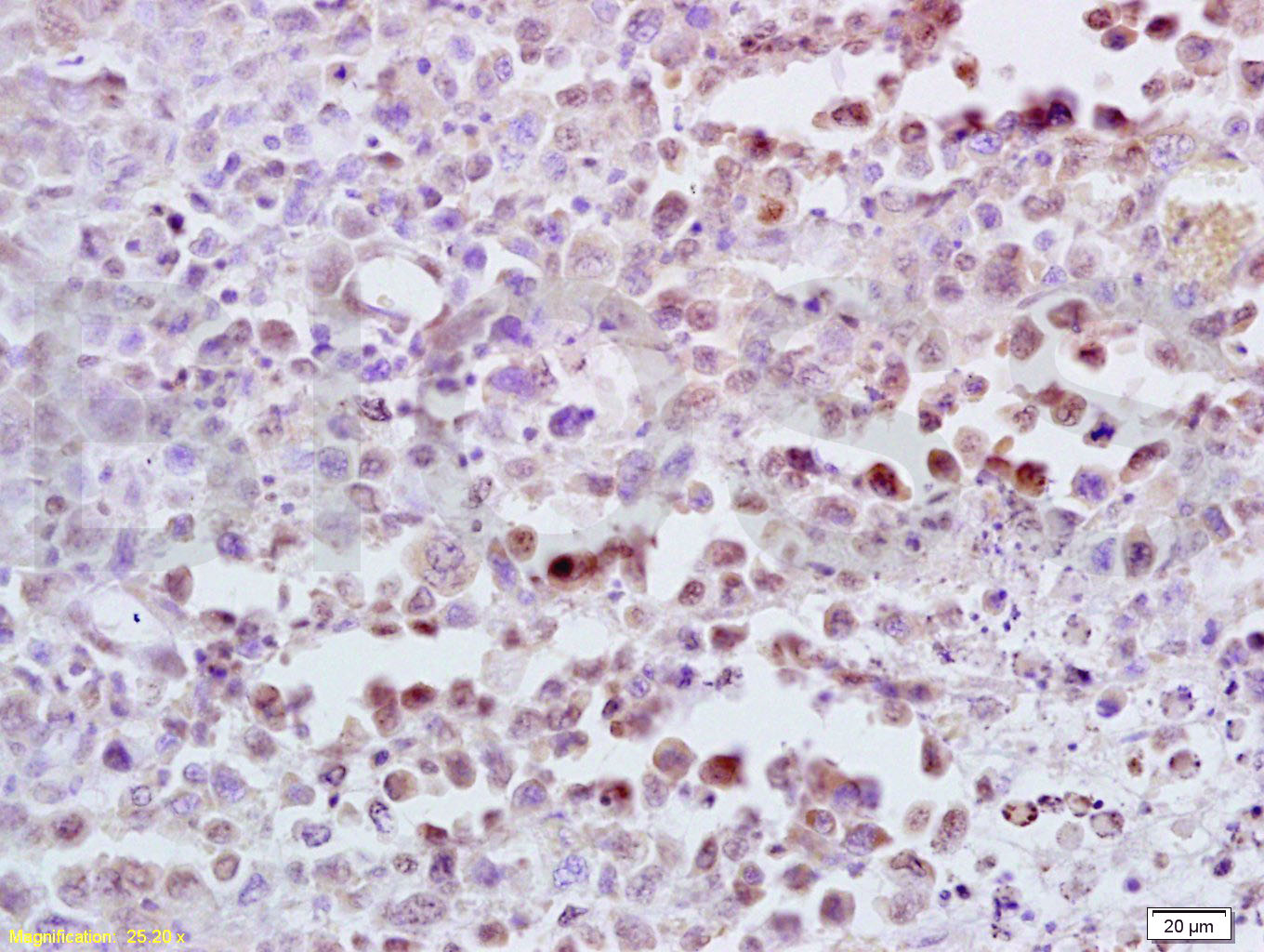
Tissue/cell: mouse lymphoma tissue; 4% Paraformaldehyde-fixed and paraffin-embedded;
Antigen retrieval: citrate buffer ( 0.01M, pH 6.0 ), Boiling bathing for 15min; Block endogenous peroxidase by 3% Hydrogen peroxide for 30min; Blocking buffer (normal goat serum,C-0005) at 37℃ for 20 min;
Incubation: Anti-CDK4 Polyclonal Antibody, Unconjugated(bs-0633R) 1:300, overnight at 4°C, followed by conjugation to the secondary antibody(SP-0023) and DAB(C-0010) staining
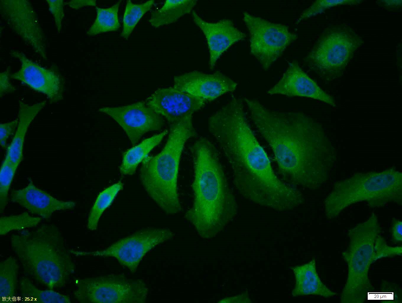
Tissue/cell:Hela cell; 4% Paraformaldehyde-fixed; Triton X-100 at room temperature for 20 min; Blocking buffer (normal goat serum,C-0005) at 37°C for 20 min; Antibody incubation with (CDK4) polyclonal Antibody, Unconjugated (bs-0633R) 1:100, 90 minutes at 37°C; followed by a FITC conjugated Goat Anti-Rabbit IgG antibody at 37°C for 90 minutes, DAPI (blue, C02-04002) was used to stain the cell nuclei.
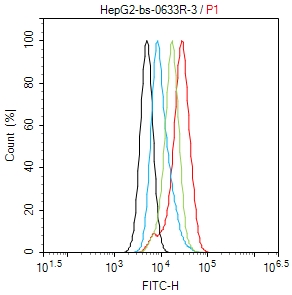
Blank control (black line): HepG2(black) (The cells were fixed with 2% paraformaldehyde (10 min) , then permeabilized with PBST for 30 min on room temperature)
Primary Antibody (Red line): Rabbit Anti-CDK4 antibody (bs-0633R) ;
Dilution: 1μg /10^6 cells;
Isotype Control Antibody (green line): Rabbit IgG .
Secondary Antibody (white blue line): Goat anti-rabbit IgG-FITC;Dilution: 1μg /test.
|
| 1、抗体溶解方法 | |
| 2、抗体修复方式 | |
| 3、常用试剂的配制 | |
| 4、免疫组化操作步骤 | |
| 5、免疫组化问题解答 | |
| 6、Western Blotting 操作步骤 | |
| 7、Western Blotting 问题解答 | |
| 8、关于肽链的设计 | |
| 9、多肽的溶解与保存 | |
| 10、酶标抗体效价测定程序 | |


