

| 产品编号 | bs-0522R |
| 英文名称 | CD45 Rabbit pAb |
| 中文名称 | 白细胞共同抗原CD45抗体 |
| 别 名 | B220; CD45; CD45R; GP180; IMD105; L-CA; LCA; LY5; T200; Ly-5; Lyt-4; loc; RT7; PTPRC_HUMAN; PTPRC; Leukocyte common antigen (L-CA); 3.1.3.48; PTPRC_MOUSE; Lymphocyte antigen 5 (Ly-5); PTPRC_RAT; protein tyrosine phosphatase receptor type C; leukocyte-common antigen; glycoprotein 180 |
 |
Specific References (20) | bs-0522R has been referenced in 20 publications.
|
| 研究领域 | 细胞生物 免疫学 神经生物学 信号转导 干细胞 转录调节因子 细胞表面分子 糖蛋白 细胞类型标志物 自然杀伤细胞 淋巴细胞 t-淋巴细胞 b-淋巴细胞 细胞膜蛋白 |
| 抗体来源 | Rabbit |
| 克隆类型 | Polyclonal |
| 交叉反应 | Human |
| 产品应用 | WB=1:500-2000,IHC-P=1:100-800,IHC-F=1:100-800,IF=1:100-800,ICC/IF=1:50-200
not yet tested in other applications. optimal dilutions/concentrations should be determined by the end user. |
| 理论分子量 | 143kDa |
| 检测分子量 | 220 |
| 细胞定位 | 细胞膜 |
| 性 状 | Liquid |
| 浓 度 | 1mg/ml |
| 免 疫 原 | KLH conjugated synthetic peptide derived from human CD45: 1210-1304/1304 <Cytoplasmic> |
| 亚 型 | IgG |
| 纯化方法 | affinity purified by Protein A |
| 缓 冲 液 | 0.01M TBS (pH7.4) with 1% BSA, 0.02% Proclin300 and 50% Glycerol. |
| 保存条件 | Shipped at 4℃. Store at -20℃ for one year. Avoid repeated freeze/thaw cycles. |
| 注意事项 | This product as supplied is intended for research use only, not for use in human, therapeutic or diagnostic applications. |
| PubMed | PubMed |
| 产品介绍 |
The protein encoded by this gene is a member of the protein tyrosine phosphatase (PTP) family. PTPs are known to be signaling molecules that regulate a variety of cellular processes including cell growth, differentiation, mitotic cycle, and oncogenic transformation. This PTP contains an extracellular domain, a single transmembrane segment and two tandem intracytoplasmic catalytic domains, and thus belongs to receptor type PTP. This gene is specifically expressed in hematopoietic cells. This PTP has been shown to be an essential regulator of T- and B-cell antigen receptor signaling. It functions through either direct interaction with components of the antigen receptor complexes, or by activating various Src family kinases required for the antigen receptor signaling. This PTP also suppresses JAK kinases, and thus functions as a regulator of cytokine receptor signaling. Four alternatively spliced transcripts variants of this gene, which encode distinct isoforms, have been reported. [provided by RefSeq, Jul 2008]. Function: Protein tyrosine-protein phosphatase required for T-cell activation through the antigen receptor. Acts as a positive regulator of T-cell coactivation upon binding to DPP4. The first PTPase domain has enzymatic activity, while the second one seems to affect the substrate specificity of the first one. Upon T-cell activation, recruits and dephosphorylates SKAP1 and FYN. Dephosphorylates LYN, and thereby modulates LYN activity. Subunit: Binds GANAB and PRKCSH. Interacts with SKAP1. Interacts with DPP4; the interaction is enhanced in a interleukin-12-dependent manner in activated lymphocytes. Contains 2 tyrosine-protein phosphatase domains. Subcellular Location: Membrane; Single-pass type I membrane protein. Membrane raft. Note=Colocalized with DPP4 in membrane rafts. Post-translational modifications: Heavily N- and O-glycosylated. DISEASE: Defects in PTPRC are a cause of severe combined immunodeficiency autosomal recessive T-cell-negative/B-cell-positive/NK-cell-positive (T(-)B(+)NK(+) SCID) [MIM:608971]. A form of severe combined immunodeficiency (SCID), a genetically and clinically heterogeneous group of rare congenital disorders characterized by impairment of both humoral and cell-mediated immunity, leukopenia, and low or absent antibody levels. Patients present in infancy recurrent, persistent infections by opportunistic organisms. The common characteristic of all types of SCID is absence of T-cell-mediated cellular immunity due to a defect in T-cell development. Genetic variations in PTPRC are involved in multiple sclerosis susceptibility (MS) [MIM:126200]. MS is a neurodegenerative disorder characterized by the gradual accumulation of focal plaques of demyelination particularly in the periventricular areas of the brain. Peripheral nerves are not affected. Onset usually in third or fourth decade with intermittent progression over an extended period. The cause is still uncertain. Similarity: Belongs to the protein-tyrosine phosphatase family. Receptor class 1/6 subfamily. Contains 2 fibronectin type-III domains. Contains 2 tyrosine-protein phosphatase domains. SWISS: P08575 Gene ID: 5788 Database links: Entrez Gene: 5788 Human Entrez Gene: 19264 Mouse Omim: 151460 Human SwissProt: P08575 Human SwissProt: P06800 Mouse Unigene: 654514 Human Unigene: 391573 Mouse Unigene: 90166 Rat CD45在活化信号转导中起到调节作用 在确定CD45为一种PTPase之前就已证实了CD45参于细胞的活化和生长调节。 抗CD45抗体可以抑制PHA或CD3交联所介导的T细胞增殖,还可抑制NK或细胞毒性T细胞对靶细胞的杀伤,抑制经CD2、CD3以及CD8膜分子介导的信号转导作用。 白细胞共同抗原是五种或更多的高分子量糖蛋白组成的蛋白家族,主要位于白细胞表面,包括T、B淋巴细胞、多形核白细胞、单核细胞等,而在红细胞、血小板及非造血系统中不表达。因此是区分淋巴瘤/白血病和非造血组织肿瘤(如未分化小细胞癌、小圆细胞肉瘤)的特异性标记物。该抗体主要用于淋巴瘤和未分化小细胞癌的鉴别诊断。 |
| 产品图片 |

25 ug total protein per lane of various lysates (see on figure) probed with CD45 polyclonal antibody, unconjugated (bs-0522R) at 1:1000 dilution and 4°C overnight incubation. Followed by conjugated secondary antibody incubation at r.t. for 60 min.
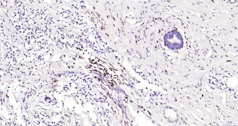
Paraformaldehyde-fixed, paraffin embedded Human Breast Cancer; Antigen retrieval by boiling in sodium citrate buffer (pH6.0) for 15 min; Antibody incubation with CD45 Polyclonal Antibody, Unconjugated (bs-0522R) at 1:200 overnight at 4°C, followed by conjugation to the bs-0295G-HRP and DAB (C-0010) staining.
Paraformaldehyde-fixed, paraffin embedded Human Thymus; Antigen retrieval by boiling in sodium citrate buffer (pH6.0) for 15 min; Antibody incubation with CD45 Polyclonal Antibody, Unconjugated (bs-0522R) at 1:200 overnight at 4°C, followed by conjugation to the bs-0295G-HRP and DAB (C-0010) staining.
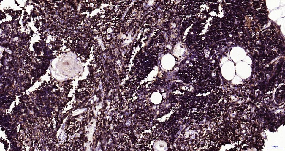
Paraformaldehyde-fixed, paraffin embedded Human Spleen; Antigen retrieval by boiling in sodium citrate buffer (pH6.0) for 15 min; Antibody incubation with CD45 Polyclonal Antibody, Unconjugated (bs-0522R) at 1:200 overnight at 4°C, followed by conjugation to the bs-0295G-HRP and DAB (C-0010) staining.
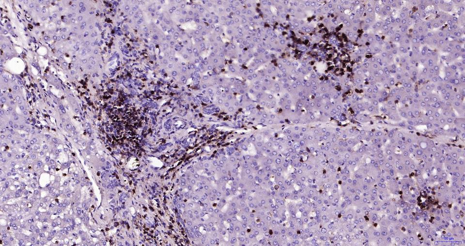
Paraformaldehyde-fixed, paraffin embedded Human Liver; Antigen retrieval by boiling in sodium citrate buffer (pH6.0) for 15 min; Antibody incubation with CD45 Polyclonal Antibody, Unconjugated (bs-0522R) at 1:200 overnight at 4°C, followed by conjugation to the bs-0295G-HRP and DAB (C-0010) staining.
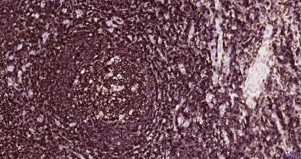
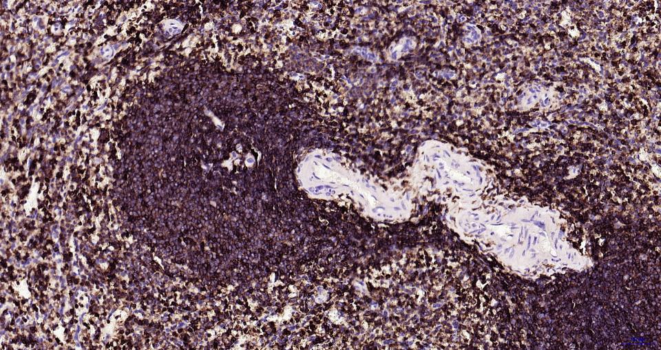
Paraformaldehyde-fixed, paraffin embedded Human Tonsil; Antigen retrieval by boiling in sodium citrate buffer (pH6.0) for 15 min; Antibody incubation with CD45 Polyclonal Antibody, Unconjugated (bs-0522R) at 1:200 overnight at 4°C, followed by conjugation to the bs-0295G-HRP and DAB (C-0010) staining.
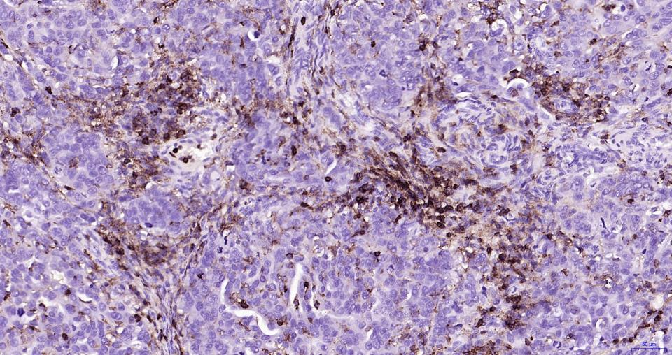
Paraformaldehyde-fixed, paraffin embedded Human Ovarian Cancer; Antigen retrieval by boiling in sodium citrate buffer (pH6.0) for 15 min; Antibody incubation with CD45 Polyclonal Antibody, Unconjugated (bs-0522R) at 1:200 overnight at 4°C, followed by conjugation to the bs-0295G-HRP and DAB (C-0010) staining.
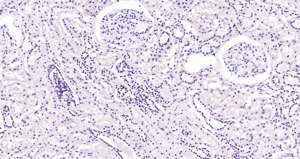
(Negative control) Paraformaldehyde-fixed, paraffin embedded Human Kidney; Antigen retrieval by boiling in sodium citrate buffer (pH6.0) for 15 min; Antibody incubation with CD45 Polyclonal Antibody, Unconjugated (bs-0522R) at 1:200 overnight at 4°C, followed by conjugation to the bs-0295G-HRP and DAB (C-0010) staining.
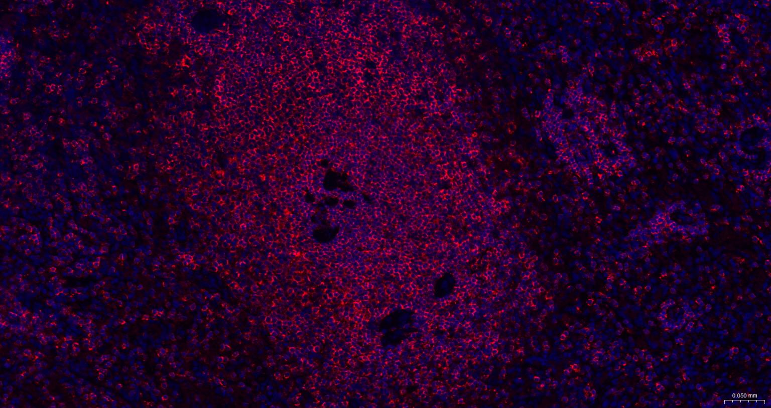
Paraformaldehyde-fixed, paraffin embedded Human Spleen; Antigen retrieval by boiling in sodium citrate buffer (pH6.0) for 15 min; The section was incubated with CD45 Polyclonal Antibody, Unconjugated (bs-0522R) at 1:200 overnight at 4°C. Followed by conjugated Goat Anti-Rabbit IgG antibody (Red, bs-0295G-BF594), DAPI (blue, C02-04002) was used to stain the cell nuclei.
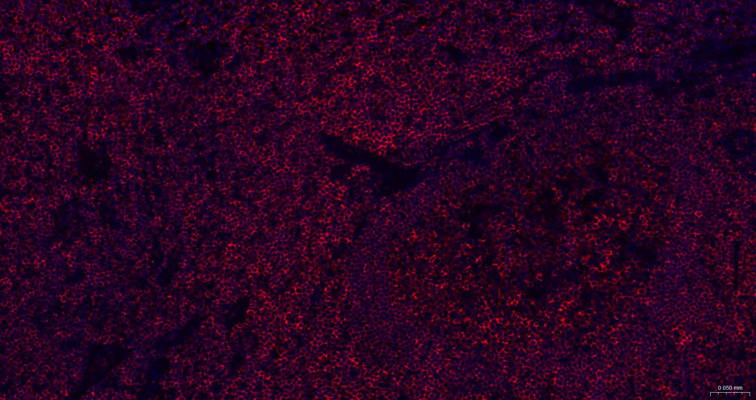
Paraformaldehyde-fixed, paraffin embedded Human Tonsil; Antigen retrieval by boiling in sodium citrate buffer (pH6.0) for 15 min; The section was incubated with CD45 Polyclonal Antibody, Unconjugated (bs-0522R) at 1:200 overnight at 4°C. Followed by conjugated Goat Anti-Rabbit IgG antibody (Red, bs-0295G-BF594), DAPI (blue, C02-04002) was used to stain the cell nuclei.
|
| 1、抗体溶解方法 | |
| 2、抗体修复方式 | |
| 3、常用试剂的配制 | |
| 4、免疫组化操作步骤 | |
| 5、免疫组化问题解答 | |
| 6、Western Blotting 操作步骤 | |
| 7、Western Blotting 问题解答 | |
| 8、关于肽链的设计 | |
| 9、多肽的溶解与保存 | |
| 10、酶标抗体效价测定程序 | |


