

| 产品编号 | bs-0646R |
| 英文名称 | CD34 Rabbit pAb |
| 中文名称 | CD34抗体 |
| 别 名 | CD34_HUMAN; CD34; CD34 molecule; CD34 antigen; Hematopoietic progenitor cell antigen CD34; CD34 sialomucin |
 |
Specific References (51) | bs-0646R has been referenced in 51 publications.
|
| 研究领域 | 肿瘤 心血管 免疫学 发育生物学 神经生物学 干细胞 细胞表面分子 糖蛋白 细胞类型标志物 血管内皮细胞 内皮细胞 |
| 抗体来源 | Rabbit |
| 克隆类型 | Polyclonal |
| 交叉反应 | Human,Mouse,Rat |
| 产品应用 | WB=1:1000-5000,IHC-P=1:200-800,IHC-F=1:200-800,IF=1:200-800
not yet tested in other applications. optimal dilutions/concentrations should be determined by the end user. |
| 理论分子量 | 39kDa |
| 检测分子量 | 80-120 |
| 细胞定位 | 细胞膜 |
| 性 状 | Liquid |
| 浓 度 | 1mg/ml |
| 免 疫 原 | KLH conjugated synthetic peptide derived from human CD34: 301-385/385 <Extracellular> |
| 亚 型 | IgG |
| 纯化方法 | affinity purified by Protein A |
| 缓 冲 液 | 0.01M TBS (pH7.4) with 1% BSA, 0.02% Proclin300 and 50% Glycerol. |
| 保存条件 | Shipped at 4℃. Store at -20℃ for one year. Avoid repeated freeze/thaw cycles. |
| 注意事项 | This product as supplied is intended for research use only, not for use in human, therapeutic or diagnostic applications. |
| PubMed | PubMed |
| 产品介绍 |
The highly glycosylated 75-120 kD antigen CD34 is possibly an adhesion molecule with a putative role in early hematopoiesis by mediating the attachment of stem cells to the bone marrow extracellular matrix or directly to stromal cells. It could act as a scaffold for the attachment of lineage specific glycans, allowing stem cells to bind to lectins expressed by stromal cells or other marrow components. CD34 is thought to have a role in presenting carbohydrate ligands to selectins. The intracellular chain of the CD34 antigen is a site of phosphorylation by activated protein kinase C, suggesting a putative role in signal transduction. Two isoforms of CD34 have been reported to be generated by alternative splicing. CD34 is highly expressed on hematopoietic progenitors, as well as on endothelial cells, brain, and testis. Staining for CD34 has been used to measure angiogenesis, which reportedly predicts tumor recurrence. Function: Possible adhesion molecule with a role in early hematopoiesis by mediating the attachment of stem cells to the bone marrow extracellular matrix or directly to stromal cells. Could act as a scaffold for the attachment of lineage specific glycans, allowing stem cells to bind to lectins expressed by stromal cells or other marrow components. Presents carbohydrate ligands to selectins. Subcellular Location: Membrane; Single-pass type I membrane protein. Tissue Specificity: Selectively expressed on hematopoietic progenitor cells and the small vessel endothelium of a variety of tissues. Post-translational modifications: Highly glycosylated. Phosphorylated on serine residues by PKC. Similarity: Belongs to the CD34 family. SWISS: P28906 Gene ID: 947 Database links: Entrez Gene: 947 Human Omim: 142230 Human SwissProt: P28906 Human Unigene: 374990 Human 造血干细胞标志物 内皮标志物 肿瘤生物标志物 细胞表面的唾液粘蛋白。 间充质干细胞(mesenchymal stem cells,MSC)也是干细胞家族的重要成员,来源于发育早期的中胚层和外胚层。MSC最初在骨髓中发现,因其具有多向分化潜能、造血支持和促进干细胞植入、免疫调控和自我复制等特点。如间充质干细胞在体内或体外特定的诱导条件下,可分化为血管内皮、脂肪、骨、软骨、肌肉、肌腱、韧带、神经、肝、心肌、等多种组织细胞。 |
| 产品图片 |
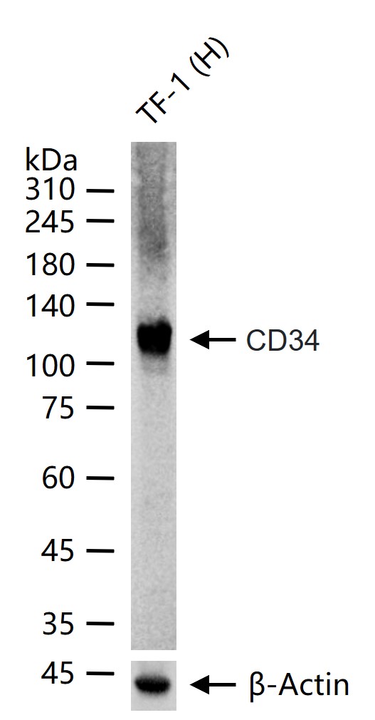
25 ug total protein per lane of various lysates (see on figure) probed with CD34 polyclonal antibody, unconjugated (bs-0646R) at 1:1000 dilution and 4°C overnight incubation. Followed by conjugated secondary antibody incubation at r.t. for 60 min.
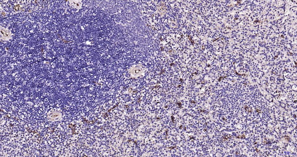
Paraformaldehyde-fixed, paraffin embedded Human Spleen; Antigen retrieval by boiling in sodium citrate buffer (pH6.0) for 15 min; Antibody incubation with CD34 Polyclonal Antibody, Unconjugated (bs-0646R) at 1:200 overnight at 4°C, followed by conjugation to the bs-0295G-HRP and DAB (C-0010) staining.
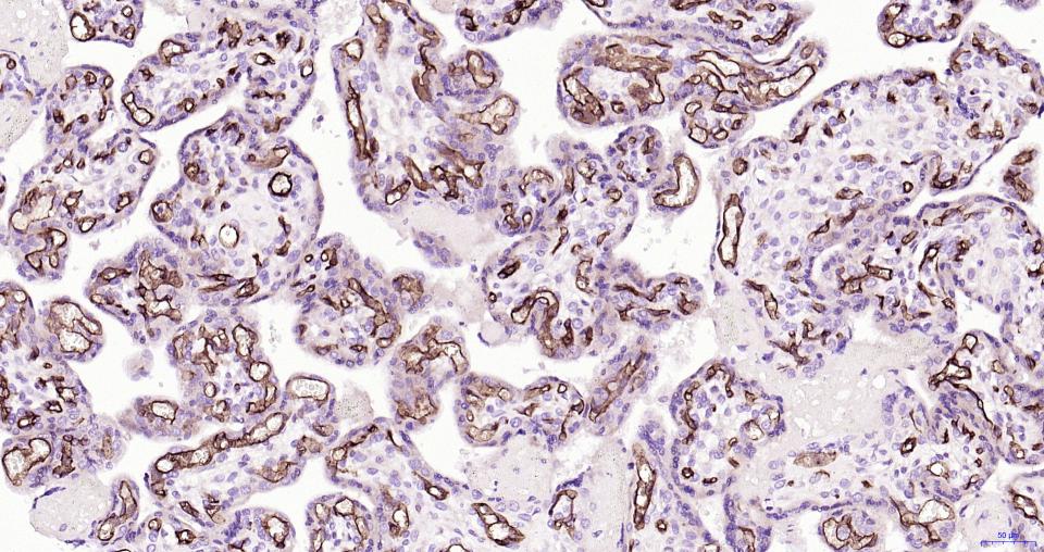
Paraformaldehyde-fixed, paraffin embedded Human Placenta; Antigen retrieval by boiling in sodium citrate buffer (pH6.0) for 15 min; Antibody incubation with CD34 Polyclonal Antibody, Unconjugated (bs-0646R) at 1:200 overnight at 4°C, followed by conjugation to the bs-0295G-HRP and DAB (C-0010) staining.

Paraformaldehyde-fixed, paraffin embedded Human Lung; Antigen retrieval by boiling in sodium citrate buffer (pH6.0) for 15 min; Antibody incubation with CD34 Polyclonal Antibody, Unconjugated (bs-0646R) at 1:200 overnight at 4°C, followed by conjugation to the bs-0295G-HRP and DAB (C-0010) staining.
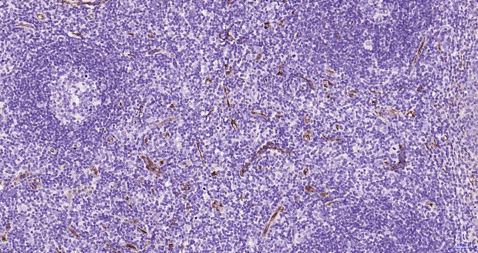
Paraformaldehyde-fixed, paraffin embedded Human Tonsil; Antigen retrieval by boiling in sodium citrate buffer (pH6.0) for 15 min; Antibody incubation with CD34 Polyclonal Antibody, Unconjugated (bs-0646R) at 1:200 overnight at 4°C, followed by conjugation to the bs-0295G-HRP and DAB (C-0010) staining.
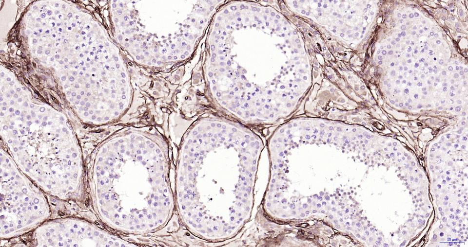
Paraformaldehyde-fixed, paraffin embedded Human Testicles; Antigen retrieval by boiling in sodium citrate buffer (pH6.0) for 15 min; Antibody incubation with CD34 Polyclonal Antibody, Unconjugated (bs-0646R) at 1:200 overnight at 4°C, followed by conjugation to the bs-0295G-HRP and DAB (C-0010) staining.
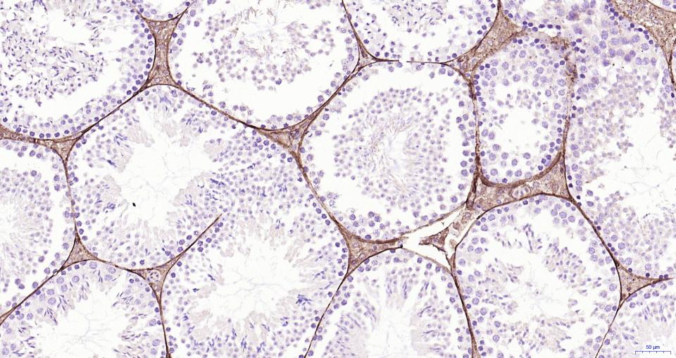
Paraformaldehyde-fixed, paraffin embedded Mouse Testicles; Antigen retrieval by boiling in sodium citrate buffer (pH6.0) for 15 min; Antibody incubation with CD34 Polyclonal Antibody, Unconjugated (bs-0646R) at 1:200 overnight at 4°C, followed by conjugation to the bs-0295G-HRP and DAB (C-0010) staining.
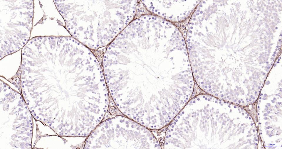
Paraformaldehyde-fixed, paraffin embedded Rat Testicles; Antigen retrieval by boiling in sodium citrate buffer (pH6.0) for 15 min; Antibody incubation with CD34 Polyclonal Antibody, Unconjugated (bs-0646R) at 1:200 overnight at 4°C, followed by conjugation to the bs-0295G-HRP and DAB (C-0010) staining.
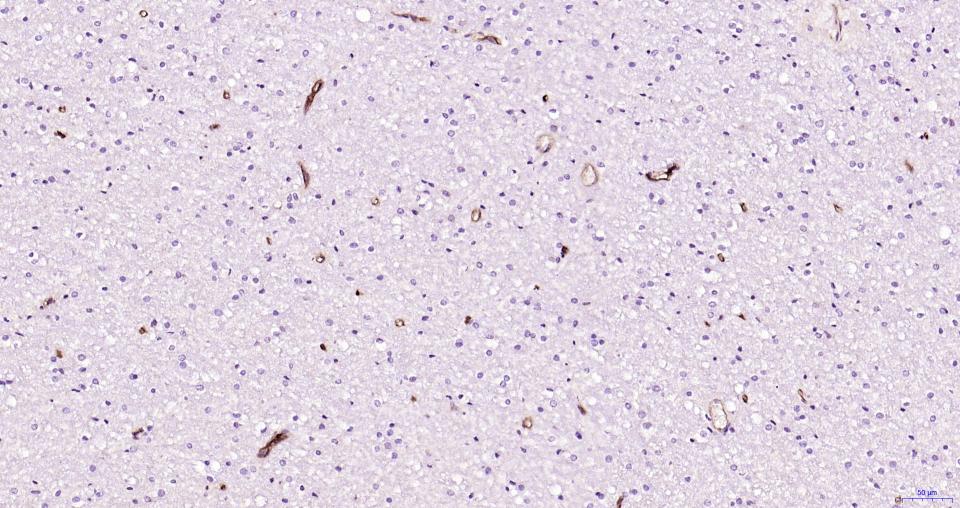
Paraformaldehyde-fixed, paraffin embedded Human Cerebrum; Antigen retrieval by boiling in sodium citrate buffer (pH6.0) for 15 min; Antibody incubation with CD34 Polyclonal Antibody, Unconjugated (bs-0646R) at 1:200 overnight at 4°C, followed by conjugation to the bs-0295G-HRP and DAB (C-0010) staining.
Paraformaldehyde-fixed, paraffin embedded Mouse Kidney; Antigen retrieval by boiling in sodium citrate buffer (pH6.0) for 15 min; Antibody incubation with CD34 Polyclonal Antibody, Unconjugated (bs-0646R) at 1:200 overnight at 4°C, followed by conjugation to the bs-0295G-HRP and DAB (C-0010) staining.

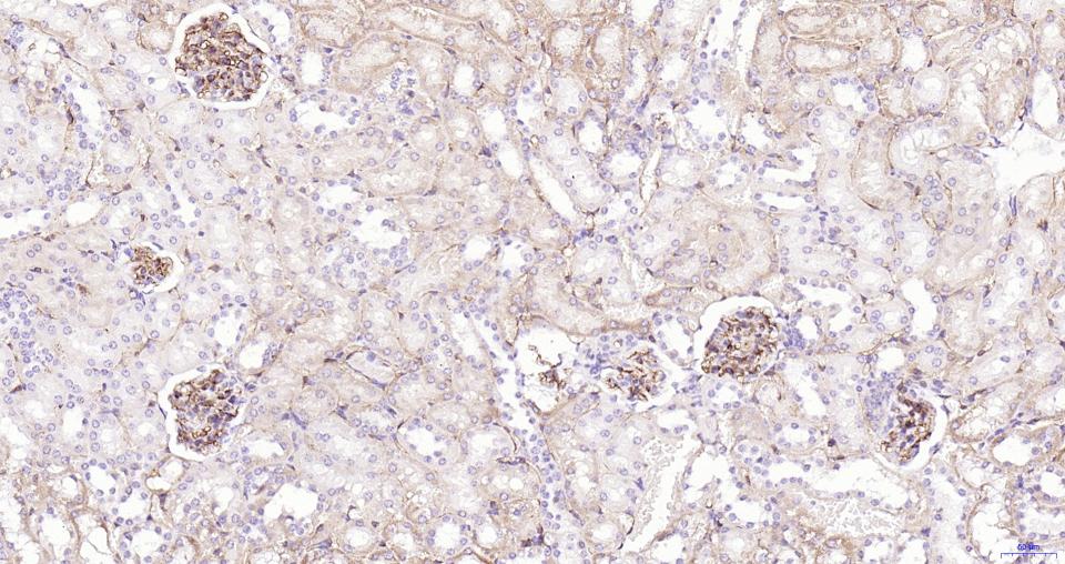
Paraformaldehyde-fixed, paraffin embedded Rat Kidney; Antigen retrieval by boiling in sodium citrate buffer (pH6.0) for 15 min; Antibody incubation with CD34 Polyclonal Antibody, Unconjugated (bs-0646R) at 1:200 overnight at 4°C, followed by conjugation to the bs-0295G-HRP and DAB (C-0010) staining.
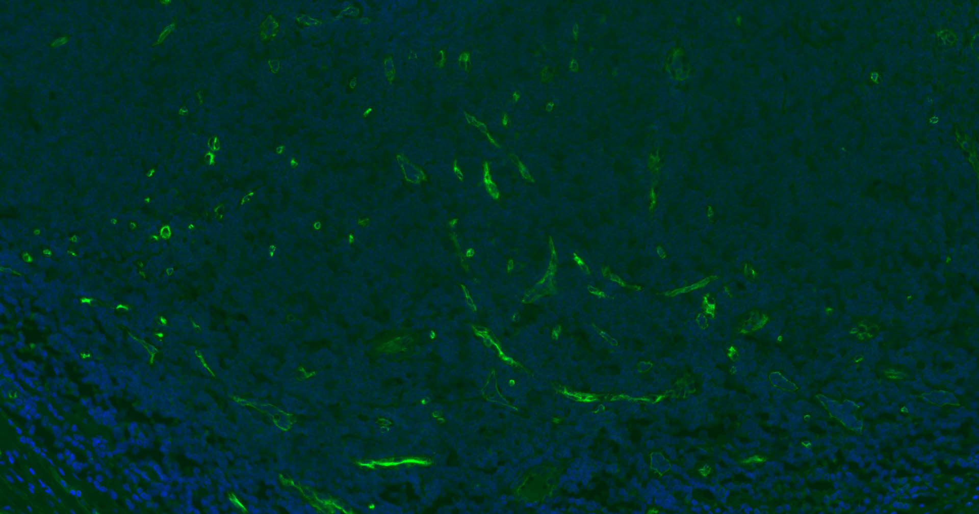
Paraformaldehyde-fixed, paraffin embedded Human Tonsil; Antigen retrieval by boiling in sodium citrate buffer (pH6.0) for 15 min; Antibody incubation with CD34 Polyclonal Antibody, Unconjugated (bs-0646R) at 1:200 overnight at 4°C. Followed by conjugated Goat Anti-Rabbit IgG antibody (green, bs-0295G-BF488), DAPI (blue, C02-04002) was used to stain the cell nuclei.
Paraformaldehyde-fixed, paraffin embedded Human Kidney; Antigen retrieval by boiling in sodium citrate buffer (pH6.0) for 15 min; Antibody incubation with CD34 Polyclonal Antibody, Unconjugated (bs-0646R) at 1:200 overnight at 4°C. Followed by conjugated Goat Anti-Rabbit IgG antibody (green, bs-0295G-BF488), DAPI (blue, C02-04002) was used to stain the cell nuclei.
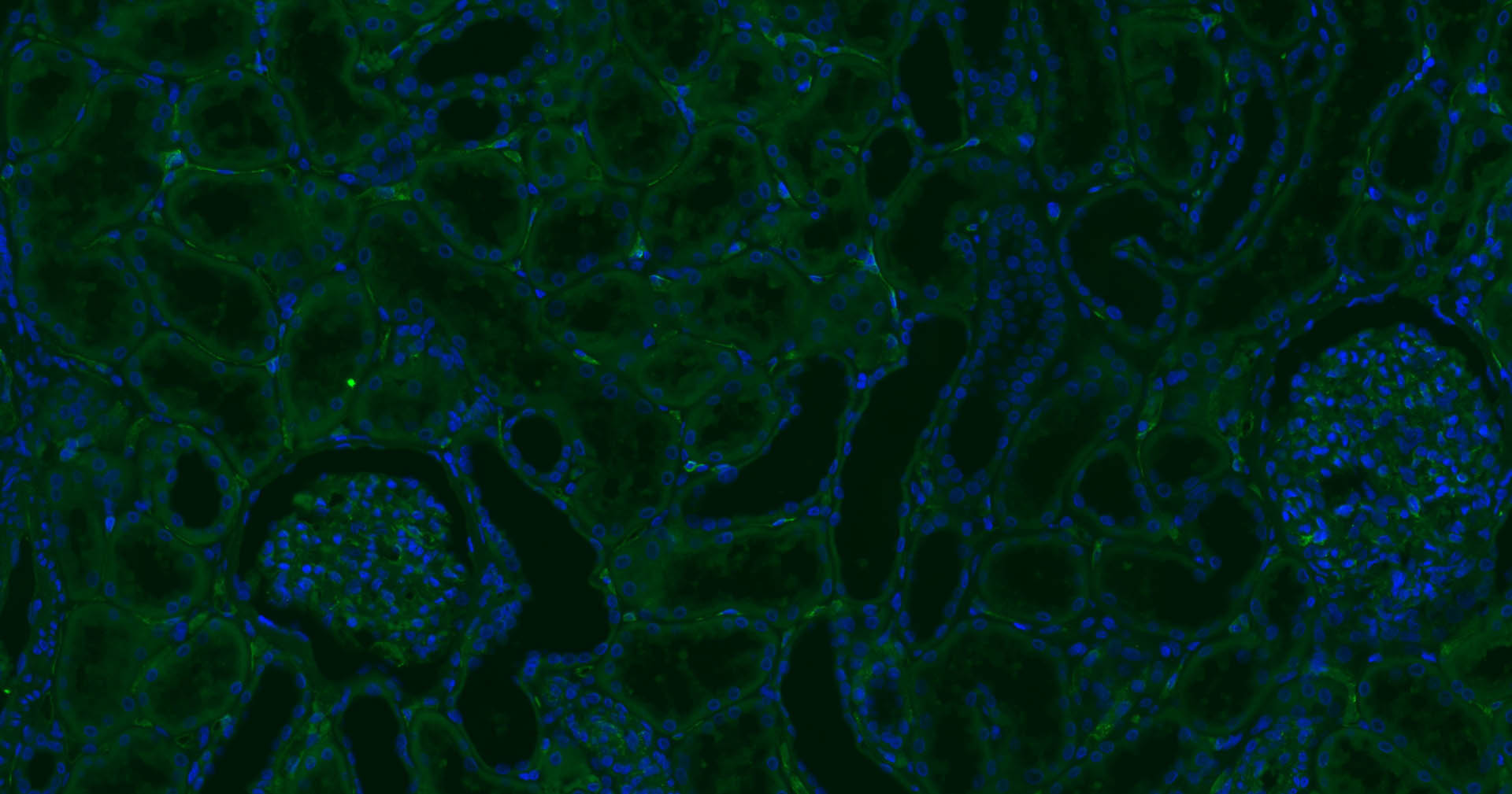
Paraformaldehyde-fixed, paraffin embedded Mouse Kidney; Antigen retrieval by boiling in sodium citrate buffer (pH6.0) for 15 min; Antibody incubation with CD34 Polyclonal Antibody, Unconjugated (bs-0646R) at 1:200 overnight at 4°C. Followed by conjugated Goat Anti-Rabbit IgG antibody (green, bs-0295G-BF488), DAPI (blue, C02-04002) was used to stain the cell nuclei.
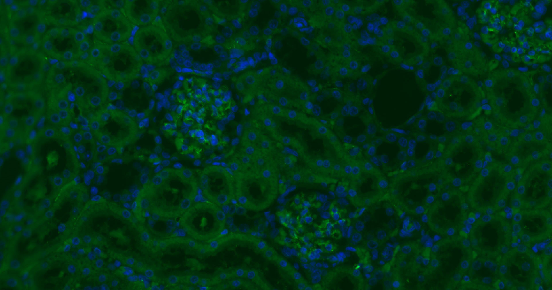
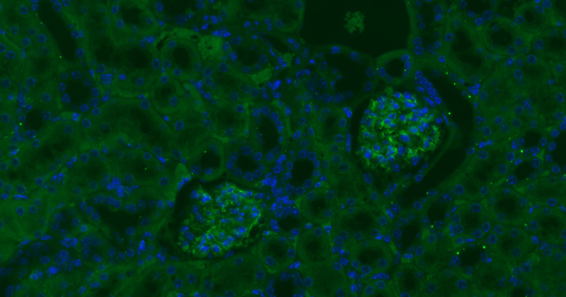
Paraformaldehyde-fixed, paraffin embedded Rat Kidney; Antigen retrieval by boiling in sodium citrate buffer (pH6.0) for 15 min; Antibody incubation with CD34 Polyclonal Antibody, Unconjugated (bs-0646R) at 1:200 overnight at 4°C. Followed by conjugated Goat Anti-Rabbit IgG antibody (green, bs-0295G-BF488), DAPI (blue, C02-04002) was used to stain the cell nuclei.
|
| 1、抗体溶解方法 | |
| 2、抗体修复方式 | |
| 3、常用试剂的配制 | |
| 4、免疫组化操作步骤 | |
| 5、免疫组化问题解答 | |
| 6、Western Blotting 操作步骤 | |
| 7、Western Blotting 问题解答 | |
| 8、关于肽链的设计 | |
| 9、多肽的溶解与保存 | |
| 10、酶标抗体效价测定程序 | |


