

| 产品编号 | bs-1134R |
| 英文名称 | RUNX2 Rabbit pAb |
| 中文名称 | 核心结合因子α1/成骨特异性转录因子/Cbfα1抗体 |
| 别 名 | AML3; CBF-alpha-1; CBFA1; CCD; CCD1; CLCD; OSF-2; OSF2; PEA2aA; PEBP2aA; Cbf; Cbfa-1; LS3; Pebp2a1; Pebpa2a; RUNX2_HUMAN; RUNX2; Acute myeloid leukemia 3 protein; Core-binding factor subunit alpha-1 (CBF-alpha-1); Oncogene AML-3; Osteoblast-specific trans |
 |
Specific References (49) | bs-1134R has been referenced in 49 publications.
|
| 研究领域 | 干细胞 转录调节因子 表观遗传学 |
| 抗体来源 | Rabbit |
| 克隆类型 | Polyclonal |
| 克 隆 号 | |
| 交叉反应 | Human,Mouse,Rat (predicted: Rabbit,Pig,Sheep,Cow,Chicken,Dog,Horse) |
| 产品应用 | WB=1:500-2000,IHC-P=1:100-500,IHC-F=1:100-500,IF=1:100-500,Flow-Cyt=1ug/Test
not yet tested in other applications. optimal dilutions/concentrations should be determined by the end user. |
| 理论分子量 | 57(hu)/67(mo,rat) kDa |
| 检测分子量 | 62 |
| 细胞定位 | 细胞核 |
| 性 状 | Liquid |
| 浓 度 | 1mg/ml |
| 免 疫 原 | KLH conjugated synthetic peptide derived from human RUNX2: 202-300/521 |
| 亚 型 | IgG |
| 纯化方法 | affinity purified by Protein A |
| 缓 冲 液 | 0.01M TBS (pH7.4) with 1% BSA, 0.02% Proclin300 and 50% Glycerol. |
| 保存条件 | Shipped at 4℃. Store at -20℃ for one year. Avoid repeated freeze/thaw cycles. |
| 注意事项 | This product as supplied is intended for research use only, not for use in human, therapeutic or diagnostic applications. |
| PubMed | PubMed |
| 产品介绍 |
This gene is a member of the RUNX family of transcription factors and encodes a nuclear protein with an Runt DNA-binding domain. This protein is essential for osteoblastic differentiation and skeletal morphogenesis and acts as a scaffold for nucleic acids and regulatory factors involved in skeletal gene expression. The protein can bind DNA both as a monomer or, with more affinity, as a subunit of a heterodimeric complex. Mutations in this gene have been associated with the bone development disorder cleidocranial dysplasia (CCD). Transcript variants that encode different protein isoforms result from the use of alternate promoters as well as alternate splicing. [provided by RefSeq, Jul 2008]. Function: Transcription factor involved in osteoblastic differentiation and skeletal morphogenesis. Essential for the maturation of osteoblasts and both intramembranous and endochondral ossification. CBF binds to the core site, 5'-PYGPYGGT-3', of a number of enhancers and promoters, including murine leukemia virus, polyomavirus enhancer, T-cell receptor enhancers, osteocalcin, osteopontin, bone sialoprotein, alpha 1(I) collagen, LCK, IL-3 and GM-CSF promoters (By similarity). Inhibits MYST4-dependent transcriptional activation. [SUBUNIT] Interaction with SATB2 results in enhanced DNA binding and transactivation by these transcription factors (By similarity). Heterodimer of an alpha and a beta subunit. Interacts with HIVEP3 (By similarity). The alpha subunit binds DNA as a monomer and through the Runt domain. DNA-binding is increased by heterodimerization. Interacts with XRCC6 (Ku70) and XRCC5 (Ku80). Interacts with MYST3 and MYST4. Subunit: Heterodimer of an alpha and a beta subunit. Interacts with HIVEP3. The alpha subunit binds DNA as a monomer and through the Runt domain. DNA-binding is increased by heterodimerization. Interacts with G22P1 (Ku70) and XRCC5 (Ku80). Interacts with MYST3 and MYST4. Subcellular Location: Nucleus. Tissue Specificity: Specifically expressed in osteoblasts. Post-translational modifications: Phosphorylated; probably by MAP kinases (MAPK). Isoform 3 is phosphorylated on Ser340. DISEASE: Defects in RUNX2 are the cause of cleidocranial dysplasia (CLCD) [MIM:119600]; also known as cleidocranial dysostosis (CCD). CLCD is an autosomal dominant skeletal disorder with high penetrance and variable expressivity. It is due to defective endochondral and intramembranous bone formation. Typical features include hypoplasia/aplasia of clavicles, patent fontanelles, wormian bones (additional cranial plates caused by abnormal ossification of the calvaria), supernumerary teeth, short stature, and other skeletal changes. In some cases defects in RUNX2 are exclusively associated with dental anomalies. Similarity: Contains 1 Runt domain. SWISS: Q13950 Gene ID: 860 Database links: Entrez Gene: 860 Human Entrez Gene: 12393 Mouse Omim: 600211 Human SwissProt: Q13950 Human SwissProt: Q9XSB7 Horse SwissProt: Q08775 Mouse Unigene: 535845 Human Unigene: 391013 Mouse Unigene: 391017 Mouse Unigene: 214214 Rat Unigene: 83672 Rat RUNX2又称:Cbfα1(Core-binding factor, alpha 3 subunit) 是新发现的一类调控间充质干细胞向成骨方向分化的特异性转录因子,参与骨形成,骨骼生长和发育的一类重要细胞,它起源于多能间充质干细胞,是间充质干细胞在体内的各种调控因素的调节下发育而成的。 |
| 产品图片 |
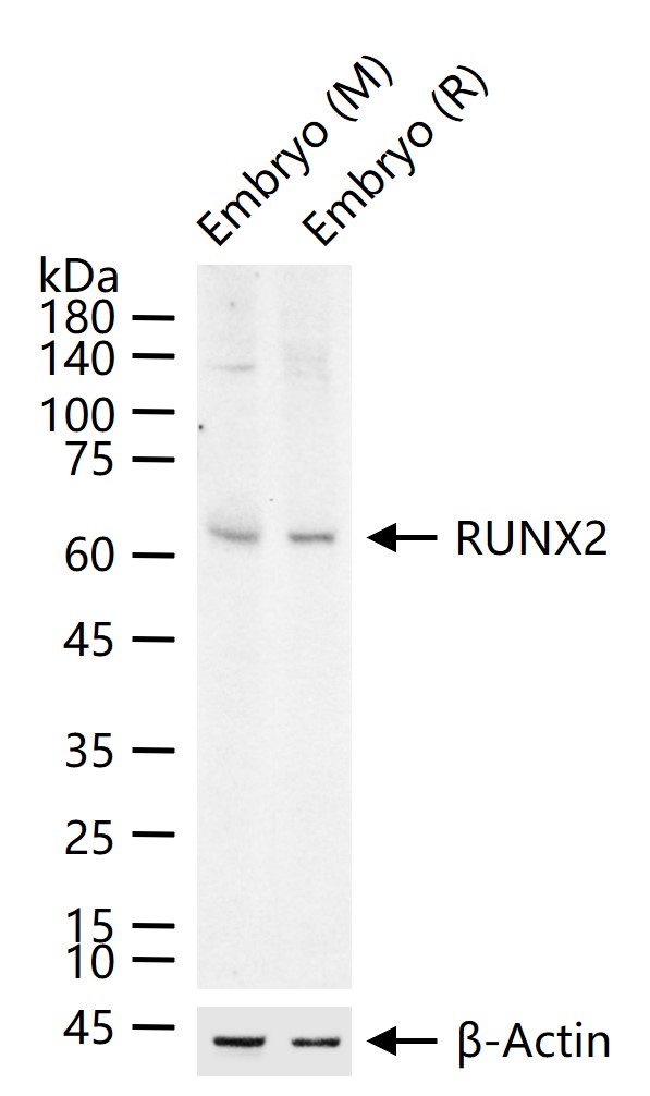
25 ug total protein per lane of various lysates (see on figure) probed with RUNX2 polyclonal antibody, unconjugated (bs-1134R) at 1:1000 dilution and 4°C overnight incubation. Followed by conjugated secondary antibody incubation at r.t. for 60 min.

Paraformaldehyde-fixed, paraffin embedded (Mouse thyroid); Antigen retrieval by boiling in sodium citrate buffer (pH6.0) for 15min; Block endogenous peroxidase by 3% hydrogen peroxide for 20 minutes; Blocking buffer (normal goat serum) at 37°C for 30min; Antibody incubation with (RUNX2) Polyclonal Antibody, Unconjugated (bs-1134R) at 1:400 overnight at 4°C, followed by operating according to SP Kit(Rabbit) (sp-0023) instructionsand DAB staining.
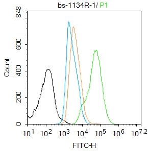
Blank control:HL-60.
Primary Antibody (green line): Rabbit Anti-RUNX2 antibody (bs-1134R)
Dilution: 1μg /10^6 cells;
Isotype Control Antibody (orange line): Rabbit IgG .
Secondary Antibody : Goat anti-rabbit IgG-AF488
Dilution: 1μg /test.
Protocol
The cells were fixed with 4% PFA (10min at room temperature)and then permeabilized with 90% ice-cold methanol for 20 min at-20℃. The cells were then incubated in 5%BSA to block non-specific protein-protein interactions for 30 min at room temperature .Cells stained with Primary Antibody for 30 min at room temperature. The secondary antibody used for 40 min at room temperature. Acquisition of 20,000 events was performed.
|
| 1、抗体溶解方法 | |
| 2、抗体修复方式 | |
| 3、常用试剂的配制 | |
| 4、免疫组化操作步骤 | |
| 5、免疫组化问题解答 | |
| 6、Western Blotting 操作步骤 | |
| 7、Western Blotting 问题解答 | |
| 8、关于肽链的设计 | |
| 9、多肽的溶解与保存 | |
| 10、酶标抗体效价测定程序 | |


