

| 产品编号 | bs-0079R |
| 英文名称 | CD19 Rabbit pAb |
| 中文名称 | CD19抗体 |
| 别 名 | B4; CVID3; CD19_HUMAN; CD19; B-lymphocyte surface antigen B4; Differentiation antigen CD19; T-cell surface antigen Leu-12; CD19_MOUSE; CD19 molecule; CD19 antigen |
 |
Specific References (12) | bs-0079R has been referenced in 12 publications.
|
| 研究领域 | 细胞生物 免疫学 干细胞 细胞表面分子 淋巴细胞 b-淋巴细胞 |
| 抗体来源 | Rabbit |
| 克隆类型 | Polyclonal |
| 交叉反应 | Human,Mouse (predicted: Rat,Pig,Cow,GuineaPig,Horse) |
| 产品应用 | IHC-P=1:100-500,IHC-F=1:100-500,IF=1:100-500,Flow-Cyt=1μg/Test
not yet tested in other applications. optimal dilutions/concentrations should be determined by the end user. |
| 理论分子量 | 59kDa |
| 检测分子量 | 95 |
| 细胞定位 | 细胞膜 |
| 性 状 | Liquid |
| 浓 度 | 1mg/ml |
| 免 疫 原 | KLH conjugated synthetic peptide derived from human CD19: 485-556/556 <Cytoplasmic> |
| 亚 型 | IgG |
| 纯化方法 | affinity purified by Protein A |
| 缓 冲 液 | 0.01M TBS (pH7.4) with 1% BSA, 0.02% Proclin300 and 50% Glycerol. |
| 保存条件 | Shipped at 4℃. Store at -20℃ for one year. Avoid repeated freeze/thaw cycles. |
| 注意事项 | This product as supplied is intended for research use only, not for use in human, therapeutic or diagnostic applications. |
| PubMed | PubMed |
| 产品介绍 |
This gene encodes a member of the immunoglobulin gene superfamily. Expression of this cell surface protein is restricted to B cell lymphocytes. This protein is a reliable marker for pre-B cells but its expression diminishes during terminal B cell differentiation in antibody secreting plasma cells. The protein has two N-terminal extracellular Ig-like domains separated by a non-Ig-like domain, a hydrophobic transmembrane domain, and a large C-terminal cytoplasmic domain. This protein forms a complex with several membrane proteins including complement receptor type 2 (CD21) and tetraspanin (CD81) and this complex reduces the threshold for antigen-initiated B cell activation. Activation of this B-cell antigen receptor complex activates the phosphatidylinositol 3-kinase signalling pathway and the subsequent release of intracellular stores of calcium ions. This protein is a target of chimeric antigen receptor (CAR) T-cells used in the treatment of lymphoblastic leukemia. Mutations in this gene are associated with the disease common variable immunodeficiency 3 (CVID3) which results in a failure of B-cell differentiation and impaired secretion of immunoglobulins. CVID3 is characterized by hypogammaglobulinemia, an inability to mount an antibody response to antigen, and recurrent bacterial infections. Alternative splicing results in multiple transcript variants encoding distinct isoforms. [provided by RefSeq, Jul 2020] Function: Assembles with the antigen receptor of B-lymphocytes in order to decrease the threshold for antigen receptor-dependent stimulation. Subunit: Forms a complex with CD21, CD81 and CD225 in the membrane of mature B-cells. Interacts with VAV. Interacts with GRB2 and SOS when phosphorylated on Tyr-348 and/or Tyr-378. Interacts with PLCG2 when phosphorylated on Tyr-409. Interacts with LYN. Subcellular Location: Membrane; Single-pass type I membrane protein. Post-translational modifications: Phosphorylated on serine and threonine upon DNA damage, probably by ATM or ATR. Phosphorylated on tyrosine following B-cell activation. Phosphorylated on tyrosine residues by LYN. DISEASE: Defects in CD19 are the cause of immunodeficiency common variable type 3 (CVID3) [MIM:613493]; also called antibody deficiency due to CD19 defect. CVID3 is a primary immunodeficiency characterized by antibody deficiency, hypogammaglobulinemia, recurrent bacterial infections and an inability to mount an antibody response to antigen. The defect results from a failure of B-cell differentiation and impaired secretion of immunoglobulins; the numbers of circulating B-cells is usually in the normal range, but can be low. Similarity: Contains 2 Ig-like C2-type (immunoglobulin-like) domains. SWISS: P15391 Gene ID: 930 Database links: Entrez Gene: 930 Human Entrez Gene: 12478 Mouse Omim: 107265 Human SwissProt: P15391 Human SwissProt: P25918 Mouse Unigene: 652262 Human Unigene: 4360 Mouse CD19是一种质膜蛋白质,参与信号传导作用。表达与前B细胞和成熟的B细胞膜表面,与B细胞的活化调节和发育调节相关,在T细胞和正常粒细胞上无表达。此抗体可以特异性识别CD19,主要用于标记正常B细胞及肿瘤性B细胞。 |
| 产品图片 |
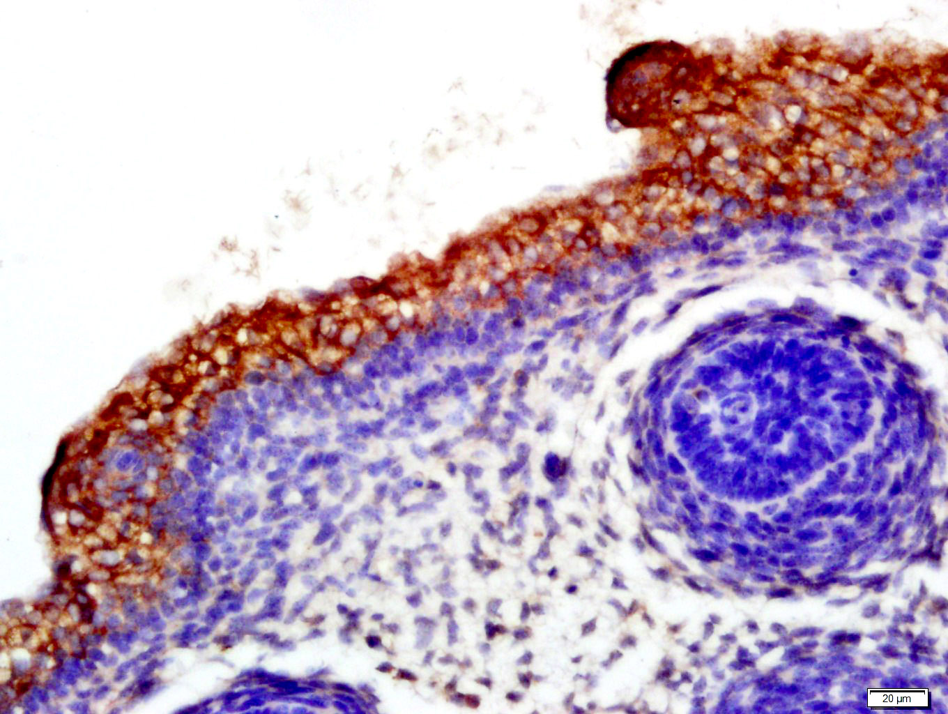
Tissue/cell: mouse fetal skin; 4% Paraformaldehyde-fixed and paraffin-embedded;
Antigen retrieval: citrate buffer ( 0.01M, pH 6.0 ), Boiling bathing for 15min; Block endogenous peroxidase by 3% Hydrogen peroxide for 30min; Blocking buffer (normal goat serum,C-0005) at 37℃ for 20 min;
Incubation: Anti-CD19 Polyclonal Antibody, Unconjugated(bs-0079R) 1:500, overnight at 4°C, followed by conjugation to the secondary antibody(SP-0023) and DAB(C-0010) staining
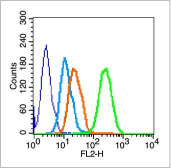
Blank control (blue line): HL60 cells (blue).
Primary Antibody (green line): Rabbit Anti-CD19 antibody (bs-0079R)
Dilution: 1μg /10^6 cells;
Isotype Control Antibody (orange line): Rabbit IgG .
Secondary Antibody (white blue line): Goat anti-rabbit IgG-PE
Dilution: 1μg /test.
Protocol
The cells were fixed with 70% methanol (Overnight at 4℃) . Cells stained with Primary Antibody for 30 min at room temperature. The cells were then incubated in 1 X PBS/2%BSA/10% goat serum to block non-specific protein-protein interactions followed by the antibody for 15 min at room temperature. The secondary antibody used for 40 min at room temperature. Acquisition of 20,000 events was performed.
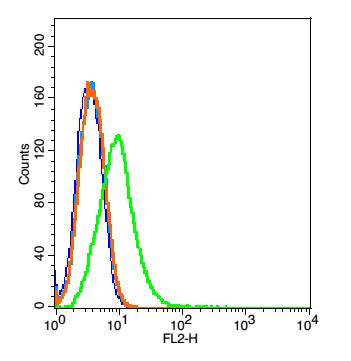
Blank control: Raji(blue). Primary Antibody: Rabbit Anti-CD19 antibody(bs-0079R), Dilution: 5μg in 100 μL 1X PBS containing 0.5% BSA; Isotype Control Antibody: Rabbit IgG (orange) ,used under the same conditions. Secondary Antibody: Goat anti-rabbit IgG-PE(white blue), Dilution: 1:200 in 1 X PBS containing 0.5% BSA.
Protocol
Primary antibody (bs-0079R, 5μg /1x10^6 cells) were incubated for 30 min on the ice, followed by 1 X PBS containing 0.5% BSA + 1 0% goat serum (15 min) to block non-specific protein-protein interactions. Then the Goat Anti-rabbit IgG/PE antibody was added into the blocking buffer mentioned above to react with the primary antibody at 1/200 dilution for 30 min on ice. Acquisition of 20,000 events was performed.
|
| 1、抗体溶解方法 | |
| 2、抗体修复方式 | |
| 3、常用试剂的配制 | |
| 4、免疫组化操作步骤 | |
| 5、免疫组化问题解答 | |
| 6、Western Blotting 操作步骤 | |
| 7、Western Blotting 问题解答 | |
| 8、关于肽链的设计 | |
| 9、多肽的溶解与保存 | |
| 10、酶标抗体效价测定程序 | |


