

| 产品编号 | bs-2202R |
| 英文名称 | VEGFR3 Rabbit pAb |
| 中文名称 | 血管内皮细胞生长因子受体3抗体 |
| 别 名 | CHTD7; FLT-4; FLT41; LMPH1A; LMPHM1; PCL; VEGFR-3; VEGFR3; Chy; VGFR3_HUMAN; FLT4; Fms-like tyrosine kinase 4 (FLT-4); Tyrosine-protein kinase receptor FLT4; 2.7.10.1; VGFR3_MOUSE; VGFR3_RAT; |
 |
Specific References (5) | bs-2202R has been referenced in 5 publications.
[IF=7.84] Zhuo, Wei, et al. "The CXCL12?CCXCR4 Chemokine Pathway: A Novel Axis Regulates Lymphangiogenesis."Clinical Cancer Research 18.19 (2012): 5387-5398.l Human, Mouse.
[IF=5.99] Wang, Zhixiong, et al. "CXCL1 from tumor-associated lymphatic endothelial cells drives gastric cancer cell into lymphatic system via activating integrin β1/FAK/AKT signaling." Cancer Letters 385 (2017): 28-38. IF(ICC) ; Human.
[IF=2.139] M Csöbönyeiová. et al. Immunohistochemical and Scanning Electron Microscopic Confirmation of the Lymphatic Lacunae in the Uterine Tube Mucosal Folds. What Are the Clinical Implications?. PHYSIOL RES. 2022 Dec 27;71(Suppl 1):S115-S123 IHC ; Human.
[IF=2.136] Hayashi KG et al. Temporal expression and localization of vascular endothelial growth factor family members in the bovine uterus during peri-implantation period. Theriogenology. 2019 Apr 24;133:56-64. IHC-P ; Cow.
[IF=2.02] Wang, Zheng, et al. "RhGH attenuates ischemia injury of intrahepatic bile ducts relating to liver transplantation." Journal of Surgical Research 171.1 (2011): 300-310. IHC-P ; Rat.
|
| 研究领域 | 肿瘤 免疫学 信号转导 生长因子和激素 激酶和磷酸酶 细胞膜受体 血管内皮细胞 |
| 抗体来源 | Rabbit |
| 克隆类型 | Polyclonal |
| 克 隆 号 | |
| 交叉反应 | Human,Mouse,Rat (predicted: Rabbit,Pig,Dog,Horse) |
| 产品应用 | IHC-P=1:100-500,IHC-F=1:100-500,IF=1:100-500
not yet tested in other applications. optimal dilutions/concentrations should be determined by the end user. |
| 理论分子量 | 151 kDa |
| 细胞定位 | 细胞核 细胞浆 细胞膜 分泌型蛋白 |
| 性 状 | Liquid |
| 浓 度 | 1mg/ml |
| 免 疫 原 | KLH conjugated synthetic peptide derived from human VEGFR-3: 901-1000/1298 <Cytoplasmic> |
| 亚 型 | IgG |
| 纯化方法 | affinity purified by Protein A |
| 缓 冲 液 | 0.01M TBS (pH7.4) with 1% BSA, 0.02% Proclin300 and 50% Glycerol. |
| 保存条件 | Shipped at 4℃. Store at -20℃ for one year. Avoid repeated freeze/thaw cycles. |
| 注意事项 | This product as supplied is intended for research use only, not for use in human, therapeutic or diagnostic applications. |
| PubMed | PubMed |
| 产品介绍 |
Vascular endothelial growth factors (VEGFs) are a family of closely related growth factors having a conserved pattern of eight cysteine esidues and sharing common VEGF receptors. VEGFs stimulate the proliferation of endothelial cells, induce angiogenesis, and increase vascular permeability in both large and small vessels. The mitogenic activity of VEGFs appears to be mediated by specific VEGF receptors. VEGF Receptor 3 is one of the five receptor tyrosine kinases (RTKs) (VEGF Receptor 1/Flt1, VEGF Receptor 2/KDR/Flk1, VEGF Receptor 3/Flt4, tie1 and tek/tie2) whose expression is almost exclusively restricted to endothelial cells Function: Tyrosine-protein kinase that acts as a cell-surface receptor for VEGFC and VEGFD, and plays an essential role in adult lymphangiogenesis and in the development of the vascular network and the cardiovascular system during embryonic development. Promotes proliferation, survival and migration of endothelial cells, and regulates angiogenic sprouting. Signaling by activated FLT4 leads to enhanced production of VEGFC, and to a lesser degree VEGFA, thereby creating a positive feedback loop that enhances FLT4 signaling. Modulates KDR signaling by forming heterodimers. Mediates activation of the MAPK1/ERK2, MAPK3/ERK1 signaling pathway, of MAPK8 and the JUN signaling pathway, and of the AKT1 signaling pathway. Phosphorylates SHC1. Mediates phosphorylation of PIK3R1, the regulatory subunit of phosphatidylinositol 3-kinase. Promotes phosphorylation of MAPK8 at 'Thr-183' and 'Tyr-185', and of AKT1 at 'Ser-473'. Subunit: Interacts with VEGFC and VEGFD. Monomer in the absence of bound VEGFC or VEGFD. Homodimer in the presence of bound VEGFC or VEGFD. Can also form a heterodimer with KDR. Interacts with PTPN14; the interaction is enhanced by stimulation with VEGFC. Interacts with CRK, GRB2, PTK2/FAK1, SHC1, PIK3R1 and PTPN11/SHP-2. Identified in a complex with SRC and ITGB1. Subcellular Location: Cell membrane; Single-pass type I membrane protein. Cytoplasm. Nucleus. Note=Ligand-mediated autophosphorylation leads to rapid internalization. Isoform 1: Cell membrane; Single-pass type I membrane protein. Note=Ligand-mediated autophosphorylation leads to rapid internalization. Isoform 2: Cell membrane; Single-pass type I membrane protein. Isoform 3: Secreted. Cytoplasm. Tissue Specificity: Detected in endothelial cells (at protein level). Widely expressed. Detected in fetal spleen, lung and brain. Detected in adult liver, muscle, thymus, placenta, lung, testis, ovary, prostate, heart, and kidney. Post-translational modifications: Autophosphorylated on tyrosine residues upon ligand binding. Autophosphorylation occurs in trans, i.e. one subunit of the dimeric receptor phosphorylates tyrosine residues on the other subunit. Phosphorylation in response to H(2)O(2) is mediated by a process that requires SRC and PRKCD activity. Phosphorylation at Tyr-1068 is required for autophosphorylation at additional tyrosine residues. Phosphorylation at Tyr-1063 and Tyr-1337 is important for interaction with CRK and subsequent activation of MAPK8. Phosphorylation at Tyr-1230, Tyr-1231 and Tyr-1337 is important for interaction with GRB2 and subsequent activation of the AKT1 and MAPK1/ERK2 and/or MAPK3/ERK1 signaling pathways. In response to endothelial cell adhesion onto collagen, can also be phosphorylated in the absence of FLT4 kinase activity by SRC. Similarity: Belongs to the protein kinase superfamily. Tyr protein kinase family. CSF-1/PDGF receptor subfamily. Contains 7 Ig-like C2-type (immunoglobulin-like) domains. Contains 1 protein kinase domain. SWISS: P35916 Gene ID: 2324 Database links: Entrez Gene: 2324 Human Entrez Gene: 14257 Mouse Omim: 136352 Human SwissProt: P35916 Human SwissProt: P35917 Mouse Unigene: 646917 Human Unigene: 3291 Mouse Unigene: 81043 Rat VEGFR3又称FLt4主要在成熟组织的淋巴管内皮细胞上表达,VEGF-R3与淋巴管内皮细胞增殖和迁移有关,有刺激淋巴管新生的作用,目前多用于肿瘤转移方面的研究。 |
| 产品图片 |
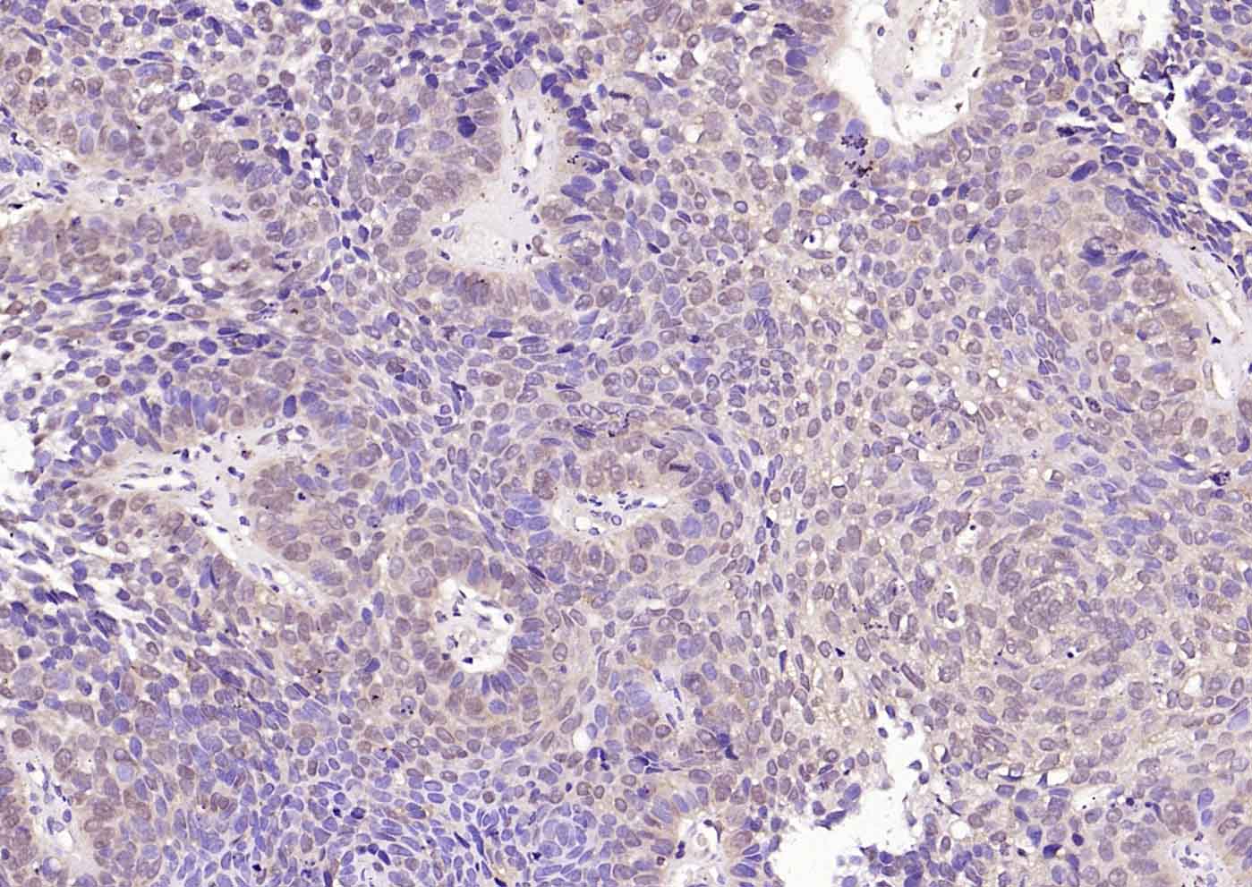
Paraformaldehyde-fixed, paraffin embedded Human Breast Cancer; Antigen retrieval by boiling in sodium citrate buffer (pH6.0) for 15 min; Antibody incubation with VEGFR3 Polyclonal Antibody, Unconjugated (bs-2202R) at 1:200 overnight at 4°C, followed by conjugation to the bs-0295G-HRP and DAB (C-0010) staining.
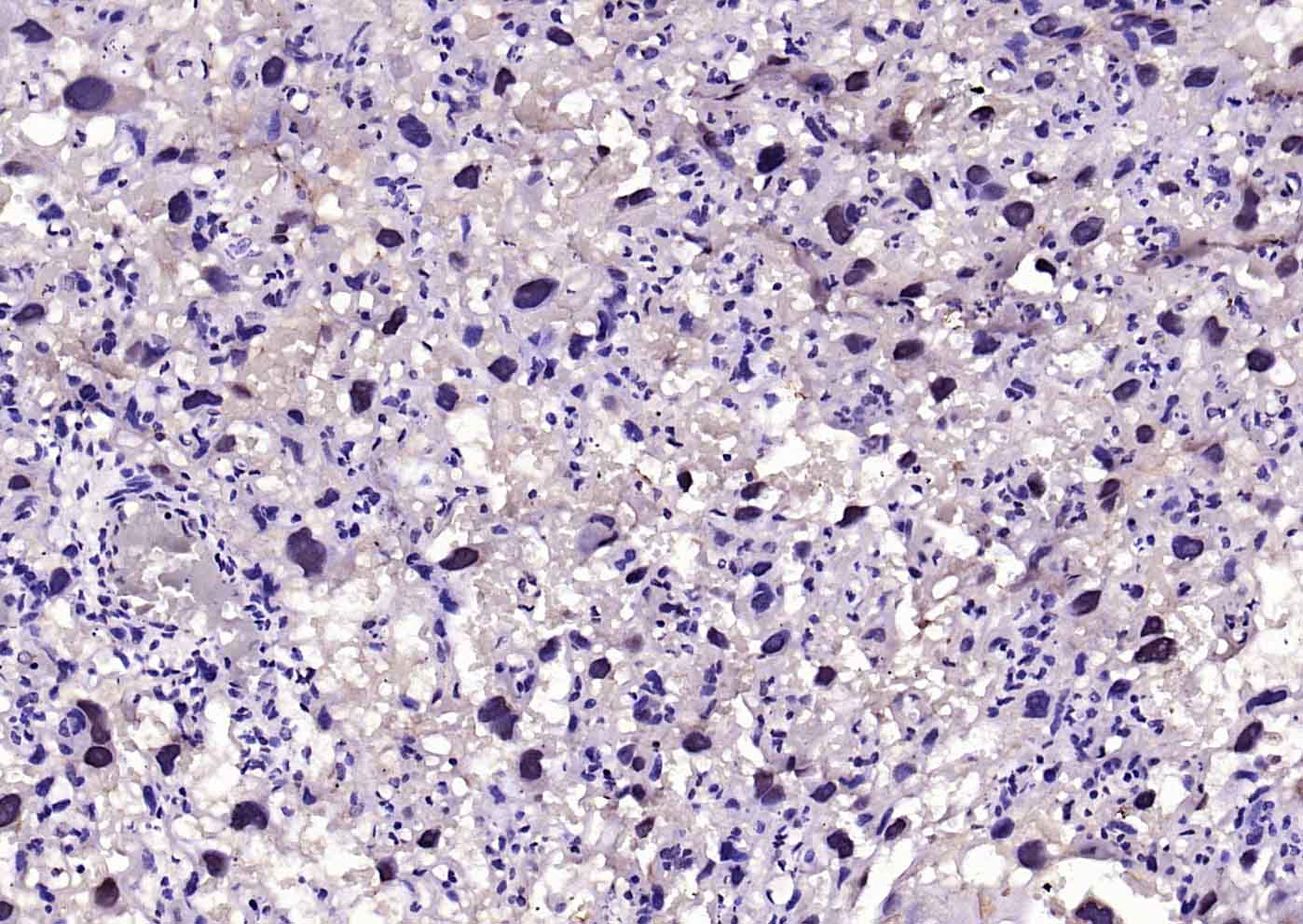
Paraformaldehyde-fixed, paraffin embedded Rat Placenta; Antigen retrieval by boiling in sodium citrate buffer (pH6.0) for 15 min; Antibody incubation with VEGFR3 Polyclonal Antibody, Unconjugated (bs-2202R) at 1:200 overnight at 4°C, followed by conjugation to the bs-0295G-HRP and DAB (C-0010) staining.
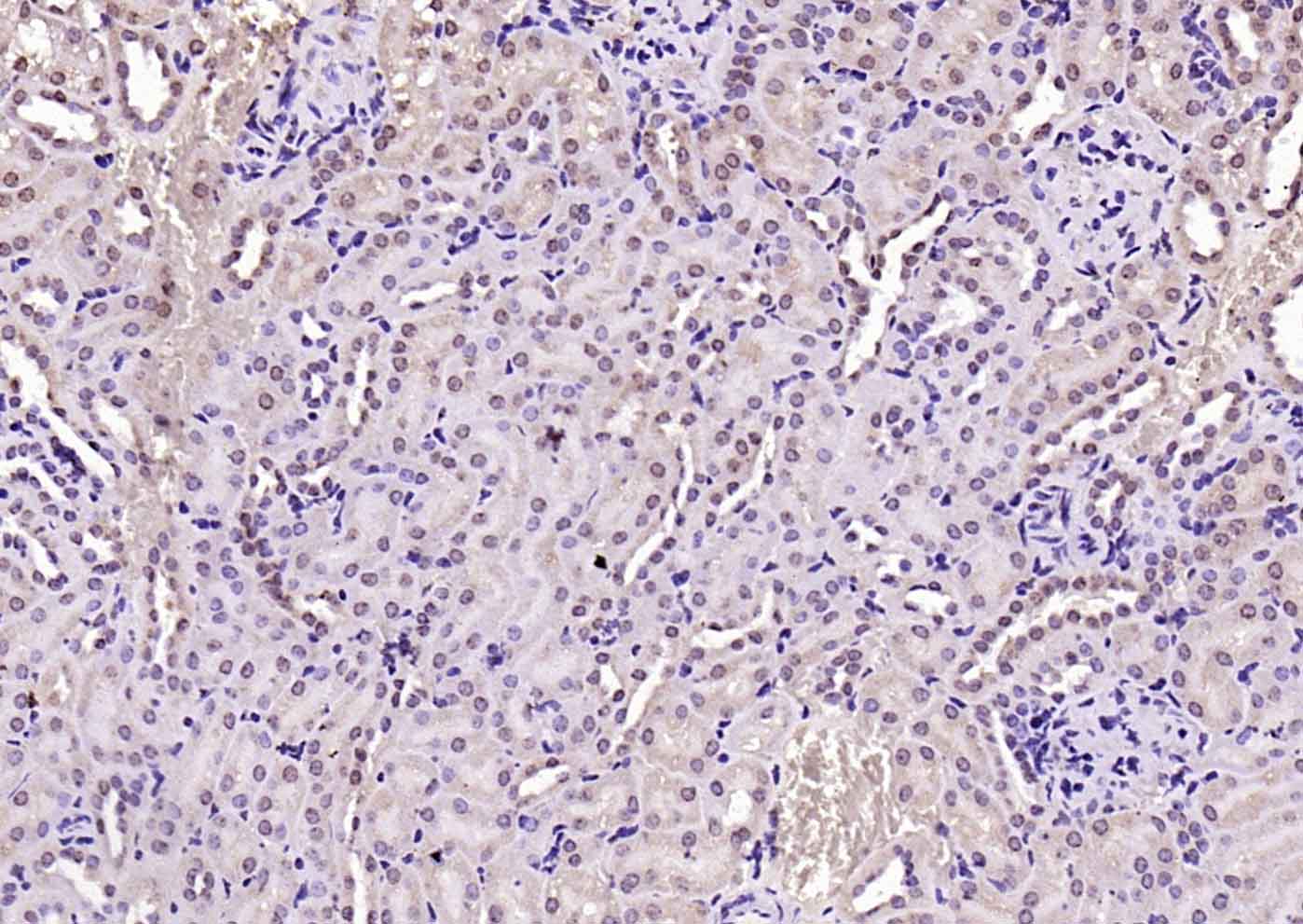
Paraformaldehyde-fixed, paraffin embedded Mouse Kidney; Antigen retrieval by boiling in sodium citrate buffer (pH6.0) for 15 min; Antibody incubation with VEGFR3 Polyclonal Antibody, Unconjugated (bs-2202R) at 1:200 overnight at 4°C, followed by conjugation to the bs-0295G-HRP and DAB (C-0010) staining.
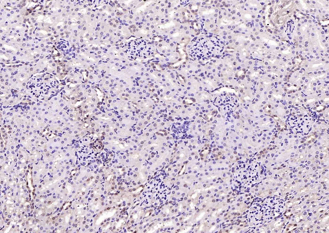
Paraformaldehyde-fixed, paraffin embedded Rat Kidney; Antigen retrieval by boiling in sodium citrate buffer (pH6.0) for 15 min; Antibody incubation with VEGFR3 Polyclonal Antibody, Unconjugated (bs-2202R) at 1:200 overnight at 4°C, followed by conjugation to the bs-0295G-HRP and DAB (C-0010) staining.
|
| 1、抗体溶解方法 | |
| 2、抗体修复方式 | |
| 3、常用试剂的配制 | |
| 4、免疫组化操作步骤 | |
| 5、免疫组化问题解答 | |
| 6、Western Blotting 操作步骤 | |
| 7、Western Blotting 问题解答 | |
| 8、关于肽链的设计 | |
| 9、多肽的溶解与保存 | |
| 10、酶标抗体效价测定程序 | |


