bs-5519R [Primary Antibody]
Rabbit  Anti-phospho-NRP1 (Thr916) Rabbit pAb  Polyclonal Antibody
www.bioss.com.cn
sales@bioss.com.cn
techsupport@bioss.com.cn
400-901-9800
DATASHEET

Host: Rabbit

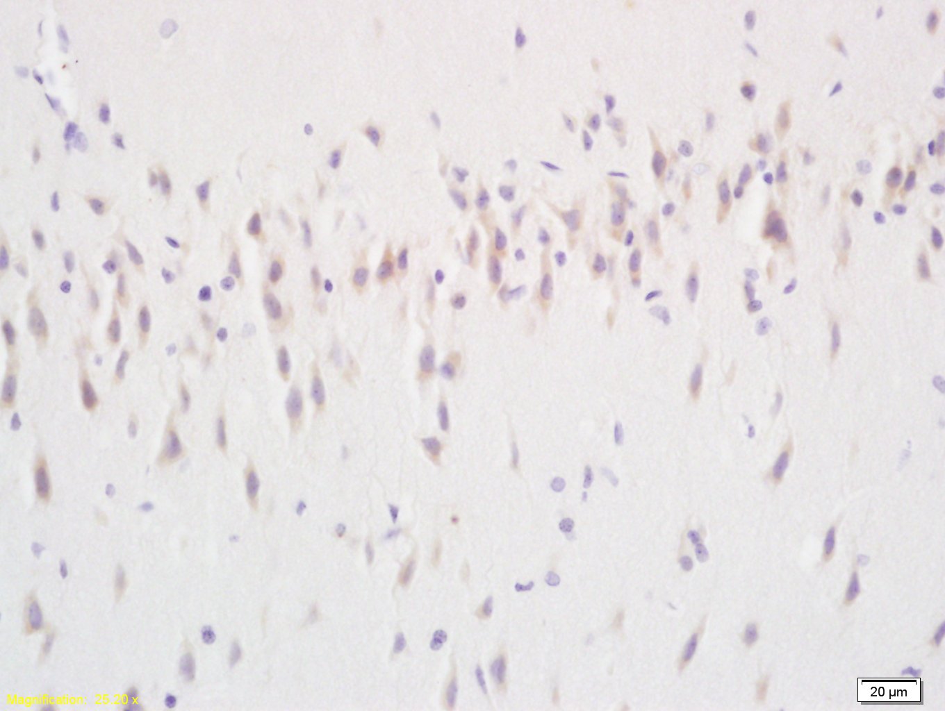
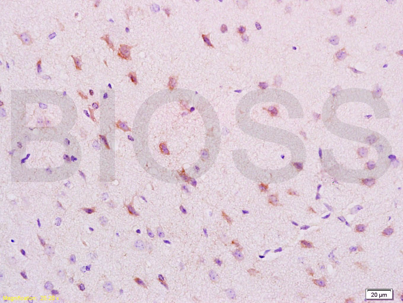
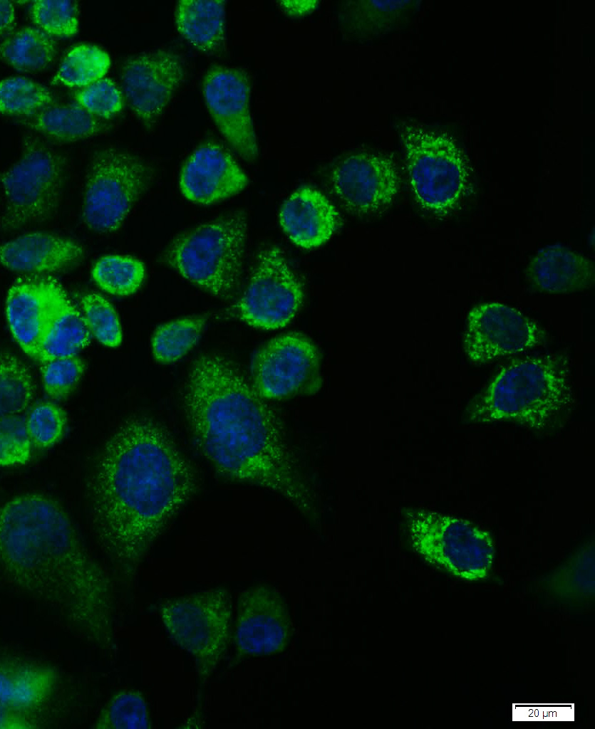
Target Protein: phospho-NRP1 (Thr916) Rabbit pAb

IR: Immunogen Range:LN(p-T)QS

Clonality: Polyclonal

Isotype: IgG

Entrez Gene: 8829

Swiss Prot: Q5T7F3

Source: KLH conjugated Synthesised phosphopeptide derived from human NRP1 around the phosphorylation site of Thr916:LN(p-T)QS 

Purification: affinity purified by Protein A

Storage: 0.01M TBS (pH7.4) with 1% BSA, 0.02% Proclin300 and 50% Glycerol. Shipped at 4℃. Store at -20℃ for one year. Avoid repeated freeze/thaw cycles.

Background: This gene encodes one of two neuropilins, which contain specific protein domains which allow them to participate in several different types of signaling pathways that control cell migration. Neuropilins contain a large N-terminal extracellular domain, made up of complement-binding, coagulation factor V/VIII, and meprin domains. These proteins also contains a short membrane-spanning domain and a small cytoplasmic domain. Neuropilins bind many ligands and various types of co-receptors; they affect cell survival, migration, and attraction. Some of the ligands and co-receptors bound by neuropilins are vascular endothelial growth factor (VEGF) and semaphorin family members. Several alternatively spliced transcript variants that encode different protein isoforms have been described for this gene. [provided by RefSeq, Oct 2011]

Size: 200ul

Concentration: 1mg/ml

Applications: IHC-P=1:100-500,IHC-F=1:100-500,IF=1:100-500,ICC/IF=1:100

Cross Reactive Species: Human,Rat (predicted: Dog)

For research use only. Not intended for diagnostic or therapeutic use.

VALIDATION IMAGES
Tissue/cell: rat brain tissue; 4% Paraformaldehyde-fixed and paraffin-embedded; Antigen retrieval: citrate buffer ( 0.01M, pH 6.0 ), Boiling bathing for 15min; Block endogenous peroxidase by 3% Hydrogen peroxide for 30min; Blocking buffer (normal goat serum,C-0005) at 37℃ for 20 min; Incubation: Anti-phospho-NRP1(Thr916) Polyclonal Antibody, Unconjugated(bs-5519R) 1:200, overnight at 4°C, followed by conjugation to the secondary antibody(SP-0023) and DAB(C-0010) staining
Hela cell; 4% Paraformaldehyde-fixed; Triton X-100 at room temperature for 20 min; Blocking buffer (normal goat serum, C-0005) at 37°C for 20 min; Antibody incubation with (phospho-NRP1 (Thr916)) polyclonal Antibody, Unconjugated (bs-5519R) 1:100, 90 minutes at 37°C; followed by a conjugated Goat Anti-Rabbit IgG antibody at 37°C for 90 minutes, DAPI (blue, C02-04002) was used to stain the cell nuclei.
 
PRODUCT SPECIFIC PUBLICATIONS