bs-5516R [Primary Antibody]
Rabbit  Anti-phospho-NFATc2 (Ser330) Rabbit pAb  Polyclonal Antibody
www.bioss.com.cn
sales@bioss.com.cn
techsupport@bioss.com.cn
400-901-9800
DATASHEET

Host: Rabbit

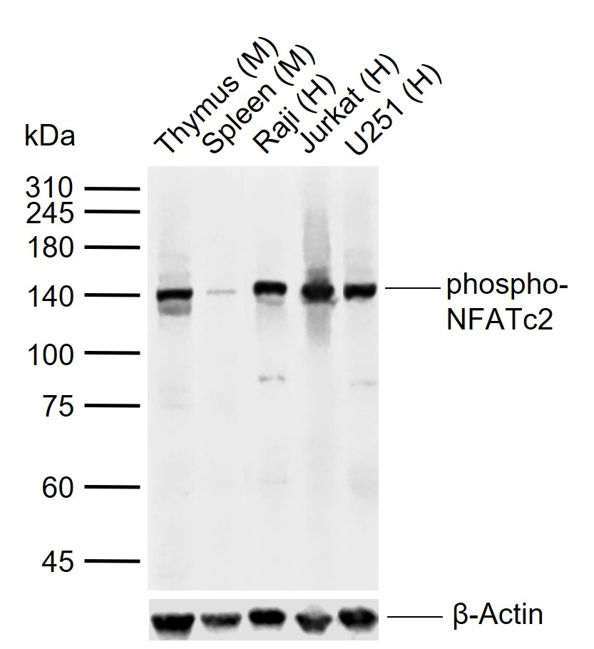
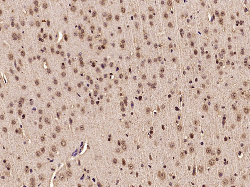
Target Protein: phospho-NFATc2 (Ser330) Rabbit pAb

IR: Immunogen Range:DP(p-S)PV

Clonality: Polyclonal

Isotype: IgG

Entrez Gene: 4773

Swiss Prot: Q13469

Source: KLH conjugated Synthesised phosphopeptide derived from human NFATc2 around the phosphorylation site of Ser330:DP(p-S)PV 

Purification: affinity purified by Protein A

Storage: 0.01M TBS (pH7.4) with 1% BSA, 0.02% Proclin300 and 50% Glycerol. Shipped at 4℃. Store at -20℃ for one year. Avoid repeated freeze/thaw cycles.

Background: This gene is a member of the nuclear factor of activated T cells (NFAT) family. The product of this gene is a DNA-binding protein with a REL-homology region (RHR) and an NFAT-homology region (NHR). This protein is present in the cytosol and only translocates to the nucleus upon T cell receptor (TCR) stimulation, where it becomes a member of the nuclear factors of activated T cells transcription complex. This complex plays a central role in inducing gene transcription during the immune response. Alternate transcriptional splice variants encoding different isoforms have been characterized. [provided by RefSeq, Apr 2012]

Size: 100ul

Concentration: 1mg/ml

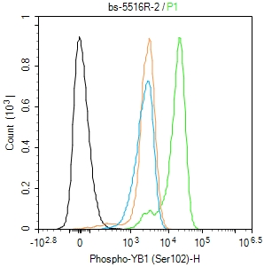
Applications: WB=1:500-2000,IHC-P=1:100-500,IHC-F=1:100-500,IF=1:100-500,Flow-Cyt=1ug/Test

Cross Reactive Species: Human,Mouse,Rat (predicted: Pig,Cow,Dog,Horse)

For research use only. Not intended for diagnostic or therapeutic use.

VALIDATION IMAGES
Sample: Lane 1: Mouse Thymus tissue lysates Lane 2: Mouse Spleen tissue lysates Lane 3: Human Raji cell lysates Lane 4: Human Jurkat cell lysates Lane 5: Human U251 cell lysates Primary: Anti-phospho-NFATc2 (Ser330) (bs-5516R) at 1/1000 dilution Secondary: IRDye800CW Goat Anti-Rabbit IgG at 1/20000 dilution Predicted band size: 100 kDa Observed band size: 140 kDa
Paraformaldehyde-fixed, paraffin embedded (Rat brain); Antigen retrieval by microwave in sodium citrate buffer (pH6.0) ; Block endogenous peroxidase by 3% hydrogen peroxide for 30 minutes; Blocking buffer (3% BSA) at RT for 30min; Antibody incubation with (phospho-NFATc2(Ser330)) Polyclonal Antibody, Unconjugated (bs-5516R) at 1:400 overnight at 4°C, followed by conjugation to the secondary antibody (labeled with HRP)and DAB staining.
Blank control(black line):Molt4. Primary Antibody (green line): Rabbit Anti-phospho-NFATc2 (Ser330) antibody (bs-5516R) Dilution:1ug/Test; Secondary Antibody(white blue line): Goat anti-rabbit IgG-AF488 Dilution: 0.5ug/Test. Isotype control(orange line): Normal Rabbit IgG Protocol The cells were fixed with 4% PFA (10min at room temperature)and then permeabilized with 90% ice-cold methanol for 20 min at -20℃, The cells were then incubated in 5%BSA to block non-specific protein-protein interactions for 30 min at room temperature .Cells stained with Primary Antibody for 30 min at room temperature. The secondary antibody used for 40 min at room temperature. Acquisition of 20,000 events was performed.
 
PRODUCT SPECIFIC PUBLICATIONS