bs-0102M [Primary Antibody]
Mouse  Anti-PKM2  Polyclonal Antibody
www.bioss.com.cn
sales@bioss.com.cn
techsupport@bioss.com.cn
400-901-9800
DATASHEET

Host: Mouse

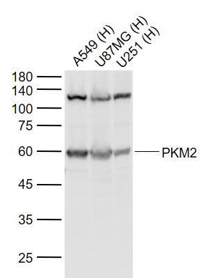
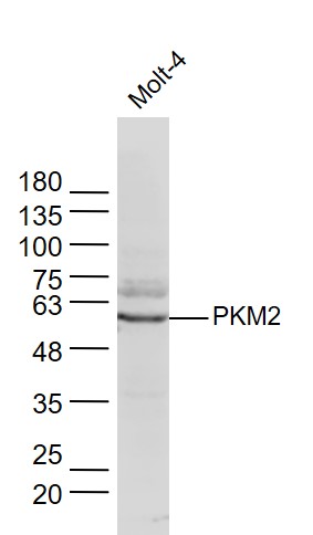
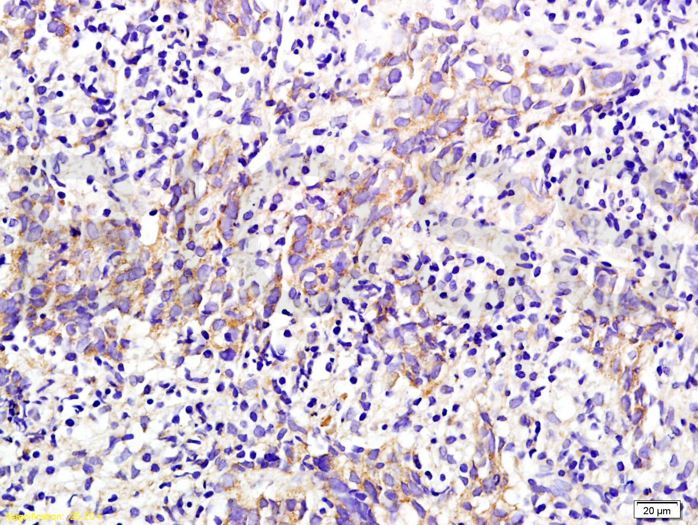
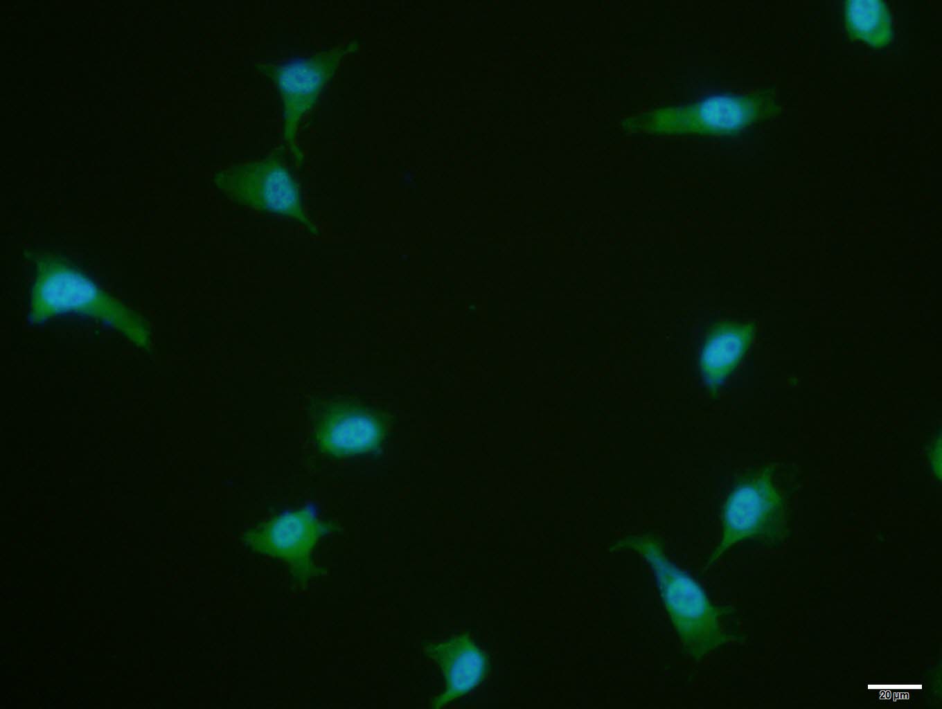
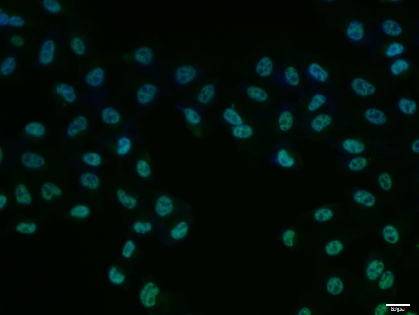
Target Protein: PKM2

IR: Immunogen Range:51-150/531

Clonality: Polyclonal

Isotype: IgG

Entrez Gene: 5315

Swiss Prot: P14618

Source: KLH conjugated synthetic peptide derived from human PKM2:51-150/531 

Purification: affinity purified by Protein A

Storage: 0.01M TBS (pH7.4) with 1% BSA, 0.02% Proclin300 and 50% Glycerol. Shipped at 4℃. Store at -20℃ for one year. Avoid repeated freeze/thaw cycles.

Background: The protein encoded by this gene is a pyruvate kinase that catalyzes the production of phosphoenolpyruvate from pyruvate and ATP. This protein has been shown to interact with thyroid hormone, and thus may mediate cellular metabolic effects induced by thyroid hormones. This protein has been found to bind Opa protein, a bacterial outer membrane protein involved in gonococcal adherence to and invasion of human cells, suggesting a role of this protein in bacterial pathogenesis. Three alternatively spliced transcript variants encoding two distinct isoforms have been reported.

Size: 100ul

Concentration: 1mg/ml

Applications: WB=1:500-2000,IHC-P=1:100-500,IHC-F=1:100-500,ICC/IF=1:100,IF=1:100-500,ELISA=1:5000-10000

Cross Reactive Species: Human,Mouse,Rat (predicted: Rabbit,Pig,Cow,Horse)

For research use only. Not intended for diagnostic or therapeutic use.

VALIDATION IMAGES
Sample: Lane 1: A549 (Human) Cell Lysate at 30 ug Lane 2: U87MG (Human) Cell Lysate at 30 ug Lane 3: U251 (Human) Cell Lysate at 30 ug Primary: Anti-PKM2 (bs-0102M) at 1/1000 dilution Secondary: IRDye800CW Goat Anti-Mouse IgG at 1/20000 dilution Predicted band size: 60 kD Observed band size: 60 kD
Sample: Hela Cell (Human) Lysate at 40 ug NIH/3T3 Cell (Mouse) Lysate at 40 ug Muscle (Mouse) Lysate at 40 ug Jurkat Cell (Human) Lysate at 40 ug Primary: Anti-PKM2 (bs-0102M) at 1/300 dilution Secondary: IRDye800CW Goat Anti-Mouse IgG at 1/20000 dilution Predicted band size: 58 kD Observed band size: 58 kD
Sample: MOLT-4(Human) Cell Lysate at 30 ug Primary: Anti-PKM2 (bs-0102M) at 1/300 dilution Secondary: IRDye800CW Goat Anti-Mouse IgG at 1/20000 dilution Predicted band size: 58 kD Observed band size: 58 kD
Tissue/cell: Human nasopharyngeal carcinoma; 4% Paraformaldehyde-fixed and paraffin-embedded; Antigen retrieval: citrate buffer ( 0.01M, pH 6.0 ), Boiling bathing for 15min; Block endogenous peroxidase by 3% Hydrogen peroxide for 30min; Blocking buffer (normal goat serum,C-0005) at 37℃ for 20 min; Incubation: Anti-M2-PK Polyclonal Antibody, Unconjugated(bs-0102M) 1:200, overnight at 4°C, followed by conjugation to the secondary antibody(SP-0024) and DAB(C-0010) staining
U-87MG cell; 4% Paraformaldehyde-fixed; Triton X-100 at room temperature for 20 min; Blocking buffer (normal goat serum, C-0005) at 37°C for 20 min; Antibody incubation with (PKM2) polyclonal Antibody, Unconjugated (bs-0102M) 1:100, 90 minutes at 37°C; followed by a conjugated Goat Anti-Mouse IgG antibody at 37°C for 90 minutes, DAPI (blue, C02-04002) was used to stain the cell nuclei.
Hela cell; 4% Paraformaldehyde-fixed; Triton X-100 at room temperature for 20 min; Blocking buffer (normal goat serum, C-0005) at 37°C for 20 min; Antibody incubation with (PKM2) polyclonal Antibody, Unconjugated (bs-0102M) 1:100, 90 minutes at 37°C; followed by a conjugated Goat Anti-Mouse IgG antibody at 37°C for 90 minutes, DAPI (blue, C02-04002) was used to stain the cell nuclei.
Tissue/cell: rat colitis tissue;4% Paraformaldehyde-fixed and paraffin-embedded; Antigen retrieval: citrate buffer ( 0.01M, pH 6.0 ), Boiling bathing for 15min; Blocking buffer (normal goat serum,C-0005) at 37℃ for 20 min; Incubation: Anti-M2-PK Polyclonal Antibody, Unconjugated(bs-0102M) 1:200, overnight at 4°C; The secondary antibody was Goat Anti-Mouse IgG, Cy5 conjugated(bs-0296G-Cy5)used at 1:200 dilution for 40 minutes at 37°C.
 
PRODUCT SPECIFIC PUBLICATIONS