bs-5447R [Primary Antibody]
Rabbit  Anti-phospho-IGF1R (Tyr980) Rabbit pAb  Polyclonal Antibody
www.bioss.com.cn
sales@bioss.com.cn
techsupport@bioss.com.cn
400-901-9800
DATASHEET

Host: Rabbit

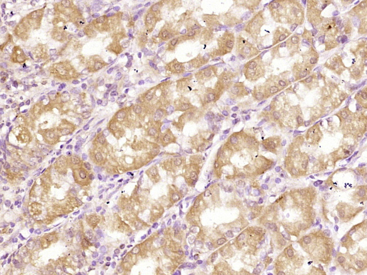
Target Protein: phospho-IGF1R (Tyr980) Rabbit pAb

IR: Immunogen Range:PE(p-Y)FS

Clonality: Polyclonal

Isotype: IgG

Entrez Gene: 3480

Swiss Prot: P08069

Source: KLH conjugated Synthesised phosphopeptide derived from human IGF1R around the phosphorylation site of Tyr980:PE(p-Y)FS 

Purification: affinity purified by Protein A

Storage: 0.01M TBS (pH7.4) with 1% BSA, 0.02% Proclin300 and 50% Glycerol. Shipped at 4℃. Store at -20℃ for one year. Avoid repeated freeze/thaw cycles.

Background: This receptor binds insulin-like growth factor with a high affinity. It has tyrosine kinase activity. The insulin-like growth factor I receptor plays a critical role in transformation events. Cleavage of the precursor generates alpha and beta subunits. It is highly overexpressed in most malignant tissues where it functions as an anti-apoptotic agent by enhancing cell survival. [provided by RefSeq, Jul 2008].

Size: 200ul

Concentration: 1mg/ml

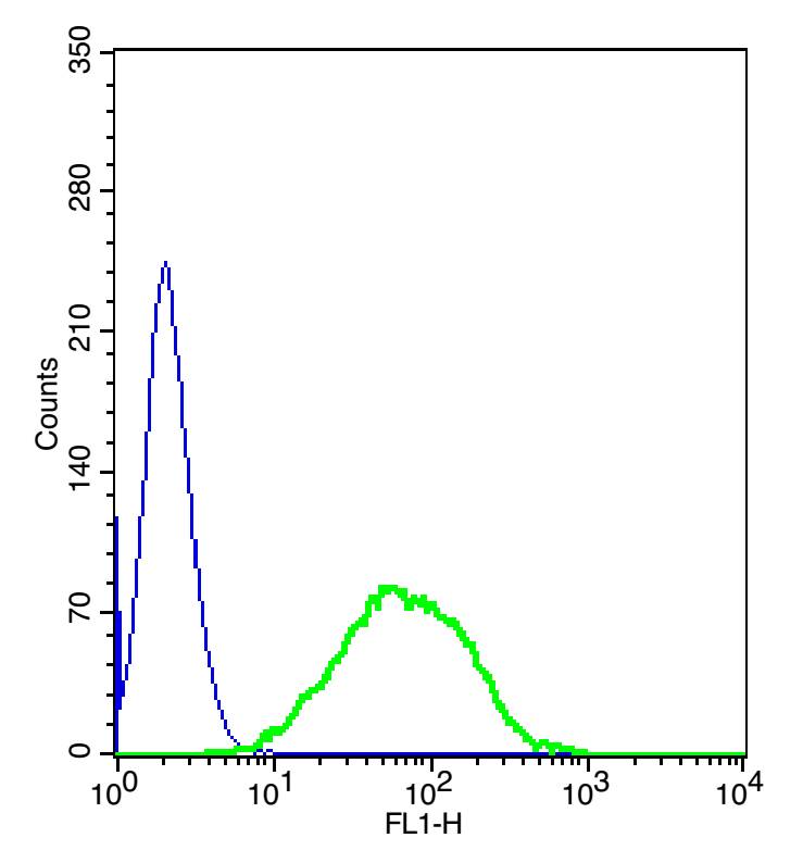
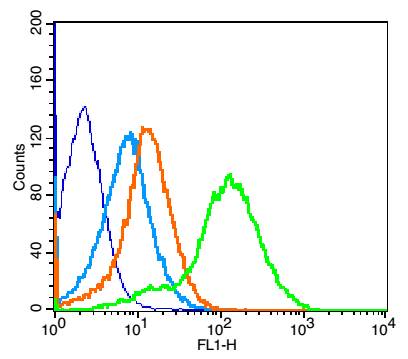
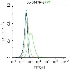
Applications: IHC-P=1:100-500,IHC-F=1:100-500,IF=1:100-500,Flow-Cyt=1μg/Test

Cross Reactive Species: Human,Mouse (predicted: Rat)

For research use only. Not intended for diagnostic or therapeutic use.

VALIDATION IMAGES
Tissue/cell: human kidney carcinoma; 4% Paraformaldehyde-fixed and paraffin-embedded; Antigen retrieval: citrate buffer ( 0.01M, pH 6.0 ), Boiling bathing for 15min; Block endogenous peroxidase by 3% Hydrogen peroxide for 30min; Blocking buffer (normal goat serum,C-0005) at 37℃ for 20 min; Incubation: Anti-phospho-IGF1R(Tyr980) Polyclonal Antibody, Unconjugated(bs-5447R) 1:200, overnight at 4°C, followed by conjugation to the secondary antibody(SP-0023) and DAB(C-0010) staining
Paraformaldehyde-fixed, paraffin embedded (human stomach); Antigen retrieval by boiling in sodium citrate buffer (pH6.0) for 15min; Block endogenous peroxidase by 3% hydrogen peroxide for 20 minutes; Blocking buffer (normal goat serum) at 37°C for 30min; Antibody incubation with (RELM beta) Polyclonal Antibody, Unconjugated (bs-5774R) at 1:400 overnight at 4°C, followed by operating according to SP Kit(Rabbit) (sp-0023) instructionsand DAB staining.
The blue histogram is unstained cells(Hepg2 cells). The green histogram is cells stained with Rabbit Anti-phospho-IGF1R (Tyr980) antibody (bs-5447R) plus secondary antibody(bs-0295G-FITC).Concentration 1:20
Blank control(blue): Mouse Kidney (fixed with 2% paraformaldehyde for 10 min at 37℃). Primary Antibody:Rabbit Anti-phospho-IGF1R (Tyr980) antibody (bs-5447R,Green); Dilution: 1μg in 100 μL 1X PBS containing 0.5% BSA; Isotype Control Antibody: Rabbit IgG(orange) ,used under the same conditions; Secondary Antibody: Goat anti-rabbit IgG-FITC(white blue), Dilution: 1:200 in 1 X PBS containing 0.5% BSA.
Blank control: MCF7. Primary Antibody (green line): Rabbit Anti-phospho-IGF1R (Tyr980) antibody (bs-5447R) Dilution: 2μg /10^6 cells; Isotype Control Antibody (orange line): Rabbit IgG . Secondary Antibody : Goat anti-rabbit IgG-FITC Dilution: 1μg /test. Protocol The cells were incubated in 5%BSA to block non-specific protein-protein interactions for 30 min at room temperature .Cells stained with Primary Antibody for 30 min at room temperature. The secondary antibody used for 40 min at room temperature. Acquisition of 20,000 events was performed.
 
PRODUCT SPECIFIC PUBLICATIONS