bs-1049R [Primary Antibody]
Rabbit  Anti-CHRNA7 Rabbit pAb  Polyclonal Antibody
www.bioss.com.cn
sales@bioss.com.cn
techsupport@bioss.com.cn
400-901-9800
DATASHEET

Host: Rabbit

Target Protein: CHRNA7 Rabbit pAb

IR: Immunogen Range:441-502/502

Clonality: Polyclonal

Isotype: IgG

Entrez Gene: 1139

Swiss Prot: Q8IUZ4

Source: KLH conjugated synthetic peptide derived from human CHRNA7:441-502/502 

Purification: affinity purified by Protein A

Storage: 0.01M TBS (pH7.4) with 1% BSA, 0.02% Proclin300 and 50% Glycerol. Shipped at 4℃. Store at -20℃ for one year. Avoid repeated freeze/thaw cycles.

Background: The Nicotinic Acetylcholine Receptors are members of a superfamily of ligand gated ion channels that mediate fast signal transmission at synapses. These receptors are thought to be hetero pentamers composed of homologous subunits. The proposed structure for each subunit is a conserved N terminal extracellular domain followed by three conserved transmembrane domains, a variable cytoplasmic loop, a fourth conserved transmembrane domain, and a short C terminal extracellular region. The Nicotinic Acetylcholine Receptor alpha 7 forms a homo oligomeric channel, displays marked permeability to calcium ions and is a major component of brain nicotinic receptors that are blocked by, and highly sensitive to, alpha bungarotoxin. Once this receptor binds acetylcholine, it undergoes an extensive change in conformation that affects all subunits and leads to opening of an ion conducting channel across the plasma membrane.

Size: 100ul

Concentration: 1mg/ml

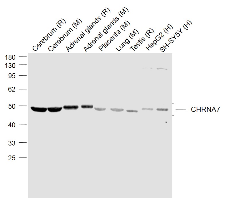
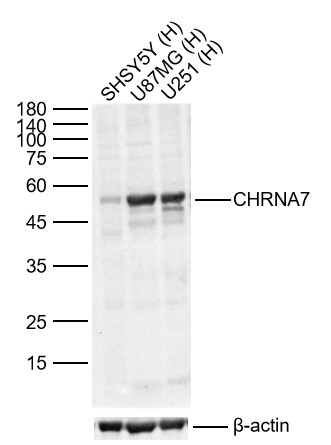
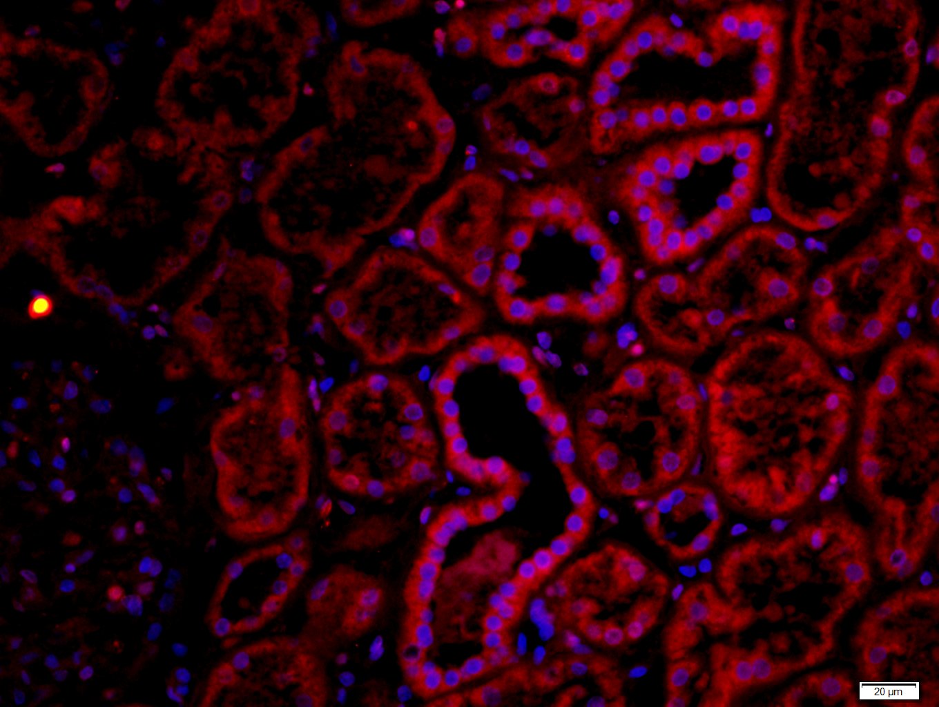
Applications: WB=1:500-2000,IHC-P=1:100-500,IHC-F=1:100-500,IF=1:100-500

Cross Reactive Species: Human,Mouse,Rat (predicted: Chicken)

For research use only. Not intended for diagnostic or therapeutic use.

VALIDATION IMAGES
Sample: Lane 1: Cerebrum (Rat) Lysate at 40 ug Lane 2: Cerebrum (Mouse) Lysate at 40 ug Lane 3: Adrenal glands (Rat) Lysate at 40 ug Lane 4: Adrenal glands (Mouse) Lysate at 40 ug Lane 5: Placenta (Mouse) Lysate at 40 ug Lane 6: Lung (Mouse) Lysate at 40 ug Lane 7: Testis (Rat) Lysate at 40 ug Lane 8: HepG2 (Human) Cell Lysate at 30 ug Lane 9: SH-SY5Y (Human) Cell Lysate at 30 ug Primary: Anti-CHRNA7 (bs-1049R) at 1/1000 dilution Secondary: IRDye800CW Goat Anti-Rabbit IgG at 1/20000 dilution Predicted band size: 56’50 kD Observed band size: 48 kD
Sample: Lane 1: Human SH-SY5Y cell Lysates Lane 2: Human U-87 MG cell Lysates Lane 3: Human U251 cell Lysates Primary: Anti-CHRNA7 (bs-1049R) at 1/1000 dilution Secondary: IRDye800CW Goat Anti-Rabbit IgG at 1/20000 dilution Predicted band size: 55kDa Observed band size: 55kDa
Tissue/cell: mouse brain tissue; 4% Paraformaldehyde-fixed and paraffin-embedded; Antigen retrieval: citrate buffer ( 0.01M, pH 6.0 ), Boiling bathing for 15min; Block endogenous peroxidase by 3% Hydrogen peroxide for 30min; Blocking buffer (normal goat serum,C-0005) at 37℃ for 20 min; Incubation: Anti-CHRNA7 Polyclonal Antibody, Unconjugated(bs-1049R) 1:200, overnight at 4°C, followed by conjugation to the secondary antibody(SP-0023) and DAB(C-0010) staining
Tissue/cell: human kidney tissue;4% Paraformaldehyde-fixed and paraffin-embedded; Antigen retrieval: citrate buffer ( 0.01M, pH 6.0 ), Boiling bathing for 15min; Blocking buffer (normal goat serum,C-0005) at 37℃ for 20 min; Incubation: Anti-CHRNA7 Polyclonal Antibody, Unconjugated(bs-1049R) 1:200, overnight at 4°C; The secondary antibody was Goat Anti-Rabbit IgG, Cy3 conjugated(bs-0295G-Cy3)used at 1:200 dilution for 40 minutes at 37°C. DAPI(5ug/ml,blue,C-0033) was used to stain the cell nuclei
 
PRODUCT SPECIFIC PUBLICATIONS