bs-6425R [Primary Antibody]
Rabbit  Anti-TRP12/TRPV4 Rabbit pAb  Polyclonal Antibody
www.bioss.com.cn
sales@bioss.com.cn
techsupport@bioss.com.cn
400-901-9800
DATASHEET

Host: Rabbit

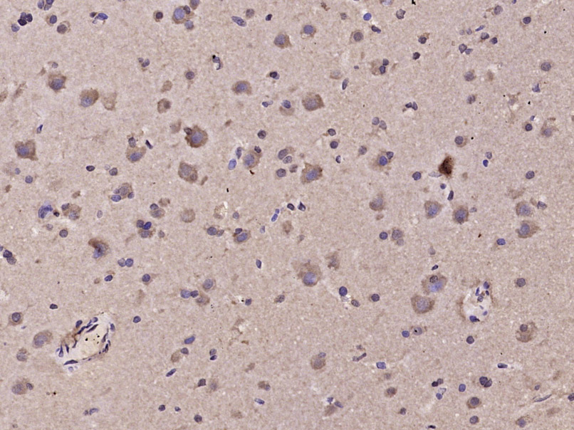
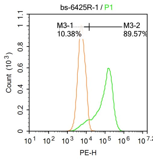
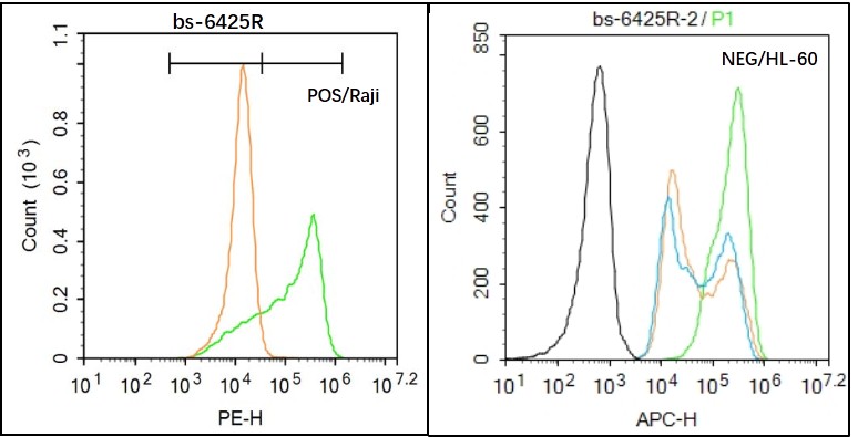
Target Protein: TRP12/TRPV4 Rabbit pAb

IR: Immunogen Range:301-400/871

Clonality: Polyclonal

Isotype: IgG

Entrez Gene: 59341

Swiss Prot: Q9HBA0

Source: KLH conjugated synthetic peptide derived from human TRPV4:301-400/871 

Purification: affinity purified by Protein A

Storage: 0.01M TBS (pH7.4) with 1% BSA, 0.02% Proclin300 and 50% Glycerol. Shipped at 4℃. Store at -20℃ for one year. Avoid repeated freeze/thaw cycles.

Background: The detection of noxious stimuli (chemical, mechanical, or thermal) occurs predominantly at the peripheral terminals of primary afferent neurons. This information is ultimately transmitted to the central nervous system to evoke appropriate protective reflexes. TRPV4 is a non selective calcium permeant, swell activated, cation channel probably involved in osmotic and mechano sensitivity. Activation by exposure to hypotonicity within the physiological range, low pH, citrate and phorbol esters exhibits an outward rectification. Once activated the channel seems to be regulated in a calmodulin dependent manner, with a negative feedback mechanism.

Size: 200ul

Concentration: 1mg/ml

Applications: IHC-P=1:100-500,IHC-F=1:100-500,IF=1:100-500,Flow-Cyt=1ug/test,ICC/IF=1:100

Cross Reactive Species: Human,Mouse (predicted: Rat,Pig,Cow,Chicken,Dog)

For research use only. Not intended for diagnostic or therapeutic use.

VALIDATION IMAGES
Paraformaldehyde-fixed, paraffin embedded (Human brain glioma); Antigen retrieval by microwave in sodium citrate buffer (pH6.0) ; Block endogenous peroxidase by 3% hydrogen peroxide for 30 minutes; Blocking buffer (3% BSA) at RT for 30min; Antibody incubation with (TRP12) Polyclonal Antibody, Unconjugated (bs-6425R) at 1:400 overnight at 4°C, followed by conjugation to the secondary antibody (labeled with HRP)and DAB staining.
NIH/3T3 cell; 4% Paraformaldehyde-fixed; Triton X-100 at room temperature for 20 min; Blocking buffer (normal goat serum, C-0005) at 37°C for 20 min; Antibody incubation with (TRP12/TRPV4) polyclonal Antibody, Unconjugated (bs-6425R) 1:100, 90 minutes at 37°C; followed by a conjugated Goat Anti-Rabbit IgG antibody at 37°C for 90 minutes, DAPI (blue, C02-04002) was used to stain the cell nuclei.
Blank control: Raji. Primary Antibody (green line): Rabbit Anti-TRP12 antibody (bs-6425R) Dilution: 1μg /10^6 cells; Isotype Control Antibody (orange line): Rabbit IgG . Secondary Antibody : Goat anti-rabbit IgG-PE Dilution: 1μg /test. Protocol The cells were fixed with 4% PFA (10min at room temperature)and then permeabilized with PBST for 20 min at room temperature. The cells were then incubated in 5%BSA to block non-specific protein-protein interactions for 30 min at at room temperature .Cells stained with Primary Antibody for 30 min at room temperature. The secondary antibody used for 40 min at room temperature. Acquisition of 20,000 events was performed.
Black line : Positive blank control (Raji); Negative blank control (HL60) Green line : Primary Antibody (Rabbit Anti-TRP12 antibody (bs-6425R) ) Orange line:Isotype Control Antibody (Rabbit IgG) . Blue line : Secondary Antibody (Goat anti-rabbit IgG-PE)/(Goat anti-rabbit IgG-AF647) Raji(Positive)and HL60(Negative control)cells (black) were fixed with 4% PFA for 10min at room temperature, permeabilized with PBST for 20 min at room temperature, and incubated in 5% BSA blocking buffer for 30 min at room temperature. Cells were then stained with TRP12 Antibody(bs-6425R)at 1:50 dilution in blocking buffer and incubated for 30 min at room temperature, washed twice with 2% BSA in PBS, followed by secondary antibody(blue) incubation for 40 min at room temperature. Acquisitions of 20,000 events were performed. Cells stained with primary antibody (green), and isotype control (orange).
 
PRODUCT SPECIFIC PUBLICATIONS