bs-0681R [Primary Antibody]
Rabbit  Anti-Insulin Receptor Rabbit pAb  Polyclonal Antibody
www.bioss.com.cn
sales@bioss.com.cn
techsupport@bioss.com.cn
400-901-9800
DATASHEET

Host: Rabbit

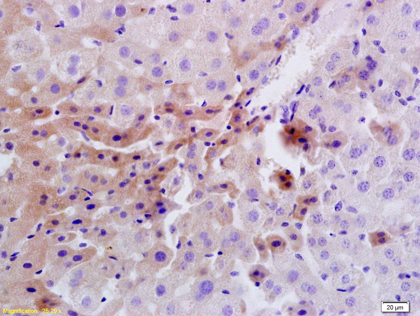
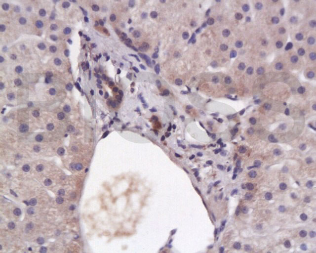
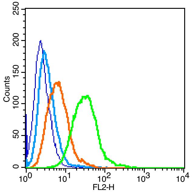
Target Protein: Insulin Receptor Rabbit pAb

IR: Immunogen Range:51-150/1384

Clonality: Polyclonal

Isotype: IgG

Entrez Gene: 3643

Swiss Prot: P06213

Source: KLH conjugated synthetic peptide derived from human Insulin Receptor:51-150/1384 

Purification: affinity purified by Protein A

Storage: 0.01M TBS (pH7.4) with 1% BSA, 0.02% Proclin300 and 50% Glycerol. Shipped at 4℃. Store at -20℃ for one year. Avoid repeated freeze/thaw cycles.

Background: The insulin receptor is a heterotetrameric membrane glycoprotein with tyrosine-protein kinase activity, consisting of disulfide-linked subunits in a beta-alpha-alpha-beta configuration. The beta subunit possesses a single transmembrane domain, whereas the alpha subunit is completely extracellular. The alpha chains contribute to the formation of the ligand-binding domain, while the beta chains carry the kinase domain. Binding of insulin to the insulin receptor stimulates its association with downstream mediators including IRS1 and phosphatidylinositol 3'-kinase (PI3K) which leads to glucose uptake. Two transcript variants encoding different isoforms have been found for this gene produced by alternative splicing.

Protein kinases are enzymes that transfer a phosphate group from a phosphate donor, generally the g phosphate of ATP, onto an acceptor amino acid in a substrate protein. By this basic mechanism, protein kinases mediate most of the signal transduction in eukaryotic cells, regulating cellular metabolism, transcription, cell cycle progression, cytoskeletal rearrangement and cell movement, apoptosis, and differentiation. With more than 500 gene products, the protein kinase family is one of the largest families of proteins in eukaryotes. The family has been classified in 8 major groups based on sequence comparison of their tyrosine (PTK) or serine/threonine (STK) kinase catalytic domains. The tyrosine kinase (TK) group is mainly involved in the regulation of cell-cell interactions such as differentiation, adhesion, motility and death. There are currently about 90 TK genes sequenced, 58 are of receptor protein TK (e.g. EGFR, EPH, FGFR, PDGFR, TRK, and VEGFR families), and 32 of cytosolic TK (e.g. ABL, FAK, JAK, and SRC families).

Size: 100ul

Concentration: 1mg/ml

Applications: WB=1:500-2000,IHC-P=1:100-500,IHC-F=1:100-500,IF=1:100-500,Flow-Cyt=1ug/Test

Cross Reactive Species: Human,Mouse,Rat (predicted: Rabbit,Sheep,Cow,Chicken,Dog,Horse)

For research use only. Not intended for diagnostic or therapeutic use.

VALIDATION IMAGES
Sample: Lane 1: MCF-7 (Human) Cell Lysate at 30 ug Lane 2: A549 (Human) Cell Lysate at 30 ug Lane 3: 293T (Human) Cell Lysate at 30 ug Lane 4: Hela (Human) Cell Lysate at 30 ug Lane 5: Liver (Rat) Lysate at 40 ug Lane 6: Lymph node (Mouse) Lysate at 40 ug Lane 7: MOLT-4 (Human) Cell Lysate at 30 ug Lane 8: NIH/3T3 (Mouse) Cell Lysate at 30 ug Primary: Anti-Insulin Receptor (bs-0681R) at 1/1000 dilution Secondary: IRDye800CW Goat Anti-Rabbit IgG at 1/20000 dilution Predicted band size: 120 kD Observed band size: 120 kD
Sample: Lane 1: HepG2 (Human) Cell Lysate at 30 ug Lane 2: 293T (Human) Cell Lysate at 30 ug Lane 3: Lovo (Human) Cell Lysate at 30 ug Lane 4: Molt-4 (Human) Cell Lysate at 30 ug Lane 5: SW480 (Human) Cell Lysate at 30 ug Lane 6: K562 (Human) Cell Lysate at 30 ug Primary: Anti-Insulin Receptor (bs-0681R) at 1/1000 dilution Secondary: IRDye800CW Goat Anti-Rabbit IgG at 1/20000 dilution Predicted band size: 120 kD Observed band size: 120 kD
Tissue/cell: rat liver tissue; 4% Paraformaldehyde-fixed and paraffin-embedded; Antigen retrieval: citrate buffer ( 0.01M, pH 6.0 ), Boiling bathing for 15min; Block endogenous peroxidase by 3% Hydrogen peroxide for 30min; Blocking buffer (normal goat serum,C-0005) at 37℃ for 20 min; Incubation: Anti-Insulin Receptor/CD220 Polyclonal Antibody, Unconjugated(bs-0681R) 1:200, overnight at 4°C, followed by conjugation to the secondary antibody(SP-0023) and DAB(C-0010) staining
Tissue/cell: human liver tissue; 4% Paraformaldehyde-fixed and paraffin-embedded; Antigen retrieval: citrate buffer ( 0.01M, pH 6.0 ), Boiling bathing for 15min; Block endogenous peroxidase by 3% Hydrogen peroxide for 30min; Blocking buffer (normal goat serum,C-0005) at 37℃ for 20 min; Incubation: Anti-Insulin Receptor/CD220 Polyclonal Antibody, Unconjugated(bs-0681R) 1:200, overnight at 4°C, followed by conjugation to the secondary antibody(SP-0023) and DAB(C-0010) staining
Blank control: Raji (blue). Primary Antibody: Rabbit Anti-Insulin Receptor alpha antibody(bs-0681R), Dilution: 1μg in 100 μL 1X PBS containing 0.5% BSA; Isotype Control Antibody: Rabbit IgG(orange),used under the same conditions ); Secondary Antibody: Goat anti-rabbit IgG-PE(white blue), Dilution: 1:200 in 1 X PBS containing 0.5% BSA. Protocol The cells were fixed with 2% paraformaldehyde (10 min) , then permeabilized with 90% ice-cold methanol for 30 min on ice. Primary antibody (bs-0681R, 1μg /1x10^6 cells) were incubated for 30 min on the ice, followed by 1 X PBS containing 0.5% BSA + 1 0% goat serum (15 min) to block non-specific protein-protein interactions. Then the Goat Anti-rabbit IgG/PE antibody was added into the blocking buffer mentioned above to react with the primary antibody at 1/200 dilution for 30 min on ice. Acquisition of 20,000 events was performed.
 
PRODUCT SPECIFIC PUBLICATIONS