bs-7535R [Primary Antibody]
Rabbit  Anti-PGC1 alpha + beta Rabbit pAb  Polyclonal Antibody
www.bioss.com.cn
sales@bioss.com.cn
techsupport@bioss.com.cn
400-901-9800
DATASHEET

Host: Rabbit

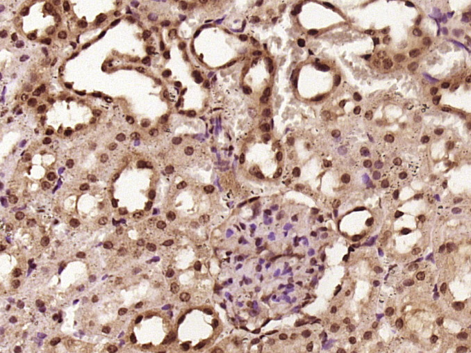
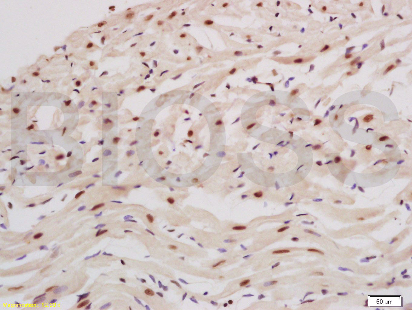
Target Protein: PGC1 alpha + beta Rabbit pAb

IR: Immunogen Range:151-250/798

Clonality: Polyclonal

Isotype: IgG

Entrez Gene: 10891

Swiss Prot: Q9UBK2

Source: KLH conjugated synthetic peptide derived from human PGC1 alpha + beta:151-250/798 

Purification: affinity purified by Protein A

Storage: 0.01M TBS (pH7.4) with 1% BSA, 0.02% Proclin300 and 50% Glycerol. Shipped at 4℃. Store at -20℃ for one year. Avoid repeated freeze/thaw cycles.

Background: The protein encoded by this gene is a transcriptional coactivator that regulates the genes involved in energy metabolism. This protein interacts with PPARgamma, which permits the interaction of this protein with multiple transcription factors. This protein can interact with, and regulate the activities of, cAMP response element binding protein (CREB) and nuclear respiratory factors (NRFs). It provides a direct link between external physiological stimuli and the regulation of mitochondrial biogenesis, and is a major factor that regulates muscle fiber type determination. This protein may be also involved in controlling blood pressure, regulating cellular cholesterol homoeostasis, and the development of obesity. [provided by RefSeq].

Size: 200ul

Concentration: 1mg/ml

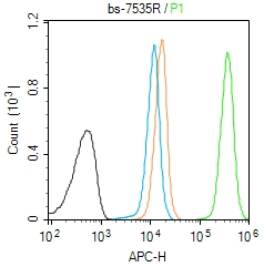
Applications: WB=1:500-2000,IHC-P=1:100-500,IHC-F=1:100-500,IF=1:100-500,Flow-Cyt=1μg/Test

Cross Reactive Species: Human,Rat (predicted: Mouse,Rabbit,Pig,Cow,Dog,Horse)

For research use only. Not intended for diagnostic or therapeutic use.

VALIDATION IMAGES
Sample: Lane 1: Molt-4 (Human) Cell Lysate at 30 ug Lane 2: A431 (Human) Cell Lysate at 30 ug Lane 3: HepG2 (Human) Cell Lysate at 30 ug Lane 4: Hela (Human) Cell Lysate at 30 ug Lane 5: Jurkat (Human) Cell Lysate at 30 ug Lane 6: HL60 (Human) Cell Lysate at 30 ug Primary: Anti-PGC1 alpha + beta (bs-7535R) at 1/1000 dilution Secondary: IRDye800CW Goat Anti-Rabbit IgG at 1/20000 dilution Predicted band size: 90/113 kD Observed band size: 113 kD
Paraformaldehyde-fixed, paraffin embedded (rat kidney tissue); Antigen retrieval by boiling in sodium citrate buffer (pH6.0) for 15min; Block endogenous peroxidase by 3% hydrogen peroxide for 20 minutes; Blocking buffer (normal goat serum) at 37°C for 30min; Antibody incubation with (PGC1 alpha + beta) Polyclonal Antibody, Unconjugated (bs-7535R) at 1:400 overnight at 4°C, followed by operating according to SP Kit(Rabbit) (sp-0023) instructionsand DAB staining.
Tissue/cell: rat heart tissue; 4% Paraformaldehyde-fixed and paraffin-embedded; Antigen retrieval: citrate buffer ( 0.01M, pH 6.0 ), Boiling bathing for 15min; Block endogenous peroxidase by 3% Hydrogen peroxide for 30min; Blocking buffer (normal goat serum,C-0005) at 37℃ for 20 min; Incubation: Anti-PGC1 alpha+beta Polyclonal Antibody, Unconjugated(bs-7535R) 1:200, overnight at 4°C, followed by conjugation to the secondary antibody(SP-0023) and DAB(C-0010) staining
Blank control (Black line): Molt4 (Black). Primary Antibody (green line):Rabbit Anti-PGC1 alpha+beta antibody (bs-7535R) Dilution:1μg /10^6 cells; Isotype Control Antibody (orange line): Rabbit IgG . Secondary Antibody (white blue line): Goat anti-rabbit IgG-AF647 Dilution: 1μg /test. Protocol The cells were fixed with 4% PFA (10min at room temperature)and then permeabilized with 90% ice-cold methanol for 20 min at room temperature. The cells were then incubated in 5%BSA to block non-specific protein-protein interactions for 30 min at room temperature .Cells stained with Primary Antibody for 30 min at room temperature. The secondary antibody used for 40 min at room temperature. Acquisition of 20,000 events was performed.
 
PRODUCT SPECIFIC PUBLICATIONS