bsm-54283R [Primary Antibody]
Rabbit  Anti-Furin Recombinant Rabbit mAb   Antibody
www.bioss.com.cn
sales@bioss.com.cn
techsupport@bioss.com.cn
400-901-9800
DATASHEET

Host: Rabbit

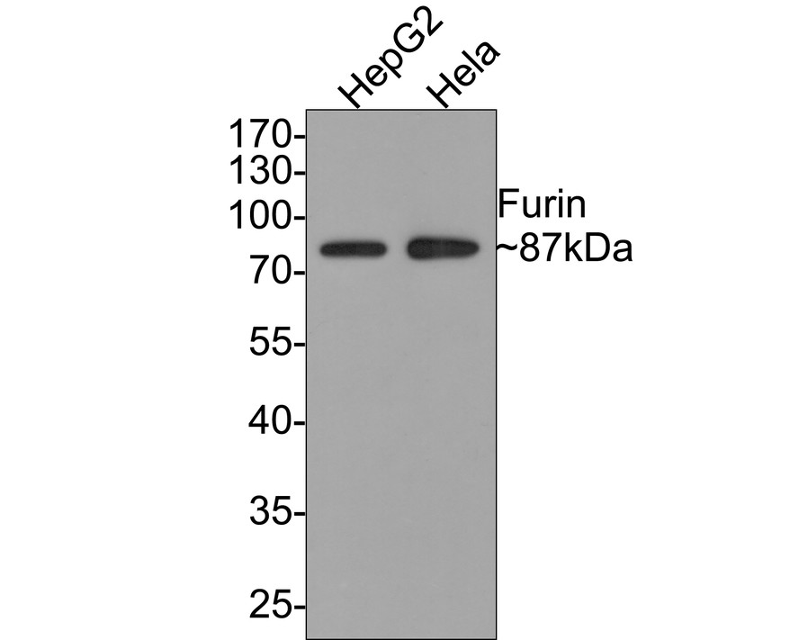
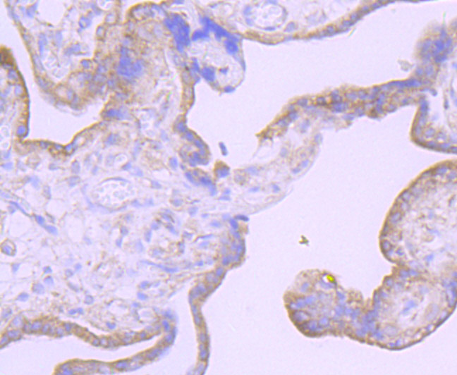
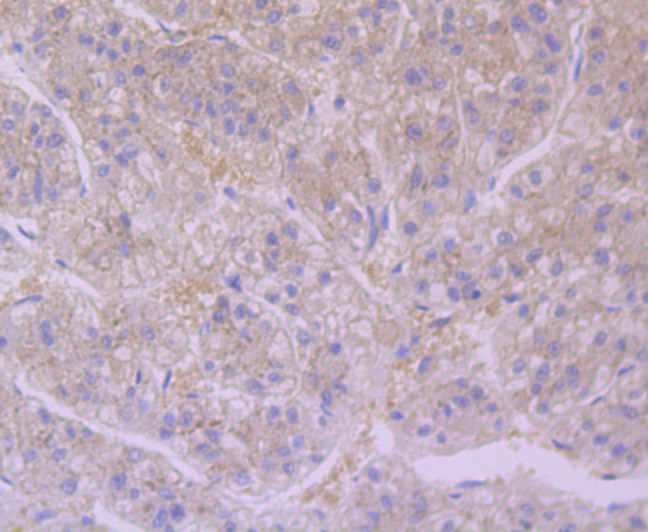
Target Protein: Furin Recombinant Rabbit mAb

IR: Immunogen Range:

Clonality:

Isotype: IgG

Entrez Gene: 5045

Swiss Prot: P09958

Source: KLH conjugated synthetic peptide derived from human Furin: 

Purification: affinity purified by Protein A

Storage: 0.01M TBS (pH7.4) with 1% BSA, 0.02% Proclin300 and 50% Glycerol. Shipped at 4℃. Store at -20℃ for one year. Avoid repeated freeze/thaw cycles.

Background: Furin is a calcium-dependent serine endoprotease that belongs to the subtilisin-like proprotein convertase family. The members of this family process latent precursor proteins into their biologically active products. Furin cleaves at paired basic amino acid processing sites within proparathyroid hormone, transforming growth factor β 1 precursor, proalbumin, pro-β-secretase, membrane type-1 matrix metalloproteinase, β subunit of pro-nerve growth factor and von Willebrand factor. Furin can directly cleave proMMP-2 within the ttrans-Golgi network leading to an inactive form of matrix metalloproteinase-2 (MMP-2). Furin is synthesized as an inactive zymogen that may minimize the occurrence of premature enzymatic activity that would lead to alternative protein activation or degradation. The inhibitory mechanism is based on the presence of an inactivating prosegment at the NH2 terminal of the Furin. After initial autocatalytic cleavage, the prosegment remains tightly associated until it reaches the trans-Golgi network where the dissociation of the prosegment and activation of furin occurs.

Size: 50ul

Concentration: 1mg/ml

Applications: WB=1:300-500,IHC-P=1:100-500,IHC-F=1:400-800,IF=1:100-500

Cross Reactive Species: Human,Mouse (predicted: Rat)

For research use only. Not intended for diagnostic or therapeutic use.

VALIDATION IMAGES
Western blot analysis of Furin on different lysates with Rabbit anti-Furin antibody (bsm-54283R) at 1/500 dilution. Lane 1: HepG2 cell lysate Lane 2: Hela cell lysate Lysates/proteins at 10 µg/Lane. Predicted band size: 87 kDa Observed band size: 87 kDa Exposure time: 1 minute; 10% SDS-PAGE gel.
Immunohistochemical analysis of paraffin-embedded human placenta tissue using anti-Furin antibody. The section was pre-treated using heat mediated antigen retrieval with Tris-EDTA buffer (pH 8.0-8.4) for 20 minutes.The tissues were blocked in 5% BSA for 30 minutes at room temperature, washed with ddH2O and PBS, and then probed with the primary antibody (ET7107-37, 1/50) for 30 minutes at room temperature. The detection was performed using an HRP conjugated compact polymer system. DAB was used as the chromogen. Tissues were counterstained with hematoxylin and mounted with DPX.
Immunohistochemical analysis of paraffin-embedded human colon tissue using anti-Furin antibody. The section was pre-treated using heat mediated antigen retrieval with Tris-EDTA buffer (pH 8.0-8.4) for 20 minutes.The tissues were blocked in 5% BSA for 30 minutes at room temperature, washed with ddH2O and PBS, and then probed with the primary antibody (bsm-54283R, 1/50) for 30 minutes at room temperature. The detection was performed using an HRP conjugated compact polymer system. DAB was used as the chromogen. Tissues were counterstained with hematoxylin and mounted with DPX.
Immunohistochemical analysis of paraffin-embedded mouse brain tissue using anti-Furin antibody. The section was pre-treated using heat mediated antigen retrieval with Tris-EDTA buffer (pH 8.0-8.4) for 20 minutes.The tissues were blocked in 5% BSA for 30 minutes at room temperature, washed with ddH2O and PBS, and then probed with the primary antibody (bsm-54283R, 1/50) for 30 minutes at room temperature. The detection was performed using an HRP conjugated compact polymer system. DAB was used as the chromogen. Tissues were counterstained with hematoxylin and mounted with DPX.
Immunohistochemical analysis of paraffin-embedded human liver tissue using anti-Furin antibody. The section was pre-treated using heat mediated antigen retrieval with Tris-EDTA buffer (pH 8.0-8.4) for 20 minutes.The tissues were blocked in 5% BSA for 30 minutes at room temperature, washed with ddH2O and PBS, and then probed with the primary antibody (bsm-54283R, 1/50) for 30 minutes at room temperature. The detection was performed using an HRP conjugated compact polymer system. DAB was used as the chromogen. Tissues were counterstained with hematoxylin and mounted with DPX.
 
PRODUCT SPECIFIC PUBLICATIONS