sales@bioss.com.cn
techsupport@bioss.com.cn
400-901-9800
Host: Rabbit
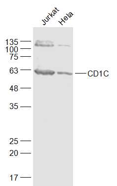
Target Protein: CD1C Rabbit pAb
IR: Immunogen Range:201-300/333
Clonality: Polyclonal
Isotype: IgG
Entrez Gene: 911
Source:
KLH conjugated synthetic peptide derived from human CD1C:201-300/333
Purification: affinity purified by Protein A
Storage: 0.01M TBS (pH7.4) with 1% BSA, 0.02% Proclin300 and 50% Glycerol. Shipped at 4℃. Store at -20℃ for one year. Avoid repeated freeze/thaw cycles.
Background: This gene encodes a member of the CD1 family of transmembrane glycoproteins, which are structurally related to the major histocompatibility complex (MHC) proteins and form heterodimers with beta-2-microglobulin. The CD1 proteins mediate the presentation of primarily lipid and glycolipid antigens of self or microbial origin to T cells. The human genome contains five CD1 family genes organized in a cluster on chromosome 1. The CD1 family members are thought to differ in their cellular localization and specificity for particular lipid ligands. The protein encoded by this gene is broadly distributed throughout the endocytic system via a tyrosine-based motif in the cytoplasmic tail. Alternatively spliced transcript variants of this gene have been observed, but their full-length nature is not known. [provided by RefSeq, Jul 2008]
Size: 200ul
Concentration: 1mg/ml
Applications: WB=1:500-2000,Flow-Cyt=1ug/Test
Cross Reactive Species: Human
For research use only. Not intended for diagnostic or therapeutic use.