bsm-33140M [Primary Antibody]
Mouse  Anti-EGFR Mouse mAb  Monoclonal Antibody
www.bioss.com.cn
sales@bioss.com.cn
techsupport@bioss.com.cn
400-901-9800
DATASHEET

Host: Mouse

Target Protein: EGFR Mouse mAb

IR: Immunogen Range:

Clonality: Monoclonal

Isotype: IgG

Entrez Gene: 1956

Swiss Prot: P00533

Source: KLH conjugated synthetic peptide derived from human EGFR: 

Purification: affinity purified by Protein G

Storage: 0.01M TBS (pH7.4) with 1% BSA, 0.02% Proclin300 and 50% Glycerol. Shipped at 4℃. Store at -20℃ for one year. Avoid repeated freeze/thaw cycles.

Background: The protein encoded by this gene is a transmembrane glycoprotein that is a member of the protein kinase superfamily. This protein is a receptor for members of the epidermal growth factor family. EGFR is a cell surface protein that binds to epidermal growth factor. Binding of the protein to a ligand induces receptor dimerization and tyrosine autophosphorylation and leads to cell proliferation. Mutations in this gene are associated with lung cancer. Multiple alternatively spliced transcript variants that encode different protein isoforms have been found for this gene. [provided by RefSeq, Jul 2010]

Size: 200ul

Concentration: 1mg/ml

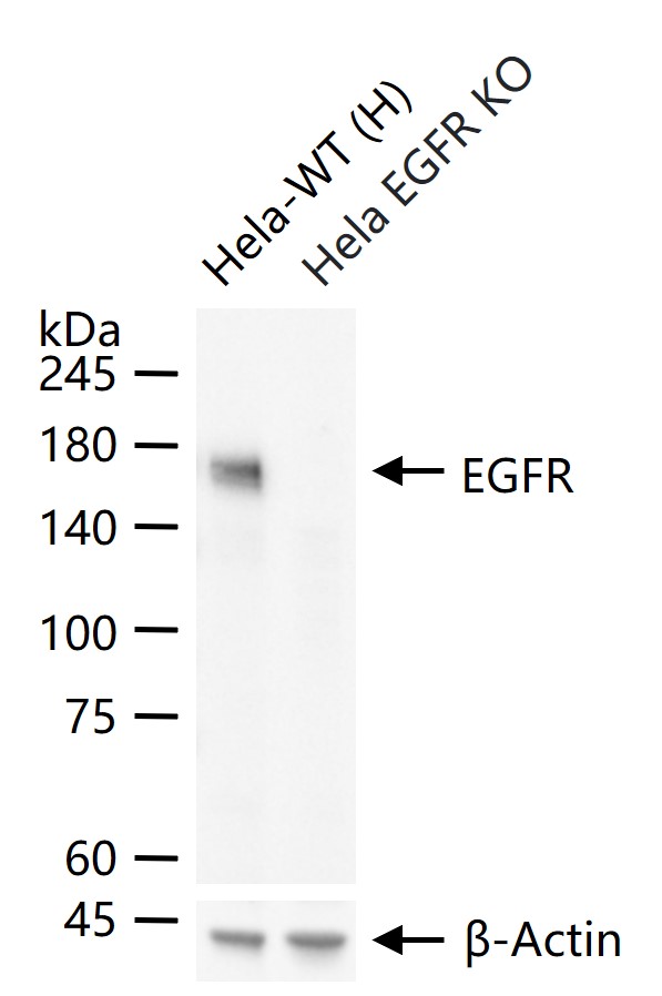
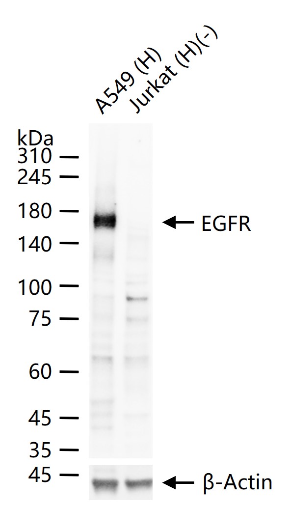
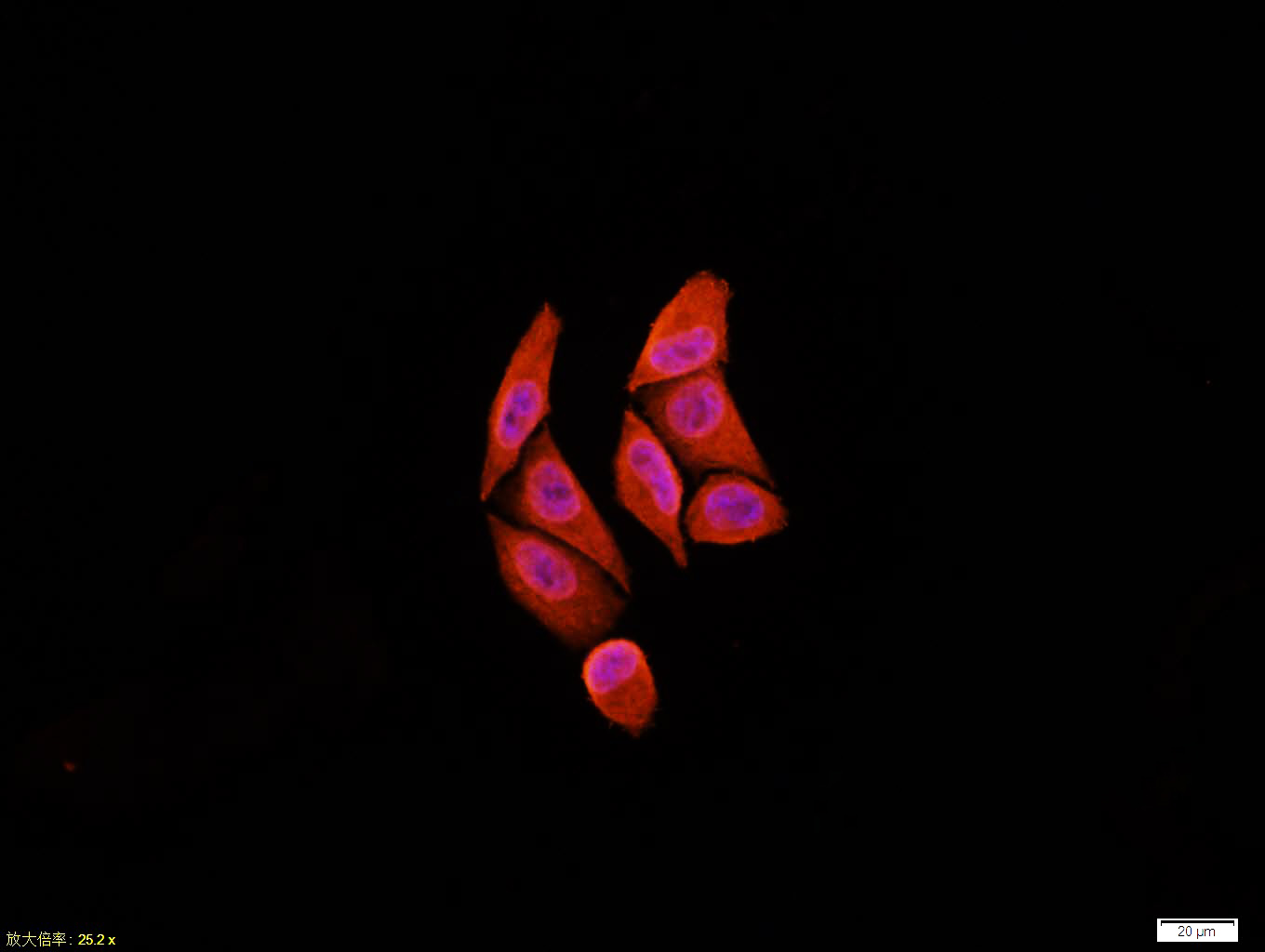
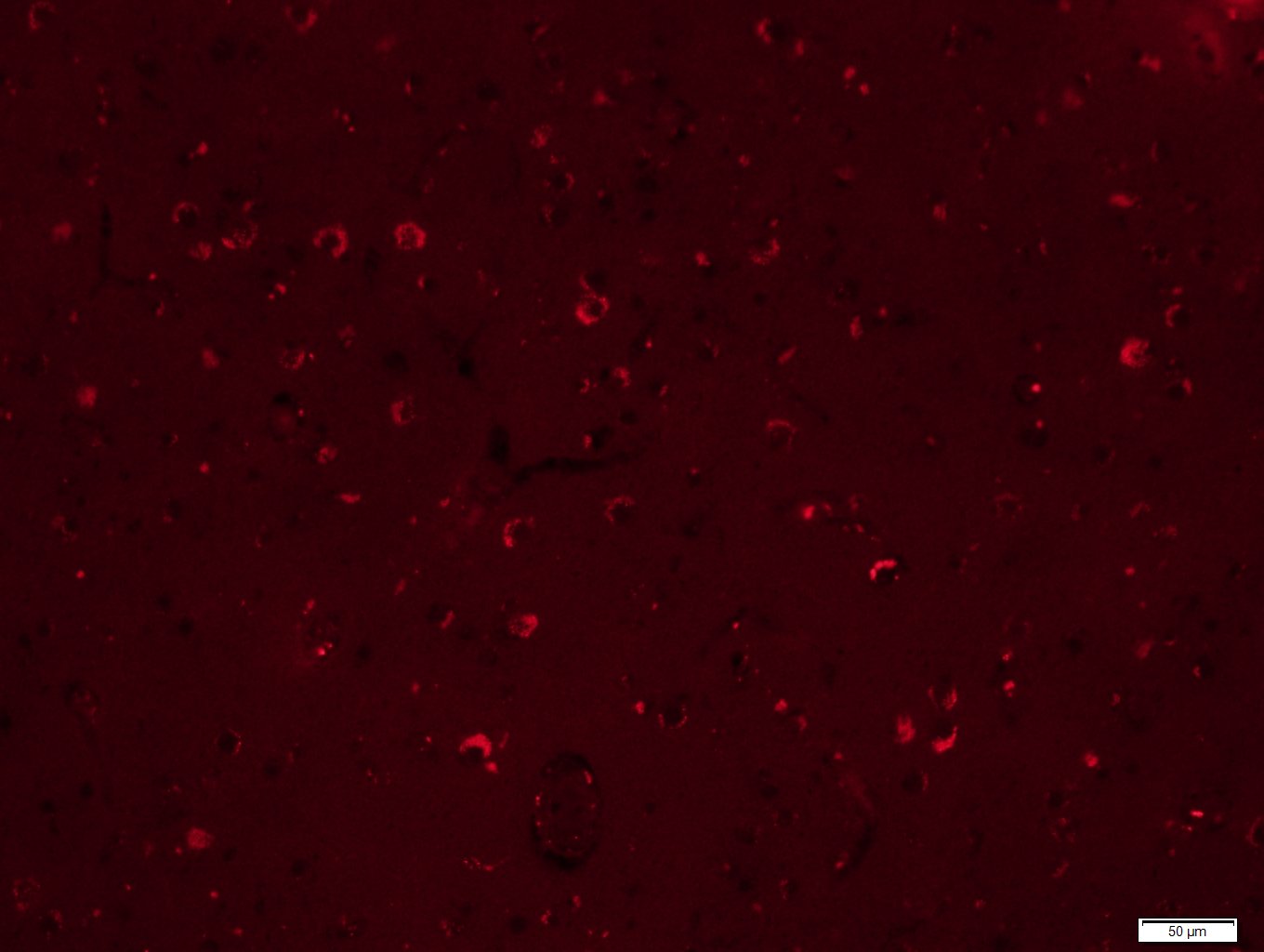
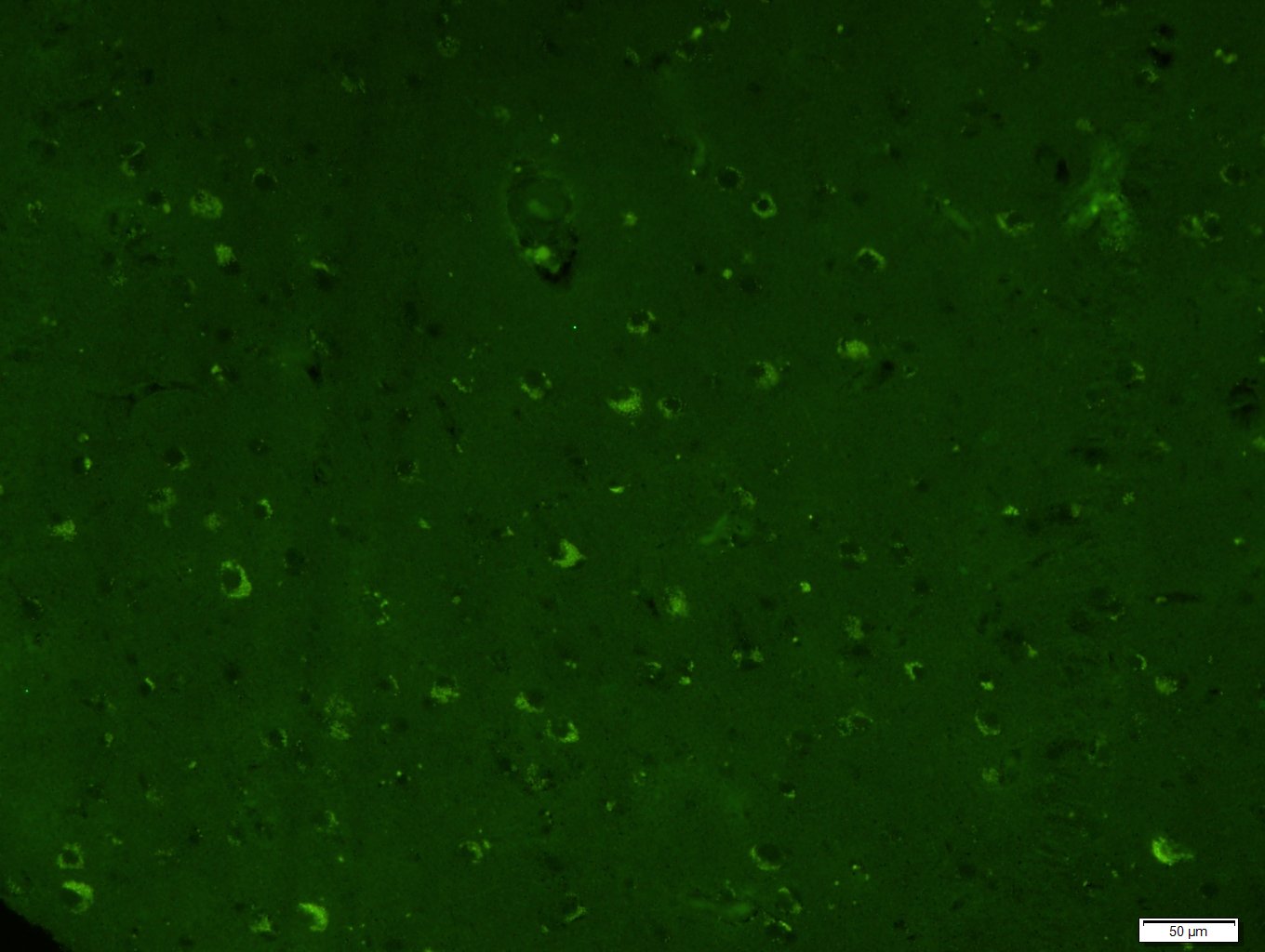
Applications: WB=1:1000-5000,IHC-P=1:100-500,IHC-F=1:100-500,IF=1:100-500,ICC/IF=1:50-200

Cross Reactive Species: Human

For research use only. Not intended for diagnostic or therapeutic use.

VALIDATION IMAGES
25 ug total protein per lane of various lysates (see on figure) probed with EGFR monoclonal antibody, unconjugated (bsm-33140M) at 1:1000 dilution and 4°C overnight incubation. Followed by conjugated secondary antibody incubation at r.t. for 60 min.
Tissue/cell:Hela cell; 4% Paraformaldehyde-fixed; Triton X-100 at room temperature for 20 min; Blocking buffer (normal goat serum,C-0005) at 37°C for 20 min; Antibody incubation with (EGFR) monoclonal Antibody, Unconjugated (bsm-33140M) 1:100, 90 minutes at 37°C; followed by a CY3 conjugated Goat Anti-Mouse IgG antibody at 37°C for 90 minutes, DAPI (blue, C02-04002) was used to stain the cell nuclei.
Paraformaldehyde-fixed, paraffin embedded (Human brain glioma); Antigen retrieval by boiling in sodium citrate buffer (pH6.0) for 15min; Block endogenous peroxidase by 3% hydrogen peroxide for 20 minutes; Blocking buffer (normal goat serum) at 37°C for 30min; Antibody incubation with (EGFR) Monoclonal Antibody, Unconjugated (bsm-33140M) at 1:400 overnight at 4°C, followed by a conjugated Goat Anti-Mouse IgG antibody (bs-0296G-CY5) for 90 minutes, and DAPI for nuclei staining.
Paraformaldehyde-fixed, paraffin embedded (Human brain glioma); Antigen retrieval by boiling in sodium citrate buffer (pH6.0) for 15min; Block endogenous peroxidase by 3% hydrogen peroxide for 20 minutes; Blocking buffer (normal goat serum) at 37°C for 30min; Antibody incubation with (EGFR) Monoclonal Antibody, Unconjugated (bsm-33140M) at 1:400 overnight at 4°C, followed by a conjugated Goat Anti-Mouse IgG antibody (bs-0296G-FITC) for 90 minutes, and DAPI for nuclei staining.
 
PRODUCT SPECIFIC PUBLICATIONS