sales@bioss.com.cn
techsupport@bioss.com.cn
400-901-9800
Host: Rabbit
Target Protein: NET1 Rabbit pAb
IR: Immunogen Range:151-250/596
Clonality: Polyclonal
Isotype: IgG
Entrez Gene: 10276
Swiss Prot: Q7Z628
Source: KLH conjugated synthetic peptide derived from human NET1:151-250/596
Purification: affinity purified by Protein A
Storage: 0.01M TBS (pH7.4) with 1% BSA, 0.02% Proclin300 and 50% Glycerol. Shipped at 4℃. Store at -20℃ for one year. Avoid repeated freeze/thaw cycles.
Background: Catecholamine, a term used for the hormone adrenaline and its sequentially hydroxylated form noradrenaline, is involved in fight or flight responses. Noradrenaline is released from the post ganglionic sympathetic nerve endings and exerts its effects locally in the immediate vicinity of its release. In the CNS, noradrenaline is involved in a number of physiological responses including mood, sleep regulation, alertness and arousal, both cognitive and non-cognitive expression of behaviors, and control of the endocrine and autonomic nervous systems. Peripherally, noradrenaline is present in sympathetic nerve endings and has full control of the sympathetic nervous system. Noradrenaline released from pre-synaptic nerve terminals is reabsorbed (70-90%) by noradrenaline transporters and its biological effects are terminated. The noradrenaline transport via noradrenaline transporters is an active, Na+/Cl- dependent transport process mediated by noradrenaline transporters. Noradrenaline transporters constitute the primary mechanism for inactivation of synaptically released noradrenaline, are targets for multiple antidepressants and psychostimulants, and are deficient in affective and autonomic disorders. In rat brain, noradrenaline transporter is expressed in noradrenergic neuronal somata, axons and dendrites, and hippocampus and cortex, but is absent from epinephrine- and dopamine-containing neurons. At least 13 genetic variations have been reported in the noradrenaline transporter protein that affect noradrenaline re-uptake and concentrations in cerebrospinal fluid in humans. The association between these genetic variations in noradrenaline transporters and several psychiatric and cardiovascular disorders is just emerging. Recently, a single amino acid mutation (hNET-A457P) showed deficiency in noradrenaline transport in an orthostatic intolerance patient. Noradrenaline transporter protein consists of 617 amino acids and has 12 trans-membrane domains, a characteristic feature of many membrane associated solute transporters.
Size: 50ul
Concentration: 1mg/ml
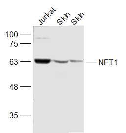
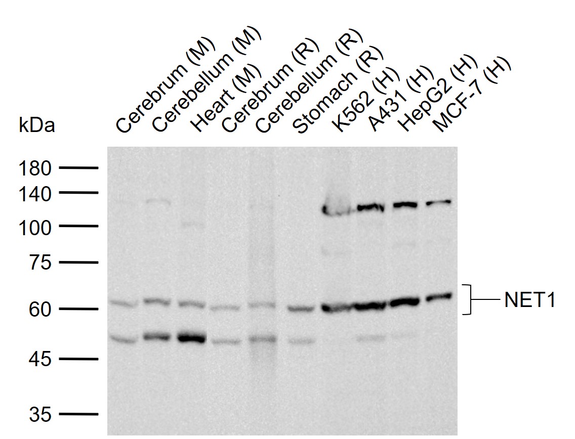
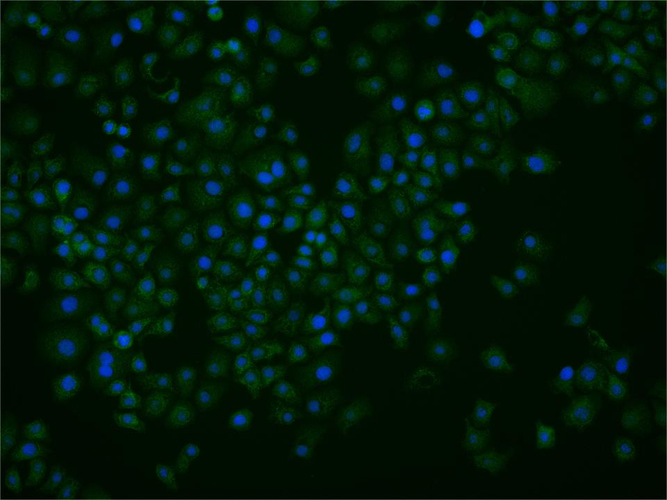
Applications: WB=1:500-2000,IHC-P=1:100-500,IHC-F=1:100-500,IF=1:100-500,ICC/IF=1:25
Cross Reactive Species: Human,Mouse,Rat (predicted: Rabbit,Pig,Sheep,Cow,Chicken,Dog,Horse)
For research use only. Not intended for diagnostic or therapeutic use.