bs-1926R [Primary Antibody]
Rabbit  Anti-UCP-2 Rabbit pAb  Polyclonal Antibody
www.bioss.com.cn
sales@bioss.com.cn
techsupport@bioss.com.cn
400-901-9800
DATASHEET

Host: Rabbit

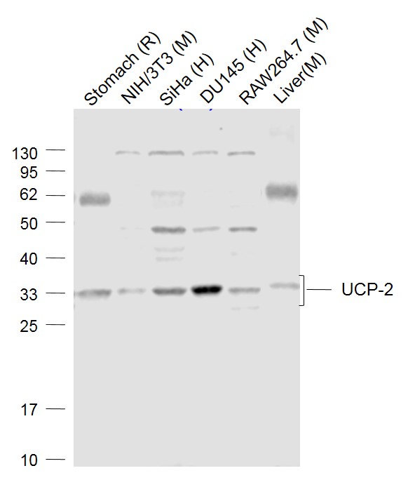
Target Protein: UCP-2 Rabbit pAb

IR: Immunogen Range:201-309/309

Clonality: Polyclonal

Isotype: IgG

Entrez Gene:

Swiss Prot:

Source: KLH conjugated synthetic peptide derived from mouse UCP-2:201-309/309 

Purification: affinity purified by Protein A

Storage: 0.01M TBS (pH7.4) with 1% BSA, 0.02% Proclin300 and 50% Glycerol. Shipped at 4℃. Store at -20℃ for one year. Avoid repeated freeze/thaw cycles.

Background: UCPs facilitate the transfer of anions from the inner to the outer mitochondrial membrane and the return transfer of protons from the outer to the inner mitochondrial membrane. They also reduce the mitochondrial membrane potential in mammalian cells. UCP2 gene is expressed in many tissues, with the greatest expression in skeletal muscle. UCP2 is thought to play a role in non shivering thermogenesis, obesity and diabetes.

Size: 200ul

Concentration: 1mg/ml

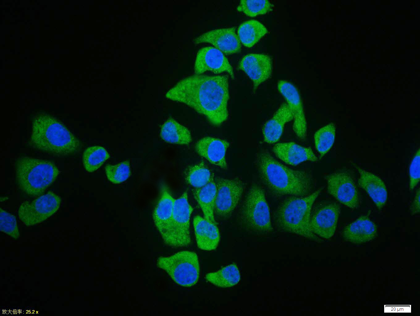
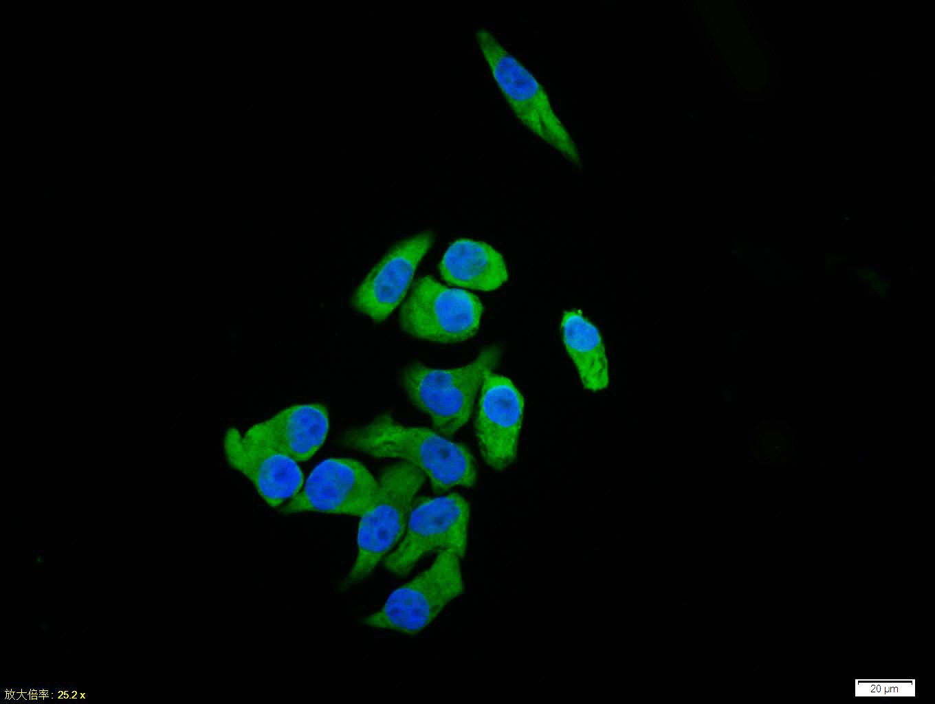
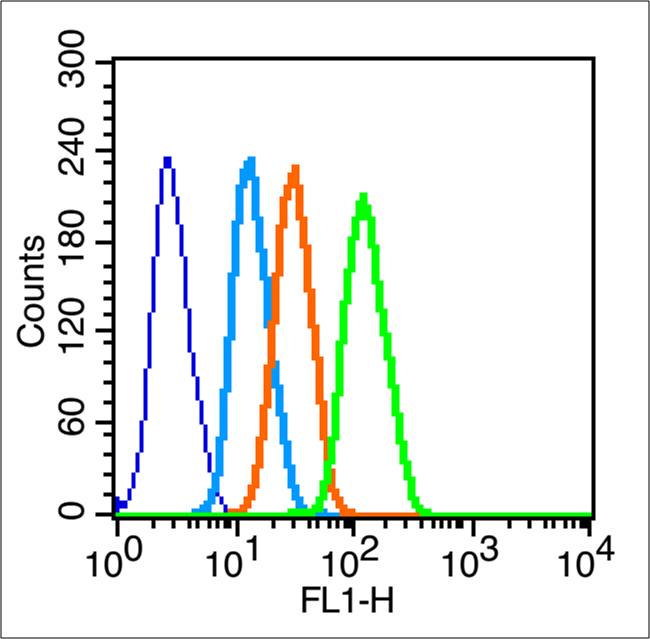
Applications: WB=1:500-2000,IHC-P=1:100-500,IHC-F=1:100-500,IF=1:100-500,Flow-Cyt=1μg /test,ICC/IF=1:100

Cross Reactive Species: Human,Mouse,Rat (predicted: Rabbit,Pig,Horse)

For research use only. Not intended for diagnostic or therapeutic use.

VALIDATION IMAGES
Sample: Lane 1: Stomach (Rat) Lysate at 40 ug Lane 2: NIH/3T3 (Mouse) Cell Lysate at 30 ug Lane 3: SiHa (Human) Cell Lysate at 30 ug Lane 4: DU145 (Human) Cell Lysate at 30 ug Lane 5: RAW264.7 (Mouse) Cell Lysate at 30 ug Lane 6: Liver (Mouse) Lysate at 40 ug Primary: Anti-UCP-2 (bs-1926R) at 1/1000 dilution Secondary: IRDye800CW Goat Anti-Rabbit IgG at 1/20000 dilution Predicted band size: 33 kD Observed band size: 33 kD
Paraformaldehyde-fixed, paraffin embedded (Mouse brain); Antigen retrieval by boiling in sodium citrate buffer (pH6.0) for 15min; Block endogenous peroxidase by 3% hydrogen peroxide for 20 minutes; Blocking buffer (normal goat serum) at 37°C for 30min; Antibody incubation with (UCP-2) Polyclonal Antibody, Unconjugated (bs-1926R) at 1:400 overnight at 4°C, followed by operating according to SP Kit(Rabbit) (sp-0023) instructionsand DAB staining.
Hela cell; 4% Paraformaldehyde-fixed; Triton X-100 at room temperature for 20 min; Blocking buffer (normal goat serum, C-0005) at 37°C for 20 min; Antibody incubation with (UCP-2) polyclonal Antibody, Unconjugated (bs-1926R) 1:100, 90 minutes at 37°C; followed by a conjugated Goat Anti-Rabbit IgG antibody at 37°C for 90 minutes, DAPI (blue, C02-04002) was used to stain the cell nuclei.
Blank control (blue line): Hela (blue). Primary Antibody (green line): Rabbit Anti-UCP-2 antibody (bs-1926R) Dilution: 1μg /10^6 cells; Isotype Control Antibody (orange line): Rabbit IgG . Secondary Antibody (white blue line): F(ab’)2 fragment goat anti-rabbit IgG-FITC Dilution: 1μg /test. Protocol The cells were fixed with 2% paraformaldehyde (10 min) , then permeabilized with 90% ice-cold methanol for 30 min on ice.Cells stained with Primary Antibody for 30 min at room temperature. The cells were then incubated in 1 X PBS/2%BSA/10% goat serum to block non-specific protein-protein interactions followed by the antibody for 15 min at room temperature. The secondary antibody used for 40 min at room temperature. Acquisition of 20,000 events was performed.
 
PRODUCT SPECIFIC PUBLICATIONS