bs-1919R [Primary Antibody]
Rabbit  Anti-TLR1 Rabbit pAb  Polyclonal Antibody
www.bioss.com.cn
sales@bioss.com.cn
techsupport@bioss.com.cn
400-901-9800
DATASHEET

Host: Rabbit

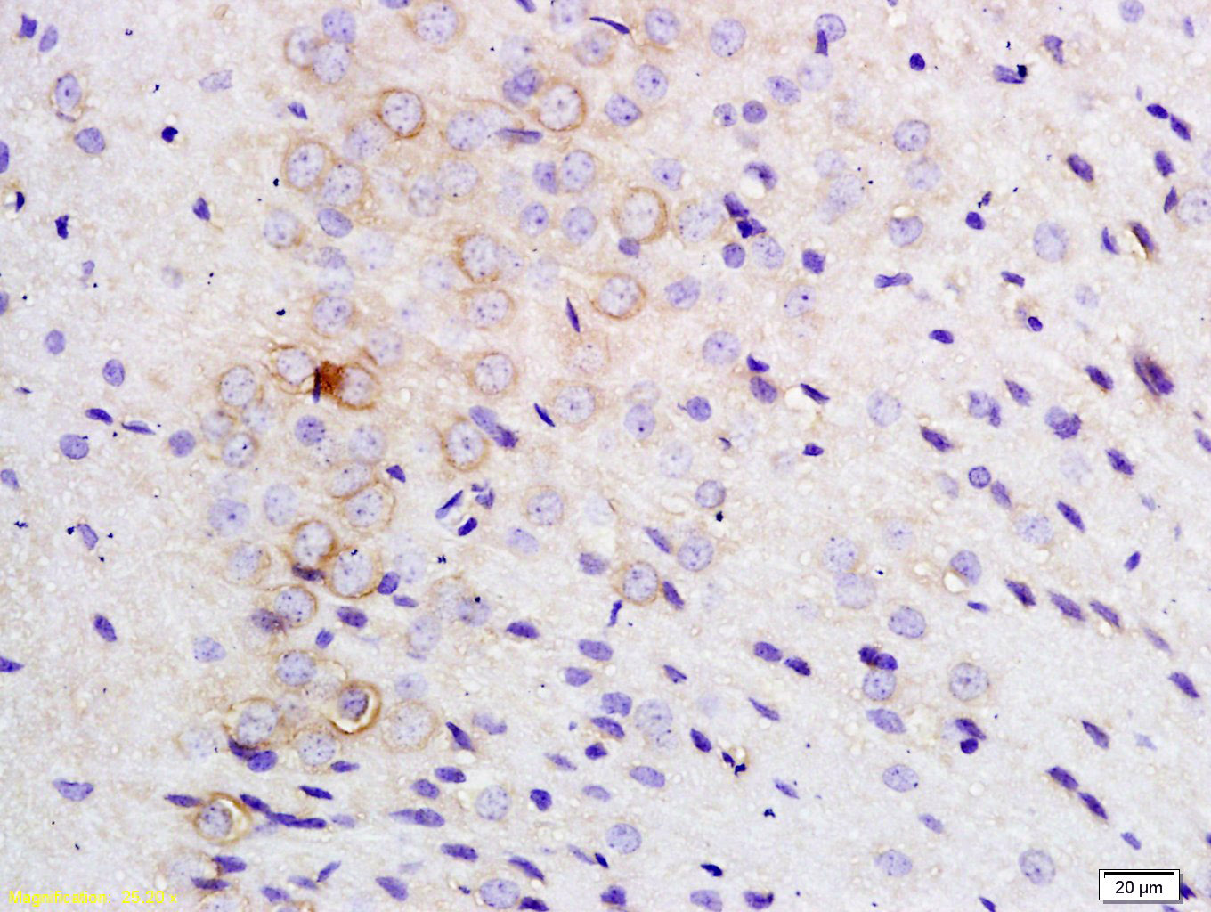
Target Protein: TLR1 Rabbit pAb

IR: Immunogen Range:101-200/786

Clonality: Polyclonal

Isotype: IgG

Entrez Gene: 7096

Swiss Prot: Q15399

Source: KLH conjugated synthetic peptide derived from human TLR1:101-200/786 

Purification: affinity purified by Protein A

Storage: 0.01M TBS (pH7.4) with 1% BSA, 0.02% Proclin300 and 50% Glycerol. Shipped at 4℃. Store at -20℃ for one year. Avoid repeated freeze/thaw cycles.

Background: TLR1 is a member of the Toll like receptor (TLR) family which plays a fundamental role in pathogen recognition and activation of innate immunity. TLRs are highly conserved from Drosophila to humans and share structural and functional similarities. They recognize pathogen associated molecular patterns (PAMPs) that are expressed on infectious agents, and mediate the production of cytokines necessary for the development of effective immunity. The various TLRs exhibit different patterns of expression. The gene encoding TLR1 is ubiquitously expressed, and at higher levels than other TLR genes. Different length transcripts presumably resulting from use of alternative polyadenylation site, and/or from alternative splicing, have been noted for this gene.

Size: 100ul

Concentration: 1mg/ml

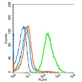
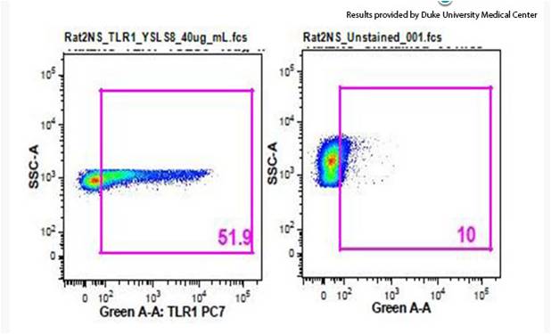
Applications: IHC-P=1:100-500,IHC-F=1:100-500,IF=1:100-500,Flow-Cyt=1μg /test

Cross Reactive Species: Human,Rat,Arctic Ground (predicted: Mouse,Cow,Dog,Horse)

For research use only. Not intended for diagnostic or therapeutic use.

VALIDATION IMAGES
Tissue/cell: rat brain tissue; 4% Paraformaldehyde-fixed and paraffin-embedded; Antigen retrieval: citrate buffer ( 0.01M, pH 6.0 ), Boiling bathing for 15min; Block endogenous peroxidase by 3% Hydrogen peroxide for 30min; Blocking buffer (normal goat serum,C-0005) at 37℃ for 20 min; Incubation: Anti-TLR1 Polyclonal Antibody, Unconjugated(bs-1919R) 1:200, overnight at 4°C, followed by conjugation to the secondary antibody(SP-0023) and DAB(C-0010) staining
Blank control: Raji(blue). Primary Antibody:Rabbit Anti-TLR1 antibody(bs-1919R), Dilution: 1μg in 100 μL 1X PBS containing 0.5% BSA; Isotype Control Antibody: Rabbit IgG(orange) ,used under the same conditions ); Secondary Antibody: Goat anti-rabbit IgG-PE(white blue), Dilution: 1:200 in 1 X PBS containing 0.5% BSA. Protocol The cells were fixed with 2% paraformaldehyde (10 min) and then permeabilized with ice-cold 90% methanol for 30 min on ice.The cells were washed twice with phosphate-buffered saline (PBS).The cells were then incubated in 1 X PBS containing 0.5% BSA + 1 0% goat serum (15 min) to block non-specific protein-protein interactions followed by the antibody (bs-1919R, 1μg /1x10^6 cells) for 30 min on ice. The secondary antibody used was Goat Anti-rabbit IgG/PE antibody at 1/200 dilution for 30 min on ice. Acquisition of 20,000 events was performed.
Arctic Ground Squirrel splenocytes stained with Anti-TLR1 Polyclonal Antibody, PE-CY7 Conjugated (bs-1919-PE-Cy7) at 1:25
 
PRODUCT SPECIFIC PUBLICATIONS