VALIDATION IMAGES
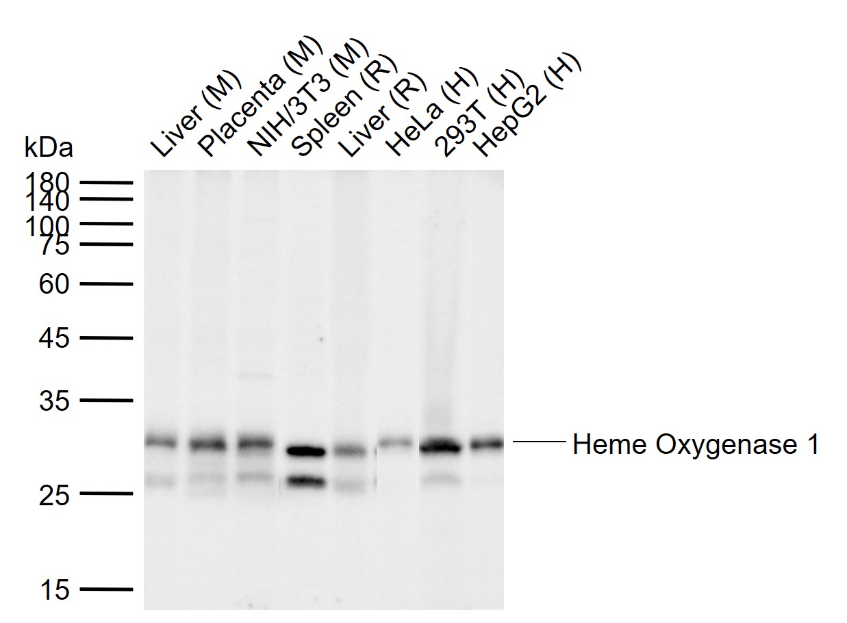
25 ug total protein per lane of various lysates (see on figure) probed with Heme Oxygenase 1/HMOX1 monoclonal antibody, unconjugated (bsm-60751R) at 1:1000 dilution and 4°C overnight incubation. Followed by conjugated secondary antibody incubation at r.t. for 60 min.
Sample:
Lane 1: Mouse Liver tissue lysates
Lane 2: Mouse Placenta tissue lysates
Lane 3: Mouse NIH/3T3 cell lysates
Lane 4: Rat Spleen tissue lysates
Lane 5: Rat Liver tissue lysates
Lane 6: Human HeLa cell lysates
Lane 7: Human 293T cell lysates
Lane 8: Human HepG2 cell lysates
Primary: Anti-Heme Oxygenase 1 (bsm-60751R) at 1/10000 dilution
Secondary: IRDye800CW Goat Anti-Rabbit IgG at 1/20000 dilution
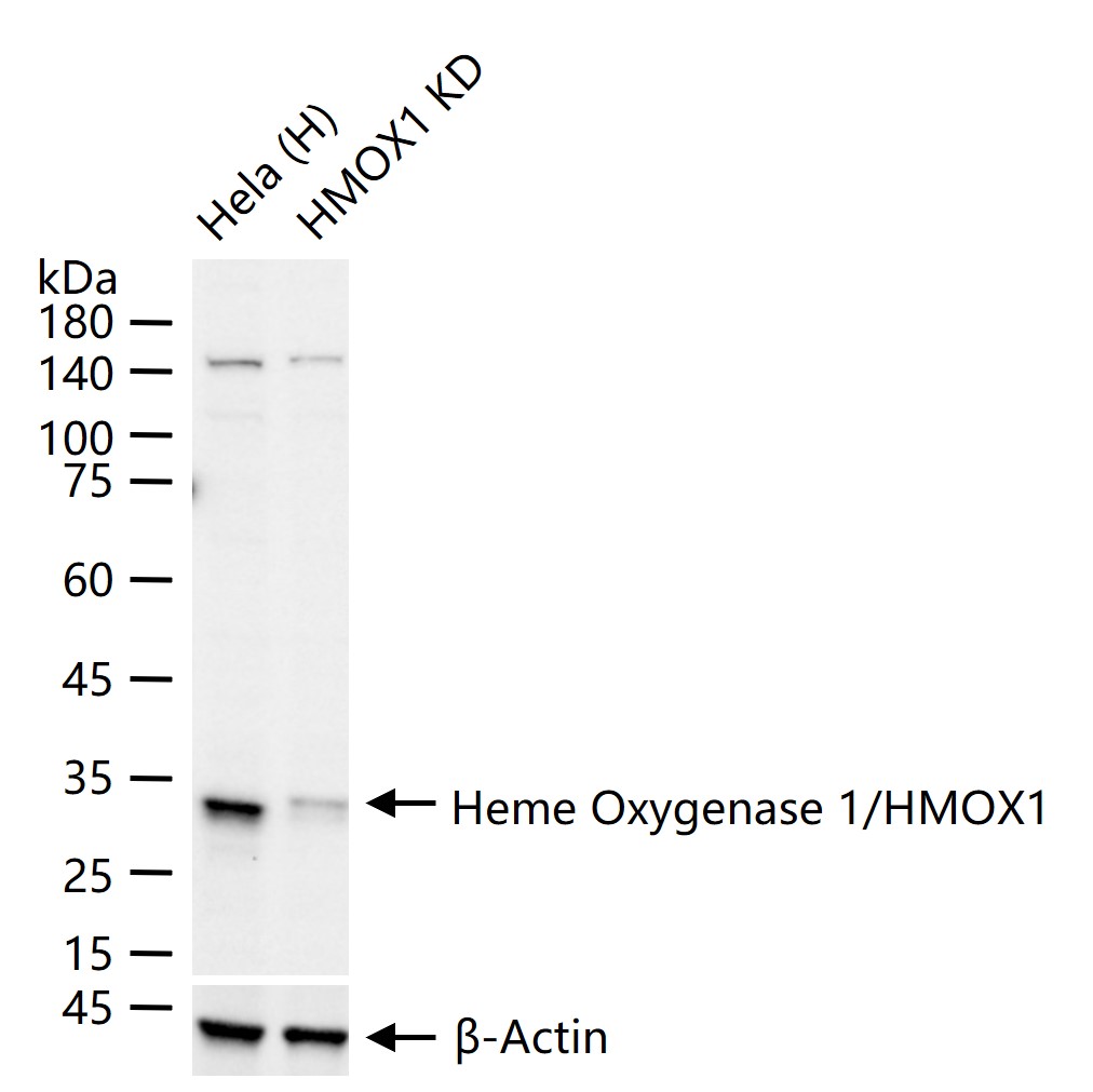
Predicted band size: 32 kDa
Observed band size: 30 kDa
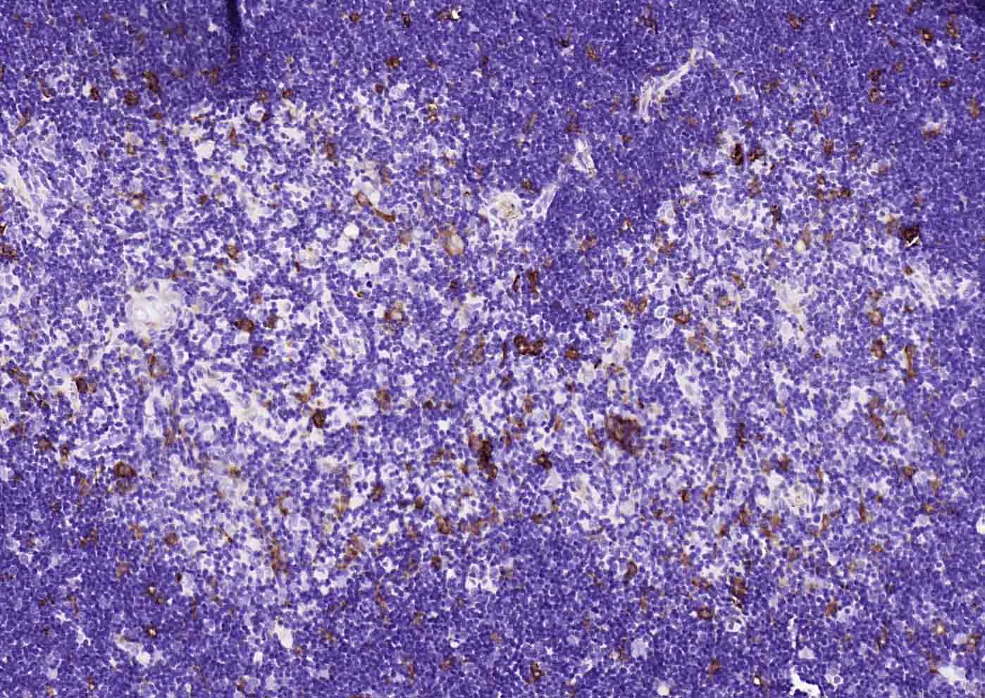
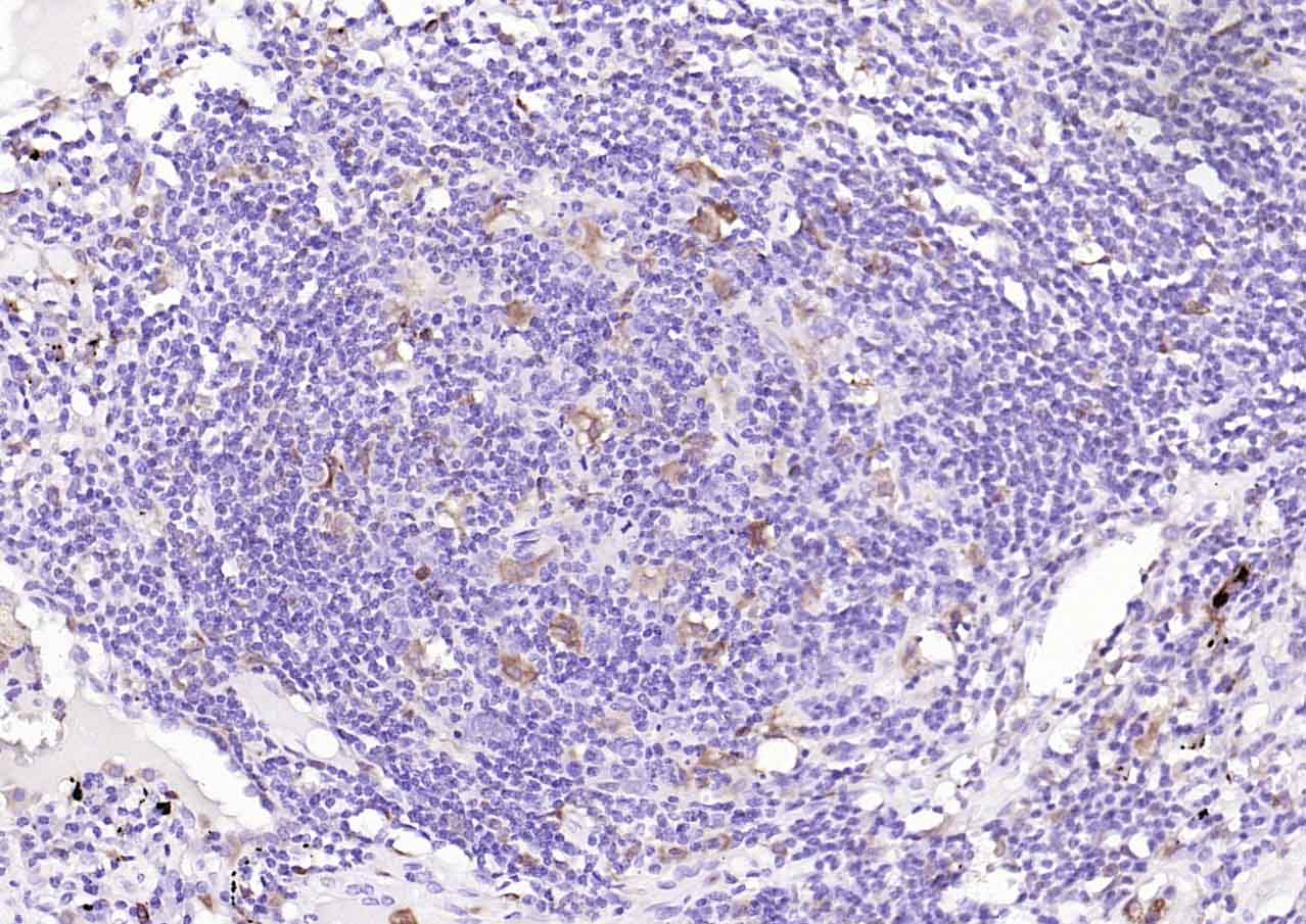
Paraformaldehyde-fixed, paraffin embedded (mouse thymus); Antigen retrieval by boiling in sodium citrate buffer (pH6.0) for 15min; Block endogenous peroxidase by 3% hydrogen peroxide for 20 minutes; Blocking buffer (normal goat serum) at 37°C for 30min; Incubation with (Heme Oxygenase 1) Monoclonal Antibody, Unconjugated (bsm-60751R) at 1:1000 overnight at 4°C, followed by operating according to SP Kit(Rabbit) (sp-0023) instructionsand DAB staining.
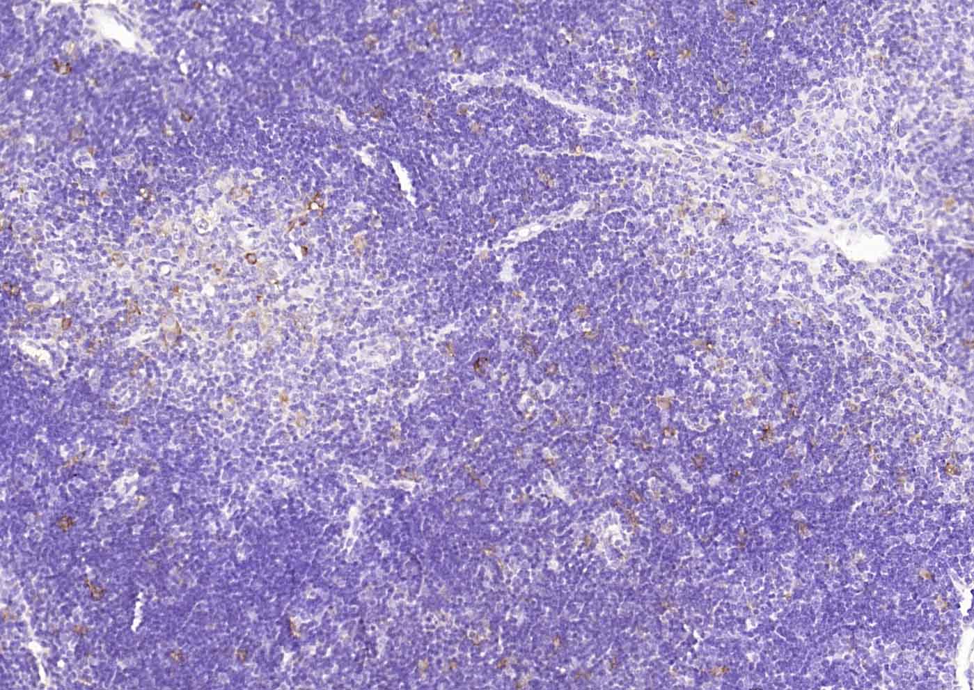
Paraformaldehyde-fixed, paraffin embedded (human spleen); Antigen retrieval by boiling in sodium citrate buffer (pH6.0) for 15min; Block endogenous peroxidase by 3% hydrogen peroxide for 20 minutes; Blocking buffer (normal goat serum) at 37°C for 30min; Incubation with (Heme Oxygenase 1) Monoclonal Antibody, Unconjugated (bsm-60751R) at 1:1000 overnight at 4°C, followed by operating according to SP Kit(Rabbit) (sp-0023) instructionsand DAB staining.
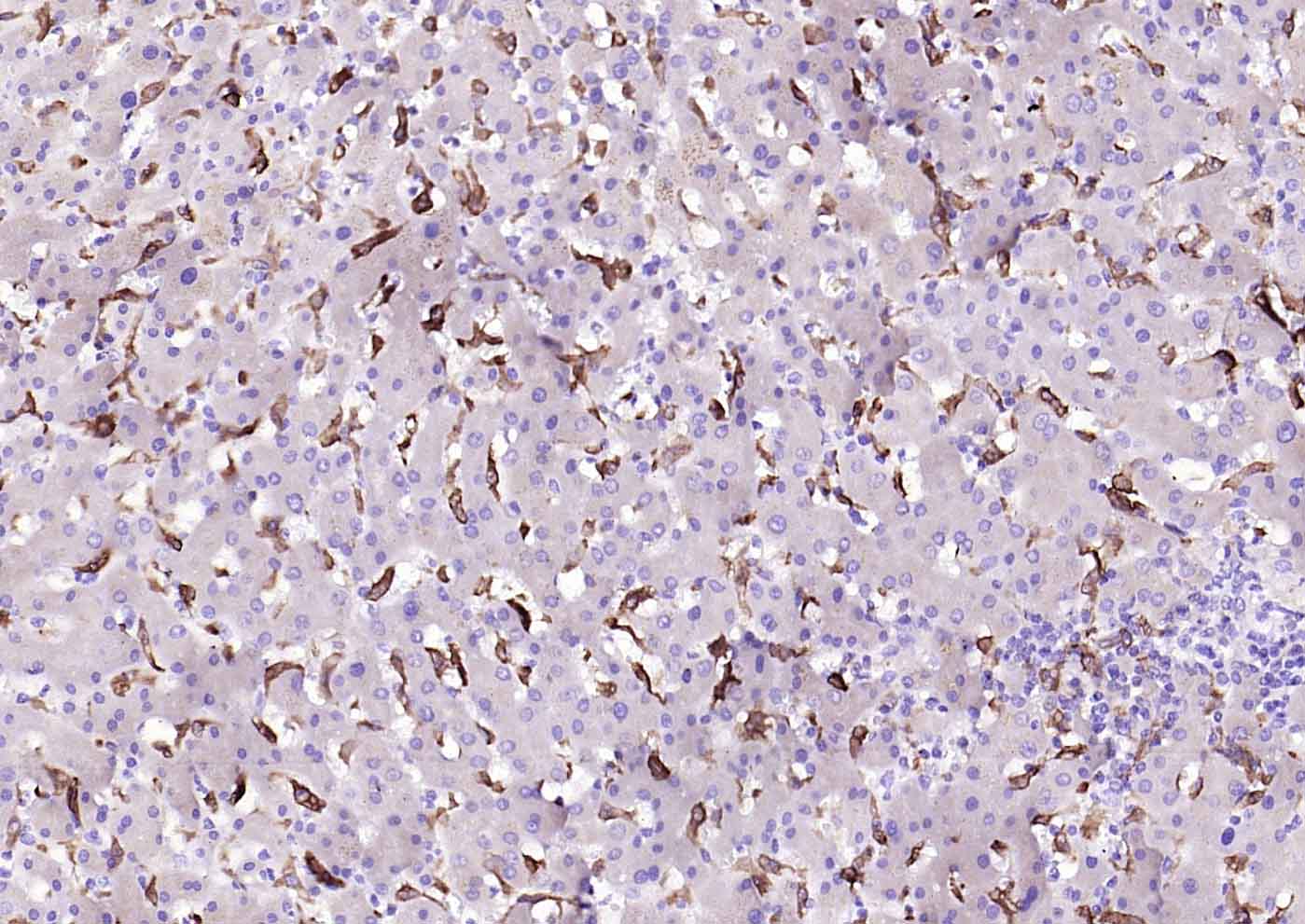
Paraformaldehyde-fixed, paraffin embedded (human liver); Antigen retrieval by boiling in sodium citrate buffer (pH6.0) for 15min; Block endogenous peroxidase by 3% hydrogen peroxide for 20 minutes; Blocking buffer (normal goat serum) at 37°C for 30min; Incubation with (Heme Oxygenase 1) Monoclonal Antibody, Unconjugated (bsm-60751R) at 1:1000 overnight at 4°C, followed by operating according to SP Kit(Rabbit) (sp-0023) instructionsand DAB staining.
Paraformaldehyde-fixed, paraffin embedded (human lung carcinoma); Antigen retrieval by boiling in sodium citrate buffer (pH6.0) for 15min; Block endogenous peroxidase by 3% hydrogen peroxide for 20 minutes; Blocking buffer (normal goat serum) at 37°C for 30min; Incubation with (Heme Oxygenase 1) Monoclonal Antibody, Unconjugated (bsm-60751R) at 1:1000 overnight at 4°C, followed by operating according to SP Kit(Rabbit) (sp-0023) instructionsand DAB staining.
Paraformaldehyde-fixed, paraffin embedded (rat thymus); Antigen retrieval by boiling in sodium citrate buffer (pH6.0) for 15min; Block endogenous peroxidase by 3% hydrogen peroxide for 20 minutes; Blocking buffer (normal goat serum) at 37°C for 30min; Incubation with (Heme Oxygenase 1) Monoclonal Antibody, Unconjugated (bsm-60751R) at 1:1000 overnight at 4°C, followed by operating according to SP Kit(Rabbit) (sp-0023) instructionsand DAB staining.
Paraformaldehyde-fixed, paraffin embedded (rat spleen); Antigen retrieval by boiling in sodium citrate buffer (pH6.0) for 15min; Block endogenous peroxidase by 3% hydrogen peroxide for 20 minutes; Blocking buffer (normal goat serum) at 37°C for 30min; Incubation with (Heme Oxygenase 1) Monoclonal Antibody, Unconjugated (bsm-60751R) at 1:1000 overnight at 4°C, followed by operating according to SP Kit(Rabbit) (sp-0023) instructionsand DAB staining.